Özet
VP şantın distal kateteri periton boşluğuna uzanır; distal kateter migrasyonu, perforasyon ve BOS psödokisti abdominal komplikasyonlar arasındadır. Abdominal BT, kateterin seyrini, bağırsak tutulumunu, serbest havayı ve eşlik eden koleksiyonları göstererek şant komplikasyonlarının değerlendirilmesine yardımcı olur. Abdominal BT, kateterin seyrini, bağırsak tutulumunu, serbest havayı ve eşlik eden koleksiyonları değerlendirmeye yardımcı olur. Bağırsak perforasyonu peritonit gibi ciddi enfeksiyöz komplikasyonlara yol açabileceğinden, BT’de kateterin sonlanma yeri ve bağırsakla ilişkisinin belirlenmesi önemlidir.
Faz ve pencere
- Kontrastsız tanısal
- Abdominal BT'de kateterin seyri ve bağırsak tutulumu, serbest hava ve eşlik eden koleksiyonlar değerlendirilir.
- Portal tanısal
- Abdominal BT'de kateterle ilişkili koleksiyonlar ve bağırsak tutulumu değerlendirilir.
Önerilen pencereler G genişlik, M merkez; değerler Hounsfield birimi (HU)
- BatınG 400 / M +50
- Yumuşak dokuG 400 / M +40
BT bulguları
- Distal kateterin rektus kası veya karın duvarı içinde sonlanması, ya da beklenen peritoneal boşluğa ulaşmadan geri çekilmesi malpozisyon/migrasyon lehinedir.
- Kateter ucunu merkez alan, sınırlı sıvı kavitesi (BOS psödokisti) veya kapsüllü koleksiyon görüntülenir; bu yapılar şant disfonksiyonuna neden olabilir.
- Kateterin bağırsak duvarını geçtiğinin ve lümen içinde devam ettiğinin farklı düzlemlerde izlenmesi visseral erozyon/perforasyon için doğrudan bulgudur.
- Kateterin bağırsak duvarını geçerek lümen içinde seyretmesi gastrointestinal perforasyonu destekler.
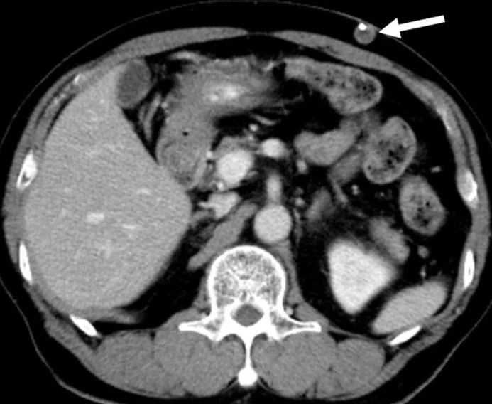
- Karın duvarında kateterin cilt dışına çıkması ya da cilt altı planda sonlanması eksternal migrasyondur; aynı alanda sıvı veya hava eşlik edebilir.
- Kateterin bağırsak ansı veya mezenter etrafında halka oluşturması, geçiş noktasındaki ans kümelenmesi ve proksimal dilatasyon mekanik obstrüksiyon/volvulus olasılığını düşündürür.
- Kateter yakınındaki sıvı koleksiyonu ve inflamatuvar değişiklikler abdominal komplikasyon kuşkusu doğurabilir; bu bulgular tek başına enfeksiyonu kanıtlamaz.
- Kronik kolon perforasyonu serbest intraperitoneal hava olmadan görülebilir; bir olguda BT’de kronik perforasyon saptanmasına rağmen serbest hava izlenmemiştir.
- Distal kateter karın duvarı subkutan dokusuna, plevral boşluğa veya karaciğer içine migrate olabilir.
Normalde
Normalde distal kateter intraperitoneal boşlukta yerleşiktir; kateterin karın duvarına, subkutan dokuya veya bağırsak lümenine doğru migrasyonu veya ekstrüzyonu patolojik kabul edilir. Aksiyel, koronal ve sagital reformasyonlar kullanılarak kateterin intraperitoneal seyri, bağırsak ilişkisi ve mevcut komplikasyonlar değerlendirilir.
Normal BT ile kıyasla
Aynı bölgenin, kaynak veri setinde patoloji içermediği belirtilen bir BT incelemesini kesit kesit kaydırın; organ sınırlarını açarak anatomiyi hasta görüntüsüyle karşılaştırın. Küçük rastlantısal bulgular tümüyle dışlanmamıştır.
Kaynak: TotalSegmentator v2.01 veri seti (Wasserthal ve ark., Radiology: Artificial Intelligence 2023), CC BY 4.0, vaka s0119; organ sınırları veri setinin otomatik segmentasyonundan, birleştirilerek sadeleştirildi. Görüntüler 2,5 mm kesit aralığına yeniden örneklendi.
Ayırıcı tanı
- Peritoneal apse
- VP şantla ilişkili abdominal apseler BT’de görülebilir.
- Asit
- VP şantlı hastada BOS psödokisti asit tablosuyla ortaya çıkabilir; bu nedenle asit görünümü psödokisti dışlamaz.
- Adezyona bağlı ince bağırsak obstrüksiyonu
- geçiş noktasında ans kalibresi değişir; şant kateteriyle ilişki ancak kateterin ans veya mezenter etrafındaki seyriyle ayrıca gösterilirse düşünülür.
Tuzaklar
- Kateterin bir bağırsak ansına dayanması penetrasyon kanıtı değildir; duvarı geçtiği ve lümen içinde seyrettiği aksiyel görüntülerle birlikte reformasyonlarda ara.
- Az miktardaki serbest peritoneal sıvı distal ucu çevreleyebilir; psödokist tanımı için sınırlı kavite ve kateter ucuyla merkezî ilişki aranır.
- İnce duvarlı veya steril BOS psödokistinde çevre inflamasyonu belirgin olmayabilir; yalnız duvar görünümüne bakarak enfeksiyonu kesinleştirme.
- Tek bir abdomen görüntü kapsamı distal kateterin tüm seyrini göstermeyebilir; kapsam dışında kalan segmentler hakkında kesintisizlik sonucu çıkarma.
Kendini dene
Abdominopelvik BT’de şant kateteri ince bağırsak duvarını geçiyor ve lümen içinde devam ediyor. En olası tanı hangisidir?
Cevabı göster
Şant ilişkili bağırsak erozyonu. Kateterin duvarı geçip bağırsak lümeninde izlenmesi erozyon/perforasyon lehine doğrudan anatomik bulgudur. Kateterin bağırsak duvarını geçerek lümen içinde seyretmesi, bağırsak perforasyonu ve intraluminal migrasyonu destekler.
Pelviste distal şant ucunu çevreleyen, kapsüllü sıvı koleksiyonu görülüyor. En uygun adlandırma hangisidir?
Cevabı göster
Abdominal BOS psödokisti. Kavitenin şant ucuna merkezlenmesi BOS psödokistini destekler. Abdominal BOS psödokisti, şant ucunu çevreleyen kapsüllü sıvı koleksiyonu olarak tanımlanabilir.
İlgili konular
Kaynaklar
Bu sayfadaki 37 cümle ve 2 quiz sorusu aşağıdaki kaynaklardan alıntılarla otomatik olarak karşılaştırıldı; 23 cümle bu karşılaştırmada düzeltildi (2026-10-09).
- Delayed transanal extrusion of a ventriculoperitoneal shunt in an adult patient: A rare delayed gastrointestinal complication following post-hemorrhagic hydrocephaluspmc.ncbi.nlm.nih.gov
- A hybrid omental-parietal peritoneal fibrous sheath: a case report of an unreported cause of recurrent ventriculoperitoneal shunt failurepmc.ncbi.nlm.nih.gov
- Perforation of the gastrointestinal tract by a ventriculoperitoneal shunt catheter with anal exteriorization in an adult patient: case reportpmc.ncbi.nlm.nih.gov
- Gastric perforation due to distal catheter migration in an adult patient with a ventriculoperitoneal shunt: A case reportpmc.ncbi.nlm.nih.gov
- Intraoperative cone-beam CT for real-time quality control in CSF shunt surgery: a prospective workflow studypmc.ncbi.nlm.nih.gov
- Spontaneous trans-anal extrusion of caudally migrated ventriculo-peritoneal shunt tip in a child: a case report.pubmed.ncbi.nlm.nih.gov
- Intrapleural Migration of Ventriculoperitoneal Shunt due to Negative Intrathoracic Pressure.pubmed.ncbi.nlm.nih.gov
- Upward Migration and Coiling of the Distal Catheter Toward the Valve Site.pubmed.ncbi.nlm.nih.gov
- Intracardial Catheter Migration of a Ventriculoperitoneal Shunt: Pathophysiology and Interdisciplinary Management.pubmed.ncbi.nlm.nih.gov
- Cross-sectional imaging of thoracic and abdominal complications of cerebrospinal fluid shunt catheters.pubmed.ncbi.nlm.nih.gov
- [Migration of a ventriculoperitoneal shunt in the liver: A rare complication].pubmed.ncbi.nlm.nih.gov
- Supradiaphragmatic intrathoracic migration of ventriculoperitoneal shunt with "double bending sign".pubmed.ncbi.nlm.nih.gov
- Intracranial migration of a ventriculoperitoneal shunt: A case report and literature review.pubmed.ncbi.nlm.nih.gov
- Abnormal Migration and Extrusion of Abdominal End of Ventriculoperitoneal Shunt: An Experience of Eight Cases.pubmed.ncbi.nlm.nih.gov
- Neuroendoscopic-assisted abdominal wall puncture technique in ventriculoperitoneal shunt surgery: implication for clinical treatment.pubmed.ncbi.nlm.nih.gov
- Bowel perforation presenting with acute abdominal pain and subcutaneous emphysema in a 14-year-old girl with an abandoned distal peritoneal shunt catheter: case report.pubmed.ncbi.nlm.nih.gov
- Evidence for increased intraabdominal pressure as a cause of recurrent migration of the distal catheter of a ventriculoperitoneal shunt: illustrative case.pubmed.ncbi.nlm.nih.gov
- Delayed transanal extrusion of a ventriculoperitoneal shunt in an adult patient: A rare delayed gastrointestinal complication following post-hemorrhagic hydrocephalus.pubmed.ncbi.nlm.nih.gov
- Protrusion of a peritoneal catheter via abdominal wall and operated myelomeningocele area: a rare complication of ventriculoperitoneal shunt.pubmed.ncbi.nlm.nih.gov
- Laparoscopic Management of Sigmoid Colon Perforation With Transanal Extrusion of a Ventriculoperitoneal Shunt Nine Weeks After Placement: A Case Report.pubmed.ncbi.nlm.nih.gov
- Transanal Extrusion of a Cystoperitoneal Shunt Catheter in a Child: A Case Report and Review of the Literature.pubmed.ncbi.nlm.nih.gov
- Intraoperative cone-beam CT for real-time quality control in CSF shunt surgery: a prospective workflow study.pubmed.ncbi.nlm.nih.gov
- Case Report: Ventriculoperitoneal Shunt Catheter Migration into the Gluteal Muscle.pubmed.ncbi.nlm.nih.gov
- Radiologic Assessment of Malpositioned Indwelling Catheters and Medical Devices: A Retrospective Case Series With Pictorial Review.pubmed.ncbi.nlm.nih.gov
- Neurologic Complications of Pregnancy and Menopause.pubmed.ncbi.nlm.nih.gov
- Delayed peritoneal shunt catheter migration into the pulmonary artery with indolent thrombosis: A case report and narrative review.pubmed.ncbi.nlm.nih.gov
- Transanal Extrusion of Ventriculoperitoneal Shunt Tubing in a Pediatric Patient.pubmed.ncbi.nlm.nih.gov
- From shunt to gut: managing a ventriculoperitoneal shunt-associated abdominal pseudocyst causing bowel obstruction - a case report.pubmed.ncbi.nlm.nih.gov
- Unraveling the enigma of sclerosing encapsulating peritonitis: a comprehensive review.pubmed.ncbi.nlm.nih.gov
- Rare case of intraperitoneal cerebrospinal fluid pseudocyst secondary to ventriculoperitoneal shunt: A case report and literature review.pubmed.ncbi.nlm.nih.gov
- Rifampin monotherapy for multidrug-resistant <i>Chryseobacterium indologenes</i> meningitis unresponsive to trimethoprim-sulfamethoxazole: a case report and review of the literature.pubmed.ncbi.nlm.nih.gov
- Lumboperitoneal shunting as a rescue strategy in the management of complex cranial wound conditions.pubmed.ncbi.nlm.nih.gov
- Complications of Gastrostomy Tube Placement.pubmed.ncbi.nlm.nih.gov
- Ventriculoperitoneal shunting: Laparoscopically assisted versus conventional open surgical approaches.pubmed.ncbi.nlm.nih.gov
- International Society of Paediatric Surgical Oncology (IPSO) Surgical Practice Guidelines.pubmed.ncbi.nlm.nih.gov
- Mechanical Shunt Catheter Injury From Repeated Insulin Injections in a Diabetic Patient.pubmed.ncbi.nlm.nih.gov
- Anal extrusion of migrated ventriculo-peritoneal shunt catheter: An unusual complication and review of literature.pubmed.ncbi.nlm.nih.gov
- The Gut-Brain Axis: A Case Report of Transanal Extrusion of a Ventriculoperitoneal Shunt and Literature Review.pubmed.ncbi.nlm.nih.gov
- Recurrent distal ventriculoperitoneal shunt migration in an obese patient with high-grade glioma: A case report.pubmed.ncbi.nlm.nih.gov
- PITCH (pediatric infratentorial tumors - hydrocephalus-related complications) registry study design: observational, prospective, multicenter study evaluating the number of surgeries associated with the treatment of hydrocephalus secondary to infratentorial tumors in childhood and adolescence.pubmed.ncbi.nlm.nih.gov
- Closed-Loop Bowel Obstruction Induced by Ventriculoperitoneal Shunt Catheter Coiling at the Sigmoid Colon: A Case Report.pubmed.ncbi.nlm.nih.gov
- A ventriculoperitoneal shunt incidentally found in the stomach.pubmed.ncbi.nlm.nih.gov
- Intraperitoneal cerebrospinal fluid pseudocyst with ventriculoperitoneal shunt.pubmed.ncbi.nlm.nih.gov
- Characteristic CT Imaging Features of Atraumatic Peritoneal and Subperitoneal Pathologies in Emergency Settings: A Pictorial Review.pubmed.ncbi.nlm.nih.gov
- A Rare Complication of Proteus Brain Abscess Due to Coccidioidomycosis Causing Hydrocephalus.pubmed.ncbi.nlm.nih.gov
- A Ventriculoperitoneal Shunt With Anal Protrusion Causing Meningitis in A Child.pubmed.ncbi.nlm.nih.gov
- Gastric perforation from a migrating ventriculoperitoneal shunt: A case report and review of literature.pubmed.ncbi.nlm.nih.gov
- Decreased Maternal Morbidity and Improved Perinatal Results of Magnesium-Free Tocolysis and Classical Hysterotomy in Fetal Open Surgery for Myelomeningocele Repair: A Single-Center Study.pubmed.ncbi.nlm.nih.gov
- Retrograde migration and subcutaneous coiling of the peritoneal catheter of a ventriculoperitoneal shunt in a cat.pubmed.ncbi.nlm.nih.gov
- Distinct Clinical Phenotypes of Infection and Mechanical Dysfunction in Ventriculoperitoneal Shunt Complications.pubmed.ncbi.nlm.nih.gov
- Cerebrospinal Fluid Shunts: An Updated Radiologic Review of Devices, Malfunctions, and Complications.pubmed.ncbi.nlm.nih.gov
- Approach to Cystic Lesions in the Abdomen and Pelvis, with Radiologic-Pathologic Correlation.pubmed.ncbi.nlm.nih.gov
- Unplanned Pediatric Surgical Readmissions: Insights from a 3-year Study at a Tertiary Care Center.pubmed.ncbi.nlm.nih.gov
- Extensive peritruncal subarachnoid hemorrhage: a risk factor for the development of spinal arachnoid cysts. Illustrative cases.pubmed.ncbi.nlm.nih.gov
- Pediatric emergency medicine point-of-care ultrasound: summary of the evidence.pubmed.ncbi.nlm.nih.gov
- A hybrid omental-parietal peritoneal fibrous sheath: a case report of an unreported cause of recurrent ventriculoperitoneal shunt failure.pubmed.ncbi.nlm.nih.gov
- Diagnostic and Therapeutic Approaches for Spinal Subarachnoid Hemorrhage Due to Spinal Aneurysms and Other Etiologies.pubmed.ncbi.nlm.nih.gov
- Patterns of injury and violence in Yaoundé Cameroon: an analysis of hospital data.pubmed.ncbi.nlm.nih.gov
- Long-term safety and effectiveness of the "OptEase" vena cava filter.pubmed.ncbi.nlm.nih.gov
- Advancing Hydrocephalus Management: Pathogenesis Insights, Therapeutic Innovations, and Emerging Challenges.pubmed.ncbi.nlm.nih.gov
- Pediatric Pulmonary Hypertension: Definitions, Mechanisms, Diagnosis, and Treatment.pubmed.ncbi.nlm.nih.gov
- Complex complications following ventriculoperitoneal shunt placement in Syria: A case report on multiple bowel perforations, migration, and obstruction.pubmed.ncbi.nlm.nih.gov
- Multiple Abdominal abscesses following ventriculoperitoneal shunt placement in an immunosuppressed patient: An illustrative case.pubmed.ncbi.nlm.nih.gov
- Perforation of the gastrointestinal tract by a ventriculoperitoneal shunt catheter with anal exteriorization in an adult patient: case report.pubmed.ncbi.nlm.nih.gov
- Cerebrospinal fluid pseudocyst presenting as ascites: A case report.pubmed.ncbi.nlm.nih.gov
- Two rare complications of cerebrospinal fluid shunting: A rare case report from Syria.pubmed.ncbi.nlm.nih.gov
- Distal catheter fixation in ventriculoperitoneal shunt by videolaparoscopy - Technical note.pubmed.ncbi.nlm.nih.gov
- Abdominal wall pseudocyst as a complication of ventriculoperitoneal shunt insertion: a case report.pubmed.ncbi.nlm.nih.gov
- Ventriculoperitoneal shunt distal dysfunction due to peritoneal malabsorption: etiologies and management strategies-An institutional review.pubmed.ncbi.nlm.nih.gov
- Congenital Chylous Ascites in a Neonate with Isolated Aqueductal Stenosis: A Case Report and Literature Review.pubmed.ncbi.nlm.nih.gov
- <i>Pseudomonas xanthomarina</i> causing ventriculoperitoneal shunt infection: a case report.pubmed.ncbi.nlm.nih.gov
- Intracolonic migration and anal protrusion of the abdominal catheter of a Ventriculoperitoneal shunt in a patient with diverticulosis: a case report.pubmed.ncbi.nlm.nih.gov
- A giant monocular intra-abdominal CSF pseudocyst mimicking mesenteric cyst in a shunted child with spina bifida and hydrocephalus: A case report of diagnostic challenge and surgical management.pubmed.ncbi.nlm.nih.gov
- Walled-Off Pancreatic Necrosis Mimicking a Pancreatic Pseudocyst: A Diagnostic Pitfall With Therapeutic Implications.pubmed.ncbi.nlm.nih.gov
- Reporting of acute pancreatitis by radiologists-time for a systematic change with structured reporting template.pubmed.ncbi.nlm.nih.gov
- Current concepts for the diagnosis of acute pancreatitis by multiparametric magnetic resonance imaging.pubmed.ncbi.nlm.nih.gov
- Lymphatic Intervention, the Frontline of Modern Lymphatic Medicine: Part II. Classification and Treatment of the Lymphatic Disorders.pubmed.ncbi.nlm.nih.gov
- Acute pancreatitis: mechanisms and therapeutic approaches.pubmed.ncbi.nlm.nih.gov
- Abdominal Pseudocyst: A Rare Complication of Ventriculoperitoneal Shunts.pubmed.ncbi.nlm.nih.gov
- Ventriculoperitoneal Shunt-Associated Cerebrospinal Fluid Pseudocyst Presenting as Abdominal Distension and Pain in an Adult Female Patient: A Case Report.pubmed.ncbi.nlm.nih.gov
- Huge abdominal cerebrospinal fluid pseudocyst following ventriculoperitoneal shunt: a case report.pubmed.ncbi.nlm.nih.gov
- Pleural effusion due to nonmalignant gastrointestinal disease.pubmed.ncbi.nlm.nih.gov
- Clinical, Radiological, and Endoscopic Features of Pancreatic Pseudocyst and Walled-Off Necrosis: How to Diagnose and How to Drain Them.pubmed.ncbi.nlm.nih.gov
- Computed tomography of pediatric abdominal trauma: optimizing utilization and enhancing diagnostic interpretation.pubmed.ncbi.nlm.nih.gov
Eğitim amaçlıdır; hasta başında tanı ve tedavi kararının yerine geçmez, tıbbi tavsiye değildir. Metinler yapay zekâ desteğiyle yazıldı ve otomatik bir adımda kaynak alıntılarıyla karşılaştırıldı; bu bir tıbbi doğrulama değildir ve içerik uzman radyolog incelemesinden geçmedi. Klinik kararlarda güncel kılavuzları, kurum protokollerini ve radyoloji raporunu esas alın.