Özet
Boş sella, BOS'un sellaya doğru uzanmasıyla hipofiz bezinin tabana yayılıp basık görünmesidir; tesadüfi primer bulgu olabileceği gibi geçirilmiş hipofiz hasarı veya tedavisi sonrasında sekonder de gelişebilir. Baş BT'si sellanın BOS yoğunluğundaki görünümünü ve belirgin kitle bulunmamasını gösterebilir, fakat küçük bez dokusunu ve infundibulumu MR kadar güvenilir değerlendirmez. Bulguyu tek başına intrakraniyal hipertansiyon tanısı gibi yorumlamak doğru değildir; eşlik eden klinik bulgular ve diğer görüntüleme işaretleri önem taşır. Sellar kitlelerin MR görünümleri örtüşebilir ve farklı lezyonların yönetim ve prognozları değişebilir; bu nedenle boş sella ile sellar kitle ayrımı dikkatle yapılmalıdır.
Okuma sırası
- Sella turcica. Sfenoid kemiğin korpusundaki ve sfenoid sinüsün üstündeki sella turcica'yı sagittal kesitte bul; tuberculum sellae ile dorsum sellae arasındaki hipofiz fossasını izle.
- Sellar boşluk. Sellada artmış BOS yoğunluğu hipofiz bezinin düzleşmesine ve sella turcica'nın genişlemesine neden olur; belirgin yumuşak doku kitlesi aranır.
- Hipofiz bezi. Sellar taban boyunca ince bir yumuşak doku şeridi kalıp kalmadığını ara; BT'de seçilemiyorsa bunu bez yokluğu olarak yorumlama.
- Suprasellar sistern ve sellar taban. Son olarak eşlik eden kitle veya hidrosefali bulunup bulunmadığını değerlendir.
Faz ve pencere
- Kontrastsız
- Kontrastsız baş BT'sinde sella içinde beyin parankiminden daha düşük, BOS'a yakın atenüasyon ve tabana yayılmış ince hipofiz dokusu görülebilir; sellar ince kesit ve koronal/sagittal reformasyonlar değerlendirmeyi kolaylaştırır. BT, özellikle parsiyel boş sellada bez dokusunu ayırt etmede sınırlıdır.
Önerilen pencereler: Beyin (G 80 / M 40), Kemik (G 1800 / M 400).
BT bulguları
- Sellanın merkezini BOS atenüasyonlu içerik kaplar; ayrı bir solid yumuşak doku kitlesi seçilmez.
- Hipofiz dokusu sellanın tabanı boyunca ince, yayvan bir bant gibi kalabilir; BT'de bu bant silik olabilir.
- Sellanın kemik konturu uzun süredir devam eden basınç/remodelling ile genişlemiş görünebilir; tek başına genişleme neden göstermez.
- Hipofiz bezinin uzantısı olan infundibulum, primer boşa normal görünürken sekonder bosta skar veya deformasyona yol açabilir.
- Parsiyel görünümde bezin tabandaki dokusu BOS ile dolu sellar bölümle birlikte izlenir.
- Sekonder olasılıkta önceki cerrahi, radyoterapi veya bilinen hipofiz lezyonuna ait klinik öykü ve selladaki skar/deformasyon aranır.
Ölçütler ve sınıflamalar
- Parsiyel ve komplet boş sella
- Yaygın morfolojik tanıma göre parsiyel boş sellada sellanın <%50’si, komplet boş sellada >%50’si BOS ile doludur. Bu oran özellikle MR tanımlamasında kullanılır; rutin BT’de hipofiz dokusunu seçmek daha sınırlıdır.
Normalde
Normal sella düzeyinde hipofiz yumuşak dokusu fossa içinde daha dolgun bir yapı olarak izlenir ve sellar boşluğun tamamı BOS atenüasyonuyla dolu değildir; bez yüksekliği kişiye ve yaşa göre değişebilir. Değerlendirmede, artmış BOS'un sellaya uzanımıyla hipofiz bezinin düzleşmesi ve sella turcica'nın yeniden şekillenmesi incelenir; belirgin yumuşak doku kitlesi bulunmaz.
Normal BT ile kıyasla
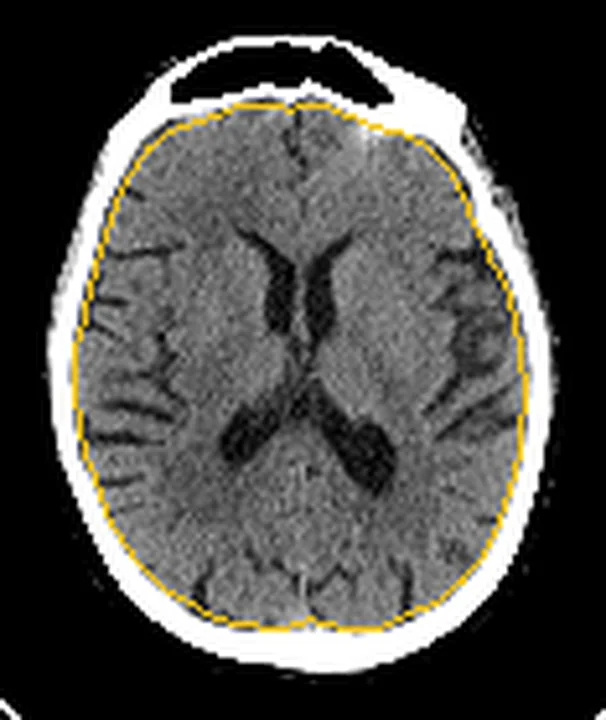
Aynı bölgenin, kaynak veri setinde patoloji içermediği belirtilen bir BT incelemesini kesit kesit kaydırın; organ sınırlarını açarak anatomiyi hasta görüntüsüyle karşılaştırın. Küçük rastlantısal bulgular tümüyle dışlanmamıştır.
Kaynak: TotalSegmentator v2.01 veri seti (Wasserthal ve ark., Radiology: Artificial Intelligence 2023), CC BY 4.0, vaka s0103; organ sınırları veri setinin otomatik segmentasyonundan, birleştirilerek sadeleştirildi. Görüntüler 2,5 mm kesit aralığına yeniden örneklendi.
Ayırıcı tanı
- Sellar/suprasellar kitle
- sellayı dolduran ayrı yumuşak doku, kitle etkisi veya suprasellar uzanım boş sella görünümüne karşıdır.
- Hipofiz atrofisi
- bez küçük olabilir; fakat sellanın BOS ile dolması ve bezin ince taban bandı oluşturması boş sella morfolojisini destekler.
- Rathke yarığı kisti
- sellar orta hatta sınırlı kistik odak olabilir; BT atenüasyonu BOS ile benzerse ayrım için MRI gerekir.
- Parsiyel hacim etkisi
- kalın aksiyel kesit sella içini BOS gibi gösterebilir; komşu kesitler ve koronal reformasyonlar görünümün devamlılığını sınar.
Tuzaklar
- Boş sella işaretini tek başına idiopatik intrakraniyal hipertansiyon tanısı sayma; bu bulgu tesadüfi olabilir ve klinik/diğer görüntüleme ölçütlerinin yerini tutmaz.
- Rutin baş BT'sinde ince hipofiz dokusu parsiyel hacim nedeniyle kaybolabilir; bez tamamen yokmuş gibi karar vermeden sagittal-koronal reformasyonları incele.
- Sekonder boş sella, hipofize zarar veren cerrahi veya radyoterapi sonrası gelişir; primer ve sekonder ayrımı görüntülemede normal bez uzantısı ile skar/deformasyon arasındaki farkla yapılır.
Kendini dene
BT’de kuşkulu boş sella görünümü MR ile çözümleniyor; MR’de sellanın <%50’si BOS ile dolu ve hipofiz tabanda ince yayvan izleniyor. En uygun tanımlama hangisidir?
Cevabı göster
Parsiyel boş sella. MR’de BOS’un sellanın <%50’sini doldurması parsiyel boş sella tanımını destekler. Primer boş sella normal hipofiz uzantısı ile karakterizedirken, sekonder boşsellada skar veya deformasyon görülür.
Boş sella morfolojisi görülen hastada yalnız bu görüntüleme bulgusuna dayanarak aşağıdaki sonuçlardan hangisi konulamaz?
Cevabı göster
İdiyopatik intrakraniyal hipertansiyon tanısı. Boş sella intrakraniyal hipertansiyonla ilişkili olabilir ancak tek başına bu tanıyı koydurmaz; klinik ve diğer tanı ölçütleri gerekir. Sellaya BOS uzanımı, bezin tabana basık görünmesi ve belirgin kitlenin olmaması morfolojik görüntüleme tanımlarıdır.
Kaynaklar
Bu sayfadaki 36 cümle ve 2 quiz sorusu aşağıdaki kaynaklardan alıntılarla otomatik olarak karşılaştırıldı; 7 cümle bu karşılaştırmada düzeltildi (2026-10-09).
- Asymptomatic Empty Sella: A Literature Review and Suggestions for Evaluation in Clinical Practicepmc.ncbi.nlm.nih.gov
- Long delay in diagnosis of a case with MEN1 due to concomitant presence of AIMAH with insulinoma: a case report and literature review.pubmed.ncbi.nlm.nih.gov
- Petrous Apex Cephaloceles: Radiology Features and Surgical Management of a Rare Entity.pubmed.ncbi.nlm.nih.gov
- High frequency of empty sella, with gender differences, in the early neuroradiology evaluation of patients with traumatic brain injury. A prospective study.pubmed.ncbi.nlm.nih.gov
- Is foramen magnum decompression for acquired Chiari I malformation like putting a finger in the dyke? - A simplistic overview of artificial intelligence in assessing critical upstream and downstream etiologies.pubmed.ncbi.nlm.nih.gov
- Ectopic secretion of GHRH by a pancreatic neuroendocrine tumor associated with an empty sella.pubmed.ncbi.nlm.nih.gov
- Partial empty sella in a woman with cerebral venous sinus thrombosis: A rare presentation of polycythaemia rubra vera.pubmed.ncbi.nlm.nih.gov
- Diagnosing hypocortisolism in disguise: clinical insights from delayed-onset endocrine disorders.pubmed.ncbi.nlm.nih.gov
- Idiopathic Intracranial Hypertension in a Non-obese Male Patient: A Case Report.pubmed.ncbi.nlm.nih.gov
- Successful Pregnancy in a Woman With Primary Infertility Associated With Isolated Hypogonadotropic Hypogonadism and Partial Empty Sella Syndrome: A Case Report.pubmed.ncbi.nlm.nih.gov
- Seizure as a Rare Initial Manifestation of Empty Sella Turcica Associated With Idiopathic Intracranial Hypertension: A Case Report.pubmed.ncbi.nlm.nih.gov
- Changes overtime in primary and secondary empty sella: a comparative real-world study.pubmed.ncbi.nlm.nih.gov
- Idiopathic intracranial hypertension imaging approaches and the implications in patient management.pubmed.ncbi.nlm.nih.gov
- Multimodal Navigation in Endoscopic Transsphenoidal Resection of Pituitary Tumors Using Image-Based Vascular and Cranial Nerve Segmentation: A Prospective Validation Study.pubmed.ncbi.nlm.nih.gov
- Comprehensive review on rhino-neurosurgery.pubmed.ncbi.nlm.nih.gov
- Acetazolamide for Idiopathic Intracranial Hypertension: An Up-to-Date Review in 2025.pubmed.ncbi.nlm.nih.gov
- Ophthalmologic assessment and intracranial pressure in children: diagnostic methods, clinical correlations, and future directions.pubmed.ncbi.nlm.nih.gov
- Idiopathic Intracranial Hypertension in Children and Adolescents with Obesity: A Narrative Review.pubmed.ncbi.nlm.nih.gov
- Diagnostic Delay in Acromegaly Due to Overlapping Features With Polycystic Ovary Syndrome: A Case Report.pubmed.ncbi.nlm.nih.gov
- Primary hypothalamic lymphoma with clinical findings mimicking pituitary apoplexy: a case report.pubmed.ncbi.nlm.nih.gov
- Italian Association of Clinical Endocrinologists (AME) and International Chapter of Clinical Endocrinology (ICCE). Position statement for clinical practice: prolactin-secreting tumors.pubmed.ncbi.nlm.nih.gov
- Imaging the brain: diagnosis aided by structural features on neuroimaging studies.pubmed.ncbi.nlm.nih.gov
- Growth hormone-releasing hormone, growth hormone, and neuroendocrine tumors.pubmed.ncbi.nlm.nih.gov
- Dual phenotype in a patient with adult-onset hyperinsulinism and diabetes caused by an activating <i>MAFA</i> variant.pubmed.ncbi.nlm.nih.gov
- A case of mixed histiocytosis (Erdheim-Chester disease and Langerhans cell histiocytosis) with STEAP3-associated anemia and type 4 hemochromatosis.pubmed.ncbi.nlm.nih.gov
- Multiple brain abscesses caused by <i>Nocardia asiatica</i> co-infection with Torque teno virus in an "immunocompetent" patient: a rare case report and literature review.pubmed.ncbi.nlm.nih.gov
- Imaging of Skull Base Tumors.pubmed.ncbi.nlm.nih.gov
- Neuroradiology for ophthalmologists.pubmed.ncbi.nlm.nih.gov
- Cranial anatomy, palaeoneurology, palaeobiology and stratigraphic age of the large-bodied ornithopod, <i>Muttaburrasaurus langdoni</i> Bartholomai and Molnar, 1981, from the mid-Cretaceous of Australia.pubmed.ncbi.nlm.nih.gov
- Commentary: an eye on PET quantification.pubmed.ncbi.nlm.nih.gov
- Neuroimaging of pediatric tumors of the sellar region-A review in light of the 2021 WHO classification of tumors of the central nervous system.pubmed.ncbi.nlm.nih.gov
- Diagnostic neuroimaging in neuro-ophthalmic disorders.pubmed.ncbi.nlm.nih.gov
- Optimizing Surgical Management of Anterior Skull Base Meningiomas: Imaging Modalities, Key Surgical Considerations, and Risk Mitigation Strategies.pubmed.ncbi.nlm.nih.gov
- Persistent Craniopharyngeal Canal (Type 3C) with Vertebrobasilar Dolichoectasia and Bilateral Sclerochoroidal Calcification.pubmed.ncbi.nlm.nih.gov
- Empty sella in somatotropic pituitary adenomas; a series of 23 cases.pubmed.ncbi.nlm.nih.gov
- Patient with two rare diseases-Renal coloboma syndrome and craniopharyngioma.pubmed.ncbi.nlm.nih.gov
- An overview of hypopituitarism's causes.pubmed.ncbi.nlm.nih.gov
- An Update on Advances in Hypopituitarism: Etiology, Diagnosis, and Current Management.pubmed.ncbi.nlm.nih.gov
- 3D Geometric Morphometric Analysis on Sexual Dimorphism in the Sella Turcica in Romanov Sheep Using CT Scan.pubmed.ncbi.nlm.nih.gov
- Pituitary MRI, Clinical and Biochemical Findings in Children With Growth Disorder: A Retrospective Comparative Study.pubmed.ncbi.nlm.nih.gov
- Pituitary Tumors in Maxillofacial Radiology and Daily Practice: A Scoping Review.pubmed.ncbi.nlm.nih.gov
- Sexual Dimorphism and Inter-Century Variability in Sella Turcica and Sphenoid Sinus Pneumatization: A Computed Tomography Study of the Romanian Population.pubmed.ncbi.nlm.nih.gov
- Three-Dimensional Volumetric Evaluation of the Sella Turcica and Sphenoid Sinus in Individuals with Unilateral Palatally Impacted Maxillary Canines Using CBCT.pubmed.ncbi.nlm.nih.gov
- Comparative Evaluation of Sella Turcica Morphology and Dimensions in Skeletal Class III Malocclusion and Cleft Lip and Palate Patients Versus Class I Individuals.pubmed.ncbi.nlm.nih.gov
- Diagnostic value of machine-learning using conventional magnetic resonance imaging markers for pediatric idiopathic intracranial hypertension: a retrospective study.pubmed.ncbi.nlm.nih.gov
- Pituitary magnetic resonance imaging abnormalities and 1-year growth response in children with idiopathic growth hormone deficiency.pubmed.ncbi.nlm.nih.gov
- Imaging and physiology across the high-low cerebrospinal fluid pressure spectrum: Navigating diagnostic uncertainty in headache practice.pubmed.ncbi.nlm.nih.gov
- Surgical management of Rathke cleft cysts in pediatric patients: a single institution experience.pubmed.ncbi.nlm.nih.gov
- Cerebrospinal fluid dynamics in idiopathic intracranial hypertension: a literature review and validation of contemporary findings.pubmed.ncbi.nlm.nih.gov
- Invited review: Recommendations for reporting intervention studies on reproductive performance in dairy cattle: Improving design, analysis, and interpretation of research on reproduction.pubmed.ncbi.nlm.nih.gov
- [<sup>68</sup>Ga]Ga-DOTATATE PET/CT and PET/MR enhances the detection of pituitary ACTH-secreting adenomas in cushing's disease.pubmed.ncbi.nlm.nih.gov
- Inter-rater reliability of MRI Neck Imaging Reporting and Data System (NI-RADS) in the follow-up of oropharyngeal squamous cell carcinoma.pubmed.ncbi.nlm.nih.gov
- Synchronous glioblastoma and pituitary adenoma: Coincidence or shared genetic predisposition? A case report and comprehensive literature review.pubmed.ncbi.nlm.nih.gov
- Sellar region neurocytomas exhibit a CIMP and neuroendocrine-like epigenetic signature distinct from other intra-axial neurocytomas.pubmed.ncbi.nlm.nih.gov
- Sellar Solitary Fibrous Tumor Mimicking Pituitary Adenoma: Diagnostic Pitfalls, Contemporary Pathological Classification, and Management Considerations.pubmed.ncbi.nlm.nih.gov
- Nonproliferative and Proliferative Lesions of the Rat and Mouse Endocrine System.pubmed.ncbi.nlm.nih.gov
- Fluid transport in the brain.pubmed.ncbi.nlm.nih.gov
- Suprasellar arachnoid cyst causing pituitary stalk compression and endocrine dysfunction in a child: MRI findings.pubmed.ncbi.nlm.nih.gov
- A Clival Mass Posing a Diagnostic Challenge: Pituitary Adenoma With Rathke Cleft Cyst Mimicking Chordoma.pubmed.ncbi.nlm.nih.gov
- Radiological Differentiation of Pituitary Adenomas From Other Sellar Masses: A Systematic Review.pubmed.ncbi.nlm.nih.gov
- Peripheral trigeminal nerve branch lesions: CT and MRI findings in a pathologically confirmed case series.pubmed.ncbi.nlm.nih.gov
- Primary and Secondary Optic Nerve Sheath Meningioma.pubmed.ncbi.nlm.nih.gov
- Endoscopic Endonasal Transsphenoidal Treatment of Pituitary Apoplexy: Outcomes in a Series of 20 Patients.pubmed.ncbi.nlm.nih.gov
- Expanding the clinical spectrum of idiopathic intracranial hypertension.pubmed.ncbi.nlm.nih.gov
- Imaging spectrum of abnormal subcutaneous and visceral fat distribution.pubmed.ncbi.nlm.nih.gov
- An ATR-PrimPol pathway confers tolerance to oncogenic KRAS-induced and heterochromatin-associated replication stress.pubmed.ncbi.nlm.nih.gov
- Guidelines for the use and interpretation of assays for monitoring autophagy (4th edition)<sup>1</sup>.pubmed.ncbi.nlm.nih.gov
- Case Report: Intracranial hypertension occurring in association with subacute thyroiditis.pubmed.ncbi.nlm.nih.gov
- Head Trauma Mechanisms and Neuroimaging Findings in Psychiatric Inpatients Referred for Neurosurgical Consultation.pubmed.ncbi.nlm.nih.gov
- CT-Based Evaluation of Sphenoid Sinus Type and Sella Turcica-Related Quantitative Parameters for Endoscopic Endonasal Transsphenoidal Approach Planning in Pediatric Patients.pubmed.ncbi.nlm.nih.gov
- Postoperative Pituitary MRI Findings in Acromegaly: A Pictorial Review.pubmed.ncbi.nlm.nih.gov
- Endoscopic endonasal treatment of Meckel's cave epidermoid cysts: case series and systematic review.pubmed.ncbi.nlm.nih.gov
- The orca (<i>Orcinus orca</i>) pituitary gland: an anatomical, immunohistochemical and ultrastructural analysis.pubmed.ncbi.nlm.nih.gov
- Invasive sellar extraventricular neurocytoma: illustrative case.pubmed.ncbi.nlm.nih.gov
- Giant Craniopharyngioma Complicated by Cerebrospinal Fluid Ascites in a 7-Year-Old Boy: A Case Report and Review of Literature.pubmed.ncbi.nlm.nih.gov
- The Evolving Role of the Endoscopic Endonasal Transplanum-Transtuberculum Approach in the Management of Craniopharyngiomas: A Systematic Review of Outcomes, Reconstruction, and Surgical Evolution.pubmed.ncbi.nlm.nih.gov
- Time is vision: a systematic review of urgent venous sinus stenting for fulminant idiopathic intracranial hypertension.pubmed.ncbi.nlm.nih.gov
- Clinical Challenges in Diagnosing Primordial Dwarfism: Insights from a MOPD II Case Study.pubmed.ncbi.nlm.nih.gov
- Prediction of Postoperative Risk of Raised Intracranial Pressure After Spontaneous Skull Base Cerebrospinal Fluid Leak Repair.pubmed.ncbi.nlm.nih.gov
- Idiopathic Intracranial Hypertension with and Without Papilledema: A Comparative Cohort Study.pubmed.ncbi.nlm.nih.gov
- Obesity and Skull Base Manifestations: Clinical Implications and Management.pubmed.ncbi.nlm.nih.gov
- Idiopathic Intracranial Hypertension Without Papilledema: A Case Emphasizing the Diagnostic Value of Optic Nerve Sheath Ultrasound.pubmed.ncbi.nlm.nih.gov
- Intracranial venous thrombosis imaging by anatomical location: an educational review with mimics and diagnostic pitfalls.pubmed.ncbi.nlm.nih.gov
- Idiopathic Intracranial Hypertension in a Child with Marfan Syndrome: Clinical, Neuroimaging, and Biomarker Findings from a Case Report.pubmed.ncbi.nlm.nih.gov
- Brain tumour post-treatment imaging and treatment-related complications.pubmed.ncbi.nlm.nih.gov
- Controversial issues in the management of hyperprolactinemia and prolactinomas - An overview by the Neuroendocrinology Department of the Brazilian Society of Endocrinology and Metabolism.pubmed.ncbi.nlm.nih.gov
- Coexistence of Autoantibodies against the Golgi Complex and Ro52 Antigen in a Patient with Nonspecific Interstitial Pneumonia.pubmed.ncbi.nlm.nih.gov
- Surgical management of sellar arachnoid cyst: state of the art and systematic review.pubmed.ncbi.nlm.nih.gov
- Pituitary Apoplexy: A Case Series.pubmed.ncbi.nlm.nih.gov
- Three-Dimensional Constructive Interference in Steady State (3D CISS) Imaging and Clinical Applications in Brain Pathology.pubmed.ncbi.nlm.nih.gov
- The challenge of diagnosing intracranial pressure elevations as an otolaryngologist.pubmed.ncbi.nlm.nih.gov
- Frontoparietal primary osteosarcoma mimicking parasagittal meningioma: A case report.pubmed.ncbi.nlm.nih.gov
- Pediatric endocrine disorders: a review of intracranial findings and appropriate imaging.pubmed.ncbi.nlm.nih.gov
- Unusual Presentation of Hypopituitarism Caused by Internal Carotid Artery Aneurysm.pubmed.ncbi.nlm.nih.gov
Eğitim amaçlıdır; hasta başında tanı ve tedavi kararının yerine geçmez, tıbbi tavsiye değildir. Metinler yapay zekâ desteğiyle yazıldı ve otomatik bir adımda kaynak alıntılarıyla karşılaştırıldı; bu bir tıbbi doğrulama değildir ve içerik uzman radyolog incelemesinden geçmedi. Klinik kararlarda güncel kılavuzları, kurum protokollerini ve radyoloji raporunu esas alın.