Özet
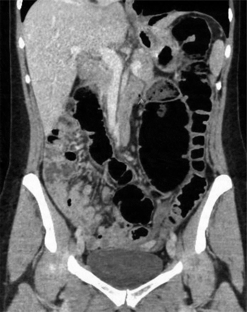
Mobil çekum, sağ kolon mezenterinin posterior pariyetal peritonla kaynaşmaması sonucu çekumun ve yükselen kolonun serbestçe hareket etmesiyle karakterizedir; bu nedenle çekum pelviste, orta hatta veya sol/üst abdomende ektopik olarak görülebilir. Tek bir BT, anormal konumu gösterir ancak organın gerçekten hareketli olduğunu kanıtlamak için önceki veya farklı pozisyondaki inceleme daha değerlidir. BT ayrıca apendiksi çekumla birlikte izleyip torsiyon, obstrüksiyon ve iskemi bulgularını araştırır. Konumsal varyantı volvulus sanmak gereksiz acil yorum doğurabilir; mobil çekumda gelişen gerçek volvulusu gözden kaçırmak ise intestinal iskemi ve gangrene yol açan kapalı ans durumunu atlatabilir.
Faz ve pencere
- Kontrastsız tanısal
- Kontrastsız veya kontrastlı BT, çekumun ektopik konumunu, terminal ileum ve apendiksin yerini, lümen genişliğini ve mezenterik yağ planındaki değişiklikleri gösterebilir; akut volvulus şüphesinde ise ince dilimli rekonstrüksiyonlar whirl işareti gibi bulguları ortaya koyar. Kontrastsız BT, çekumun konumsal anomalilerini ve gaz dilatasyonunu gösterebilir; kontrast destekli ince dilimli incelemeler ise mezenterik vasküler yapıları ve duvar perfüzyonunu ayrıntılı olarak değerlendirmeyi sağlar.
Önerilen pencereler: Batın (G 400 / M 50), Yumuşak doku (G 400 / M 40).
BT bulguları
- Ektopik çekum — çekum sağ alt kadran dışında, pelviste, orta hatta veya sol/üst abdomen düzeyinde izlenebilir.
- İleoçekal bileşkenin birlikte yer değiştirmesi — terminal ileum ve ileoçekal valv çekuma uzanır; ektopik konumu tanımaya yardım eder.
- Apendiksin çekuma bağlı seyri — apendiks çekum tabanından kör sonlanan tübüler yapı olarak çıkar ve çekumla aynı tarafa taşınır.
- Fiksasyon kaybı — sağ kolonun posterior duvara yapışık olmaması ve serbestçe yer değiştirmesi mobiliteyi düşündürür; tek pozisyonlu görüntüde hareketlilik kesin olarak kanıtlanamaz, ancak ektopik konum ve mezenterin gevşekliği buna işaret eder.
- Volvulus lehine dönme yok — mezenter damarlarında whirl, belirgin kapalı ans, keskin geçiş veya proksimal ince bağırsak/kolon dilatasyonu bulunmaz.
- Duvar perfüzyon bulgusu yok — duvar kalınlaşması, pnömatozis, portal venöz gaz veya çevresel sıvı saptanmaz.
- Önceki görüntüyle yer değişimi — farklı tarihte çekumun başka bir abdominal kadranda görülmesi sabit ektopik çekumdan ziyade mobiliteyi destekler.
Normalde
Normal yerleşimli çekum sağ iliak fossada bulunur; terminal ileum ile birleşir ve apendiks tabanı çekumun posteromedial bölümünden çıkar. Kıyaslamada çekumun tek başına konumundan ziyade, terminal ileum ve apendisin aynı anda yer değiştirdiğini, sağ kolonun fiksasyonunun yetersiz olduğunu ve mezenterde torsiyon veya obstrüksiyon bulgusu olup olmadığını değerlendirmek önceliklidir.
Normal BT ile kıyasla
Aynı bölgenin, kaynak veri setinde patoloji içermediği belirtilen bir BT incelemesini kesit kesit kaydırın; organ sınırlarını açarak anatomiyi hasta görüntüsüyle karşılaştırın. Küçük rastlantısal bulgular tümüyle dışlanmamıştır.
Kaynak: TotalSegmentator v2.01 veri seti (Wasserthal ve ark., Radiology: Artificial Intelligence 2023), CC BY 4.0, vaka s0119; organ sınırları veri setinin otomatik segmentasyonundan, birleştirilerek sadeleştirildi. Görüntüler 2,5 mm kesit aralığına yeniden örneklendi.
Ayırıcı tanı
- Çekum volvulusu
- ektopik çekumda belirgin distansiyon, mezenterik whirl, geçiş noktası ve distal kolon dekompresyonu bulunabilir.
- Çekum bascule
- çekum öne ve yukarı katlanarak obstrüksiyon yapar; belirgin eksenel mezenter girdabı olmayabilir.
- Malrotasyon
- ince bağırsak ve kolonun daha yaygın anormal yerleşimi, SMA-SMV ilişkisi değişikliği veya duodenum seyri bulguları eşlik edebilir.
- Sol yerleşimli normal çekum
- situs inversus veya bağırsak rotasyon anomalisi bağlamında değerlendirilir; yalnızca sağ fossa dışında bulunması, tek başına mobilite kanıtı olarak kabul edilmez.
- Başka segmentteki obstrüksiyona bağlı kolon dilatasyonu
- proximal tıkanıklık nedeniyle oluşan aşırı gaz ve içerik birikimi, çekumun yer değiştirmesine veya üst abdomende izlenmesine neden olabilir.
Tuzaklar
- Tek incelemede pelviste görülen çekum için hareketliliği kesinleştirmeyin; önceki BT ile konumu ve sağ kolon fiksasyonunu karşılaştırın.
- Çekumun yer değiştirmesi tek başına volvulus değildir; mezenter kökünde dönme, ani geçiş ve kapalı ansı birlikte arayın.
- Apendiksin ektopik çekumla birlikte yer değiştirmesi beklenebilir; inflamasyonu dışlamak için kör ucu, duvarı ve çevre yağını kendi konumunda takip edin.
Kendini dene
Pelviste çekum, ona giren terminal ileum ve çekumdan çıkan ince apendiks görülüyor; lümen açık, mezenterde dönme yok. En uygun yorum?
Cevabı göster
Ektopik çekum (tek BT mobiliteyi kanıtlamaz). Tek BT çekumun ektopik konumunu gösterir, ancak hareketliliği kanıtlamaz; farklı tarih veya pozisyondaki görüntüde yer değişimi mobiliteyi destekler. Volvulusta dönme ve obstrüksiyon, kanserde kitle, apandisitte duvar/çevre inflamasyonu aranır.
Önceki BT'de sağ alt kadranda olan çekum bu kez sol üst kadranda; mezenter damarları girdap oluşturuyor ve ince bağırsaklar geniş. Hangi komplikasyon düşünülür?
Cevabı göster
Çekum volvulusu. Yer değiştirmiş çekumla birlikte mezenterik girdap ve proksimal ince bağırsak dilatasyonu volvulus/obstrüksiyon lehinedir. Uzun ve ektopik çekum tek başına volvulus anlamına gelmez; bir olguda önceki BT'de distansiyon veya volvulus olmadan uzun, ektopik çekum görülmüştür. Dolikokolon uzamış kolon seyri yapar ama tek başına dönme ve geçiş açıklamaz; diğer seçenekler bu akut bulguları açıklamaz.
Kaynaklar
Bu sayfadaki 37 cümle ve 2 quiz sorusu aşağıdaki kaynaklardan alıntılarla tek tek karşılaştırıldı; 11 cümle bu karşılaştırmada düzeltildi (2026-10-08).
- Acute Volvulus of the Cecumpmc.ncbi.nlm.nih.gov
- Surgery to Treat Symptomatic Mobile Cecum Syndrome Is Safe and Associated with Good Recovery Outcomespmc.ncbi.nlm.nih.gov
- Laparoscopic Management of Mobile Cecumpmc.ncbi.nlm.nih.gov
- Laparoscopic Management of Mobile Cecum.pubmed.ncbi.nlm.nih.gov
- Cecal Volvulus Resolved Spontaneously.pubmed.ncbi.nlm.nih.gov
- A Case of Intussusception Associated with Pneumatosis Cystoides Intestinalis.pubmed.ncbi.nlm.nih.gov
- Cecal bascule - A rare cause of cecal volvulus after cesarean section.pubmed.ncbi.nlm.nih.gov
- Cecal volvulus in giant ventral hernia.pubmed.ncbi.nlm.nih.gov
- Cecal Volvulus a Rare Cause of Intestinal Obstruction. A Case Report.pubmed.ncbi.nlm.nih.gov
- Cecal bascule herniation into the lesser sac.pubmed.ncbi.nlm.nih.gov
- Laparoscopic right hemicolectomy for ascending colon cancer with persistent mesocolon.pubmed.ncbi.nlm.nih.gov
- Cecal volvulus caused by a large uterine leiomyoma.pubmed.ncbi.nlm.nih.gov
- Cecal volvulus caused by internal herniation after roux-en-y gastric bypass surgery.pubmed.ncbi.nlm.nih.gov
- Three Surgical Cases of Cecal Volvulus.pubmed.ncbi.nlm.nih.gov
- Internal hernia with incarceration of the cecum through a loop created by an elongated fallopian tube.pubmed.ncbi.nlm.nih.gov
- Patterns of injury and violence in Yaoundé Cameroon: an analysis of hospital data.pubmed.ncbi.nlm.nih.gov
- The British technique of purely robotic Whipple's pancreatoduodenectomy a systematic and stepwise ('clockwise') approach.pubmed.ncbi.nlm.nih.gov
- Imaging Modalities for Evaluation of Intestinal Obstruction.pubmed.ncbi.nlm.nih.gov
- Special Issue: EACR 2026 Congress: Innovative Cancer Science, 8-11 June 2026.pubmed.ncbi.nlm.nih.gov
- Torsion of the Vermiform Appendix in an 18-Day-Old Neonate: A Case Report from Romania and Review of the Literature.pubmed.ncbi.nlm.nih.gov
- Appendico-Ileal Knotting (Appendiceal Tourniquet Syndrome): A Systematic Review and Clinicopathological Classification.pubmed.ncbi.nlm.nih.gov
- Amyand's hernia associated with acute appendicitis and incidental sessile serrated lesion in a recurrent incarcerated inguinal hernia: a case report.pubmed.ncbi.nlm.nih.gov
- Long-Segment Right Colon Volvulus in a Young Adult With Severe Developmental Disability: An Unusual Presentation and a Diagnostic Challenge.pubmed.ncbi.nlm.nih.gov
- Emergency surgery for malignant large bowel obstruction: Assessing management options and outcomes.pubmed.ncbi.nlm.nih.gov
- Complete common mesentery revealed by a cecal bascule: Two case reports.pubmed.ncbi.nlm.nih.gov
- Diagnostic Dilemma in Pediatric Subhepatic Appendicitis: A Report of Two Cases.pubmed.ncbi.nlm.nih.gov
- Gastrointestinal Ultrasound: Measurements and Normal Findings - What Do You Need to Know?pubmed.ncbi.nlm.nih.gov
- Small Bowel Obstruction and Appendicitis in Patient With Fitz-Hughes-Curtis Syndrome.pubmed.ncbi.nlm.nih.gov
- Gangrenous cecal volvulus with 720° torsion and prolonged intraoperative hypotension in a resource-limited setting: a case report.pubmed.ncbi.nlm.nih.gov
- Cecal volvulus and nonrotation: two rare pathologic entities as culprits of intestinal obstruction-a case report.pubmed.ncbi.nlm.nih.gov
- An extremely rare case of malignant jejunal mesenteric inflammatory myofibroblastic tumor in a 61-year-old male patient: A case report and literature review.pubmed.ncbi.nlm.nih.gov
- Case Report: Midgut malrotation presenting as left-sided appendicitis with secondary omental torsion: a diagnostic challenge in a child.pubmed.ncbi.nlm.nih.gov
- International Society of Paediatric Surgical Oncology (IPSO) Surgical Practice Guidelines.pubmed.ncbi.nlm.nih.gov
- Pneumatosis intestinalis revisited: associated imaging findings, clinical correlation, management, risk stratification and future perspectives.pubmed.ncbi.nlm.nih.gov
- Colonic Ischemia Without Mesenteric Occlusion: A Case Implicating Interferon-β.pubmed.ncbi.nlm.nih.gov
- When It Is Not NEC: Recognizing Mimics of Necrotizing Enterocolitis in Preterm Infants.pubmed.ncbi.nlm.nih.gov
- Alpha-ketoglutarate mitigates insulin resistance and metabolic inflexibility in a mouse model of Ataxia-Telangiectasia.pubmed.ncbi.nlm.nih.gov
- Ammonium ion binding to DNA G-quadruplexes: do electrospray mass spectra faithfully reflect the solution-phase species?pubmed.ncbi.nlm.nih.gov
- Twist in the cecum: A case series of mobile cecum, cecal volvulus and cecal bascule.pubmed.ncbi.nlm.nih.gov
- Caecal volvulus around a Ladd's band in undiagnosed adult malrotation.pubmed.ncbi.nlm.nih.gov
- Incomplete reversed midgut rotation with 540° volvulus in an adult: a case report.pubmed.ncbi.nlm.nih.gov
- Radiologic Hallmarks of Colonic Volvulus: A Narrative Review.pubmed.ncbi.nlm.nih.gov
- Diagnostic Challenges in Cecal Bascule: A Case Report.pubmed.ncbi.nlm.nih.gov
- Acute cecal volvulus and large bowel obstruction in a patient with persistent Ladd bands and chronic malrotation: case report and review of literature.pubmed.ncbi.nlm.nih.gov
- Intestinal Malrotation With Chronic Midgut Volvulus, Chylous Ascites, and Severe Malnutrition in an Adolescent: A Case Report.pubmed.ncbi.nlm.nih.gov
- Diagnosis of adult midgut malrotation in CT: sign of absent retromesenteric duodenum reliable.pubmed.ncbi.nlm.nih.gov
- Gut Malrotation in a Human Cadaver: Exploration Into the Prevention and Screening of Undiagnosed Malrotation in Adults.pubmed.ncbi.nlm.nih.gov
- Rare Association of Duodenal Obstruction and Situs Inversus Abdominis: A Two Case Report.pubmed.ncbi.nlm.nih.gov
- Pitfall of Diagnosis Left-Sided Appendicitis Due to Undiagnosed Intestinal Malrotation in Adulthood - a Rare Case Report from Syria and Literature Review.pubmed.ncbi.nlm.nih.gov
- Epigastric Pain Caused by Left Upper Quadrant Acute Appendicitis in the Setting of Undiagnosed Intestinal Malrotation.pubmed.ncbi.nlm.nih.gov
- A Rare Case of Poorly Differentiated Neuroendocrine Carcinoma of the Descending Colon with Regional Lymph Node Involvement Presenting in a Young Adult Nigerian Male: A Case Report.pubmed.ncbi.nlm.nih.gov
- Acute Colonic Pseudo-obstruction and Volvulus: Pathophysiology, Evaluation, and Treatment.pubmed.ncbi.nlm.nih.gov
- Charge/stiffness-tunable nano-microsphere overcomes intestinal mucosal barrier for rheumatoid arthritis treatment.pubmed.ncbi.nlm.nih.gov
- Incidentally discovered, intentionally managed: a narrative review of the last decade's updates on the management of neuroendocrine tumor of the appendix.pubmed.ncbi.nlm.nih.gov
- Imaging Approach to Diverticular Disease of the Small Bowel.pubmed.ncbi.nlm.nih.gov
- Acute appendicitis due to appendiceal endometriosis: Two case report and literature review.pubmed.ncbi.nlm.nih.gov
- Iliopsoas hydatid cyst mimicking an ovarian tumor: a diagnostic pitfall in an endemic setting-a case report.pubmed.ncbi.nlm.nih.gov
- Pediatric Abdominal Ultrasound Training Program: Standard Views.pubmed.ncbi.nlm.nih.gov
- Pediatric emergency medicine point-of-care ultrasound: summary of the evidence.pubmed.ncbi.nlm.nih.gov
- Appendicitis on the Wrong Side: A Case of Left-Sided Acute Appendicitis With Intestinal Nonrotation.pubmed.ncbi.nlm.nih.gov
- Mechanical Small Bowel Obstruction Secondary to Low-Grade Appendiceal Mucinous Neoplasm: A Case Report.pubmed.ncbi.nlm.nih.gov
- A Rare Presentation of Small Bowel Volvulus Secondary to Meckel's Diverticulum-Induced Intussusception in an Adolescent Female.pubmed.ncbi.nlm.nih.gov
- Early Post-cesarean Section Gangrenous Ileocecal Volvulus in a Primigravida: A Rare Postpartum Complication Case Report.pubmed.ncbi.nlm.nih.gov
- Cecal volvulus with acute appendicitis in a 33-year-old male managed by manual detorsion, cecopexy, and appendectomy: A case report.pubmed.ncbi.nlm.nih.gov
- Validating the Cattell-Braasch maneuver with a case of inherent atypical malrotation of the bowel: a case report.pubmed.ncbi.nlm.nih.gov
- Midgut volvulus caused by intestinal malrotation; A rare cause of acute abdomen in adults. Case report.pubmed.ncbi.nlm.nih.gov
- Rare Implantation Sites of Ectopic Pregnancy: A Case Series of Ovarian and Hepatic Pregnancy and Review of Diagnostic Challenges.pubmed.ncbi.nlm.nih.gov
- Management of colonic volvulus.pubmed.ncbi.nlm.nih.gov
- Small-Bowel Obstruction Secondary to Ileal Trichobezoar in a Patient with Rapunzel Syndrome.pubmed.ncbi.nlm.nih.gov
- Laparoscopic Fixation of the Proximal Jejunum and Gastrojejunostomy for Small Bowel Volvulus and Recurrent Duodenal Obstruction Caused by Incomplete Fixation and Abnormal Mobility of the Ligament of Treitz: A Case Report.pubmed.ncbi.nlm.nih.gov
- Cecal volvulus revealing previously undiagnosed intestinal non-rotation in adulthood: intraoperative diagnosis of a rare congenital anomaly.pubmed.ncbi.nlm.nih.gov
- Small bowel obstruction due to herniation through the foramen of Winslow in an elderly patient: a case report.pubmed.ncbi.nlm.nih.gov
- Caught in the Whirl: Computed Tomography Diagnosis of Transverse Colon Volvulus With Proximal Jejunal Involvement and Cecocolic Pneumatosis.pubmed.ncbi.nlm.nih.gov
- Intraluminal Appendiceal Migration of an Intrauterine Device Without Appendicitis: A Case Report.pubmed.ncbi.nlm.nih.gov
- Diagnostic Challenge of Sequential Appendicitis in a Patient With Appendiceal Duplication: A Case Report.pubmed.ncbi.nlm.nih.gov
- Appendiceal Neuroendocrine Tumor Presenting as Acute Appendicitis: A Case Report.pubmed.ncbi.nlm.nih.gov
- Laparoscopic Appendectomy for Left-Sided Acute Appendicitis in Situs Inversus Totalis: A Case Report and Technical Considerations.pubmed.ncbi.nlm.nih.gov
- Living Donor Liver Transplantation in Pediatric Situs Inversus: A Case Series and Systematic Review.pubmed.ncbi.nlm.nih.gov
Eğitim amaçlıdır; hasta başında tanı ve tedavi kararının yerine geçmez. İçerik yapay zekâ destekli bir süreçle güncel literatüre göre cümle cümle kaynaklarla karşılaştırıldı, ancak uzman radyolog incelemesinden geçmedi. Klinik kararlarda güncel kılavuzları, kurum protokollerini ve radyoloji raporunu esas alın.