Özet
Cryptococcus inhalasyonla akciğere ulaşır; enfeksiyon hem bağışıklığı normal kişilerde sınırlı nodül olarak hem de bağışıklığı baskılanmış kişilerde yaygın akciğer veya sistemik hastalık olarak görülebilir. Toraks BT tek ya da kümelenmiş nodülleri, kitle benzeri konsolidasyonu ve eşlik edebilecek buzlu cam, kavite, lenf nodu veya plevra bulgularını tanımlar. Görüntüleme kanserden güvenilir biçimde ayırmayabilir; BT dağılımı ve yayılımı gösterir, kesin tanı mikrobiyoloji veya doku değerlendirmesine dayanır. Nodülün malignite sanılması gereksiz gecikmeye, enfeksiyonun veya eşlik eden santral sinir sistemi yayılımının gözden kaçması ise ciddi sonuçlara yol açabilir.
Faz ve pencere
- HRCT tanısal
- Kontrastsız ince kesit toraks BT'de tek, kümelenmiş veya dağınık yuvarlak nodül/kitleler, subplevral yerleşim, kitle benzeri ya da hava bronkogramlı konsolidasyon; eşlik eden buzlu cam, kavite ve küçük hava yolu opasiteleri incelenir.
Önerilen pencereler G genişlik, M merkez; değerler Hounsfield birimi (HU)
- AkciğerG 1500 / M −600
- MediastenG 350 / M +50
BT bulguları
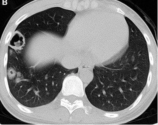
- Tek veya çok sayıda, iyi ya da kısmen sınırlı parankimal nodül; bazıları kümelenmiş, bazıları ayrı dağılmıştır
- Subplevral yerleşimli tek veya çok sayıda nodül veya kitle.
- Kitle benzeri fokal konsolidasyon; içinde hava bronkogramı ve çevresinde buzlu cam bulunabilir
- İmmün baskılanmış hastada nodül ya da kitle içinde kaviteleşme
- Nodül çevresinde halo biçimli buzlu cam; etkeni tek başına tanımlamaz
- Pnömoni tipi pulmoner kriptokokkozda iki taraflı dağılım görülebilir.
- Mediastinal/hiler lenf nodu büyümesi ve plevral sıvı eşlik edebilir, ancak her hastada beklenmez
- Pulmoner kriptokokkozda baskın bir nodüle başka loblarda ek nodüller eşlik edebilir; bu dağılım metastazı düşündürebilir.
Normalde
Normal akciğer kesitinde sekonder pulmoner lobüller homojen havalı görünür; subplevral alanlarda yuvarlak yumuşak doku nodülü veya fokal hava boşluğu konsolidasyonu bulunmaz. Aynı seviyede karşı lobun plevral yüzeyini, segmental bronşların açıklığını ve mediastinal penceredeki hiler düğümlerin boyut/şekil benzerliğini lezyonlu tarafla karşılaştır.
Normal BT ile kıyasla
Aynı bölgenin, kaynak veri setinde patoloji içermediği belirtilen bir BT incelemesini kesit kesit kaydırın; organ sınırlarını açarak anatomiyi hasta görüntüsüyle karşılaştırın. Küçük rastlantısal bulgular tümüyle dışlanmamıştır.
Kaynak: TotalSegmentator v2.01 veri seti (Wasserthal ve ark., Radiology: Artificial Intelligence 2023), CC BY 4.0, vaka s1278; organ sınırları veri setinin otomatik segmentasyonundan, birleştirilerek sadeleştirildi. Görüntüler 2,5 mm kesit aralığına yeniden örneklendi.
Ayırıcı tanı
- Primer akciğer kanseri
- spikülasyon, bronş tıkanması ve aynı lobda sekonder değişiklikler görülebilir; kriptokokkozdan BT ile kesin ayrılamayan tek kitle olabilir.
- Metastaz
- rastgele dağılan çoklu yuvarlak nodüller ve bilinen primer tümör öyküsü ayırıcıda öne çıkar.
- Tüberküloz
- üst lob kavitesi, sentrilobüler ağaç tomurcuk opasiteleri veya nekrotik lenf nodları destekleyebilir.
- İnvaziv aspergilloz
- nötropeni zemininde halo, nodül ve kavite yapabilir; konak durumu ve seri değişim önem taşır.
- Bakteriyel bronkopnömoni
- segmental/lobüler konsolidasyon ve bronş çevresi sentrilobüler opasiteler belirgin olabilir.
- Organize pnömoni ve septik emboli gibi inflamatuvar veya enfeksiyöz süreçler, çoklu pulmoner nodüllerin ayırıcı tanısında düşünülebilir; bu nodüllerin görüntüleme özellikleri tek başına özgül değildir.
Tuzaklar
- Kriptokok nodülleri yüksek FDG tutulumu göstererek maligniteyi taklit edebilir; PET veya tek BT bulgusuyla kesin malignite tanısı konulamaz.
- Kavite ve halo immün baskılanmış hastada daha görünür olabilir; bu işaretlerin yokluğu kriptokokkoz tanısını dışlamaz.
- Soliter odak bulunan hastada eşlik eden küçük nodüller ince kesitlerde gözden kaçabilir; her iki akciğeri aynı pencerede tarayarak dağılımı belirle.
- Hiler/mediastinal lenf nodu ve plevral sıvı değişkendir; yoklukları akciğer kriptokokkozuna karşı kanıt değildir.
- Pulmoner kriptokokkoz santral sinir sistemine yayılabilir; bu yayılımı dışlamak için ayrıca değerlendirme gerekir.
Kendini dene
Bağışıklığı normal hastada alt lobda kümelenmiş subplevral nodüller ve fokal hava bronkogramlı konsolidasyon görülüyor. Bu BT bulguları için en doğru yorum hangisidir?
Cevabı göster
Kriptokokkoz ayırıcı tanıda yer alır; BT etkeni kesinleştirmez ve klinik-mikrobiyolojik korelasyon gerekir.. Pulmoner kriptokokkoz nodül veya kitle şeklinde görülebilir ve görüntüleme bulguları özgül değildir; tanı serum kriptokok antijeni ve histopatolojik inceleme gibi yöntemlerle desteklenebilir.
Tek kriptokok benzeri nodül FDG tutuyor ve kenarları düzensiz. Bu PET/BT bulgusu için en doğru yorum hangisidir?
Cevabı göster
Enfeksiyon ile kanseri ayıramaz. Kriptokok nodülleri FDG tutulumu ve düzensiz sınırla maligniteyi taklit edebilir; PET görünümü etkeni kesinleştirmez. Doku veya mikrobiyolojik doğrulama gerekir; diğer seçenekler görüntüleme bulgusuna aşırı tanısal kesinlik yükler.
Kaynaklar
Bu sayfadaki 38 cümle ve 2 quiz sorusu aşağıdaki kaynaklardan alıntılarla otomatik olarak karşılaştırıldı; 6 cümle bu karşılaştırmada düzeltildi (2026-10-09).
- Pulmonary cryptococcosis mimicking metastatic progression despite systemic disease stability in advanced colon cancerpmc.ncbi.nlm.nih.gov
- Ectopic adrenocorticotropic hormone syndrome with Pneumocystis jirovecii pneumonia and pulmonary cryptococcosis: A case report and literature reviewpmc.ncbi.nlm.nih.gov
- Lung adenocarcinoma concurrent with pulmonary cryptococcosis: a case report and literature reviewpmc.ncbi.nlm.nih.gov
- Pulmonary cryptococcosis complicated with pulmonary aspergillosis: a series of studies and a literature reviewpmc.ncbi.nlm.nih.gov
- Two cases of refractory Cryptococcus gatti ( C gatti ) infection and literature review: Case reportpmc.ncbi.nlm.nih.gov
- [Imaging Findings of Pulmonary Cryptococcosis].pubmed.ncbi.nlm.nih.gov
- Clinicopathological features of isolated pulmonary cryptococcosis in HIV-negative patients.pubmed.ncbi.nlm.nih.gov
- Clinical analysis of pulmonary cryptococcosis in non-HIV patients in south China.pubmed.ncbi.nlm.nih.gov
- Clinical diversity of invasive cryptococcosis in AIDS patients from central China: report of two cases with review of literature.pubmed.ncbi.nlm.nih.gov
- An unusual case of reactivated latent pulmonary cryptococcal infection in a patient after short-term steroid and azathioprine therapy: a case report.pubmed.ncbi.nlm.nih.gov
- Cryptococcal pleuritis with pleural effusion as the only clinical presentation in a patient with hepatic cirrhosis: A case report and literature review.pubmed.ncbi.nlm.nih.gov
- Pulmonary cryptococcosis with trachea wall invasion in an immunocompetent patient: a case report and literature review.pubmed.ncbi.nlm.nih.gov
- Pulmonary cryptococcosis coexisting with adenocarcinoma: a case report and review of the literature.pubmed.ncbi.nlm.nih.gov
- Possible environmental exposure-associated pulmonary cryptococcosis in a patient with rheumatoid arthritis: a case report and literature review.pubmed.ncbi.nlm.nih.gov
- Pleural fluid secondary to pulmonary cryptococcal infection: a case report and review of the literature.pubmed.ncbi.nlm.nih.gov
- Cryptococcosis with pulmonary cavitation in an immunocompetent child: a case report and literature review.pubmed.ncbi.nlm.nih.gov
- Lung adenocarcinoma concurrent with pulmonary cryptococcosis: a case report and literature review.pubmed.ncbi.nlm.nih.gov
- Pulmonary cryptococcosis coexisting with central type lung cancer in an immuocompetent patient: a case report and literature review.pubmed.ncbi.nlm.nih.gov
- Successful Treatment of Pulmonary Cryptococcosis in a Liver Transplant Recipient Before and After Liver Transplant: Case Report and Literature Review.pubmed.ncbi.nlm.nih.gov
- The Diagnostic Challenge in Pulmonary Cryptococcosis Among Immunocompetent Patients: The 'False-Negative' Serum Cryptococcal Antigen.pubmed.ncbi.nlm.nih.gov
- Pulmonary cryptococcosis with cavitation in an immunocompetent child: a case report and literature review.pubmed.ncbi.nlm.nih.gov
- Basal ganglia cryptococcomas presenting as parkinsonism in an immunocompetent patient.pubmed.ncbi.nlm.nih.gov
- Clinical features and chest CT findings of <i>Chlamydia pneumoniae</i> pneumonia.pubmed.ncbi.nlm.nih.gov
- Pulmonary cryptococcosis mimicking metastatic progression despite systemic disease stability in advanced colon cancer: A case report and literature review.pubmed.ncbi.nlm.nih.gov
- Concurrent <i>Nocardia</i>, <i>Cryptococcus</i> and <i>Mycobacterium</i> Infections Unmask Anti-GM-CSF Antibody-Associated Immunodeficiency and Pulmonary Alveolar Proteinosis: A Case Report.pubmed.ncbi.nlm.nih.gov
- Diagnostic Failure in Invasive Fungal Infections: Causes, Clinical Consequences, and Mitigation Strategies.pubmed.ncbi.nlm.nih.gov
- Severe Disseminated Cryptococcosis Leading to Multi-organ Failure in a Renal Transplant Patient: A Case Reportpubmed.ncbi.nlm.nih.gov
- Unusual Sporotrichosis: A New Concept Proposal on the Unexpected Faces of <i>Sporothrix</i> spp. Infection.pubmed.ncbi.nlm.nih.gov
- Diagnostic accuracy of cryptococcal antigen test in pulmonary cryptococcosis: a protocol for a systematic review and meta-analysis.pubmed.ncbi.nlm.nih.gov
- Imaging Approach to Pulmonary Infections in the Immunocompromised Patient.pubmed.ncbi.nlm.nih.gov
- Chinese expert consensus on imaging diagnosis of drug-resistant pulmonary tuberculosis.pubmed.ncbi.nlm.nih.gov
- The use of surgery in a real-world clinic to diagnose and treat pulmonary cryptococcosis in immunocompetent patients.pubmed.ncbi.nlm.nih.gov
- Brain miliary enhancement.pubmed.ncbi.nlm.nih.gov
- COVID-19 or non-COVID viral pneumonia: How to differentiate based on the radiologic findings?pubmed.ncbi.nlm.nih.gov
- Clinicoradiological Features of Pulmonary Cryptococcosis in Immunocompetent Patients.pubmed.ncbi.nlm.nih.gov
- Clinical and Computed Tomography Characteristics of Solitary Pulmonary Nodules Caused by Fungi: A Comparative Study.pubmed.ncbi.nlm.nih.gov
- Clinical-imaging and therapeutic evaluation of nodule-mass pulmonary cryptococcosis and pneumonia-type pulmonary cryptococcosis.pubmed.ncbi.nlm.nih.gov
- Pulmonary mycoses: a clinical perspective.pubmed.ncbi.nlm.nih.gov
- Clinical and radiological characteristics of Mpox pneumonia in immunosuppressed HIV-infected patients: a multicenter study from China.pubmed.ncbi.nlm.nih.gov
- A polymicrobial opportunistic infection leading to osteolytic destruction and acute respiratory failure: a case report.pubmed.ncbi.nlm.nih.gov
- <i>Cryptococcus neoformans</i> infection presenting as a mediastinal mass in an immunocompetent child with parrot exposure: a case report and literature review.pubmed.ncbi.nlm.nih.gov
- Acute mediastinitis, mediastinal granuloma, and fibrosing mediastinitis: a narrative review.pubmed.ncbi.nlm.nih.gov
- Subglottic Mucormycosis and Invasive Candidiasis in Uncontrolled Diabetes Mellitus - a Case Report with Review of the Literature.pubmed.ncbi.nlm.nih.gov
- Non-metastatic causes of multiple pulmonary nodules.pubmed.ncbi.nlm.nih.gov
- Proceedings of the 2013 Joint JSTP/NTP Satellite Symposium.pubmed.ncbi.nlm.nih.gov
- Ultrasound Findings After Breast Cancer Radiation Therapy: Cutaneous, Pleural, Pulmonary, and Cardiac Changes.pubmed.ncbi.nlm.nih.gov
- Fleischner Society: Glossary of Terms for Thoracic Imaging.pubmed.ncbi.nlm.nih.gov
- Consensus statement on thoracic radiology terminology in Portuguese used in Brazil and in Portugal.pubmed.ncbi.nlm.nih.gov
- State-of-the-Art Mediastinal Staging in Non-Small-Cell Lung Cancer: Integration of Combined Endosonographic Techniques with Updated IASLC TNM 9th Classification.pubmed.ncbi.nlm.nih.gov
- Clinical TNM Lung Cancer Staging: A Diagnostic Algorithm with a Pictorial Review.pubmed.ncbi.nlm.nih.gov
- A Rare Case of Giant Gangrenous Lung Abscess.pubmed.ncbi.nlm.nih.gov
- Sarcoidosis with hepatic involvement in a 60-year-old patient.pubmed.ncbi.nlm.nih.gov
- Pathologic and Radiologic Correlation of Adult Cystic Lung Disease: A Comprehensive Review.pubmed.ncbi.nlm.nih.gov
- A practical approach to high-resolution CT of diffuse lung disease.pubmed.ncbi.nlm.nih.gov
- Differential diagnosis of pulmonary sarcoidosis: a review.pubmed.ncbi.nlm.nih.gov
- COVID-19 pneumonia: the great radiological mimicker.pubmed.ncbi.nlm.nih.gov
- Imaging of community-acquired pneumonia: Roles of imaging examinations, imaging diagnosis of specific pathogens and discrimination from noninfectious diseases.pubmed.ncbi.nlm.nih.gov
- A framework for exclusion of alternative diagnoses in sarcoidosis.pubmed.ncbi.nlm.nih.gov
- Approach to a solid solitary pulmonary nodule in two different settings-"Common is common, rare is rare".pubmed.ncbi.nlm.nih.gov
- Commentary: an eye on PET quantification.pubmed.ncbi.nlm.nih.gov
- Drug-resistant tuberculosis and pulmonary co-infections in immunocompromised patients: from multi-omics to precision therapy.pubmed.ncbi.nlm.nih.gov
- Antimicrobial prescribing guidelines for horses in Australia.pubmed.ncbi.nlm.nih.gov
- Pulmonary cryptococcosis with or without immunocompromise: a retrospective comparative study of clinical and CT features and influential factors for lesion absorption.pubmed.ncbi.nlm.nih.gov
- A multicenter prospective clinical cohort study of pulmonary cryptococcosis in adult non-HIV-infected patients in a southeastern province of China.pubmed.ncbi.nlm.nih.gov
- AWMF S2k Guideline: Diagnosis and Assessment of Lung Disease Caused by Occupational Silica Dust Exposure (Silicosis) - Disease No. 4101 as per Occupational Diseases Ordinance (BKV) Annex 1.pubmed.ncbi.nlm.nih.gov
- A CT-based radiomics integrated model for discriminating pulmonary cryptococcosis granuloma from lung adenocarcinoma-a diagnostic test.pubmed.ncbi.nlm.nih.gov
- Cryptococcosis in Pediatric Renal Transplant Recipients: Comparative Insights from Adult Cases.pubmed.ncbi.nlm.nih.gov
- Pulmonary cryptococcosis in non-HIV-infected individuals: HRCT characteristics in 58 patients.pubmed.ncbi.nlm.nih.gov
- Radiologists' diagnostic confidence in surgically referred pulmonary nodules: a routine computed tomography report retrospective analysis.pubmed.ncbi.nlm.nih.gov
- Comparison of radiomics handcrafted features and deep features in <sup>18</sup>F-FDG PET/CT for differentiating benign and malignant persistent pulmonary ground-glass nodules.pubmed.ncbi.nlm.nih.gov
- Cannonball appearance, a tricky yet underrated imaging manifestation of COVID-19.pubmed.ncbi.nlm.nih.gov
- Venous Plexus of Batson and Its Important Role in the Development of Cryptococcal Meningitis.pubmed.ncbi.nlm.nih.gov
- Identification of imaging-based pulmonary and extrapulmonary treatable traits in COPD: a review.pubmed.ncbi.nlm.nih.gov
- The intersection of liver cirrhosis and pulmonary fibrosis.pubmed.ncbi.nlm.nih.gov
- Clinical Factors Associated with hrCT-Confirmed Interstitial Lung Disease in Rheumatoid Arthritis: A Retrospective Case-Control Study.pubmed.ncbi.nlm.nih.gov
- Contemporary 0.55 T MRI for Lung Disease Assessment in Children and Young Adults with Cystic Fibrosis: A Proof-of-Concept Study.pubmed.ncbi.nlm.nih.gov
- Localized Pulmonary Amyloidosis Associated With Sjögren's Syndrome, Coexisting Lymphoid Interstitial Pneumonia, and a Severe Double Aortic Lesion: A Case Report and Literature Review.pubmed.ncbi.nlm.nih.gov
- Ultra-high resolution photon-counting CT of the lung after lung transplantation: should we go for optimal image quality or reduced radiation dose?pubmed.ncbi.nlm.nih.gov
- Multidisciplinary Approach to the Diagnosis of Idiopathic Interstitial Pneumonias: Focus on the Pathologist's Key Role.pubmed.ncbi.nlm.nih.gov
- The blind spots on chest computed tomography: what do we miss.pubmed.ncbi.nlm.nih.gov
- Is it possible to differentiate pulmonary sarcoidosis in tumor patients and pulmonary lymphangitic carcinomatosis caused by extrapulmonary tumors on 18F-FDG PET/CT images?pubmed.ncbi.nlm.nih.gov
- Modern radiopharmaceuticals for lung cancer imaging with positron emission tomography/computed tomography scan: A systematic review.pubmed.ncbi.nlm.nih.gov
- Noninfectious Granulomatous Diseases of the Chest.pubmed.ncbi.nlm.nih.gov
- Symptomatic diffuse pulmonary meningotheliomatosis with persistent diffusing-capacity impairment.pubmed.ncbi.nlm.nih.gov
- CT Imaging Features of Pulmonary Sarcoidosis: Typical and Atypical Radiological Features and Their Differential Diagnosis.pubmed.ncbi.nlm.nih.gov
- Incidental pulmonary findings on CT in daily practice: the nodule and the interstitial lung abnormalities - what's old, what's new.pubmed.ncbi.nlm.nih.gov
- Ectopic adrenocorticotropic hormone syndrome with <i>Pneumocystis jirovecii</i> pneumonia and pulmonary cryptococcosis: A case report and literature review.pubmed.ncbi.nlm.nih.gov
- Real-Time AI-Augmented Fluoroscopic Navigation for Intraoperative Pulmonary Nodule Localization: Prospective Observational Pilot Study.pubmed.ncbi.nlm.nih.gov
- Non-thrombotic pulmonary emboli: imaging findings and differential diagnoses.pubmed.ncbi.nlm.nih.gov
- Postoperative Pulmonary Complications: Clinical and Imaging Insightspubmed.ncbi.nlm.nih.gov
- Imaging of Coronavirus Disease 2019 Infection From Head to Toe: A Primer for the Radiologist.pubmed.ncbi.nlm.nih.gov
- FDG-PET/CT of COVID-19 and Other Lung Infections.pubmed.ncbi.nlm.nih.gov
- Cannon ball appearance on radiology in a middle-aged diabetic female.pubmed.ncbi.nlm.nih.gov
- Recurrent Pulmonary Nodules, from Pulmonary Cryptococcosis to ANCAAssociated Vasculitis: A Case Reportpubmed.ncbi.nlm.nih.gov
- Pulmonary cryptococcosis beyond human immunodeficiency virus: a five-year cohort study of diagnostic challenges, radiologic mimics, and clinical predictors.pubmed.ncbi.nlm.nih.gov
- Intralobar pulmonary sequestration coexisting with pulmonary cryptococcosis: A case report and literature review.pubmed.ncbi.nlm.nih.gov
- Isolated pulmonary Cryptococcus neoformans infection presenting with severe pulmonary involvement: a case report.pubmed.ncbi.nlm.nih.gov
- Based on a radiomics-clinical nomogram to predict the therapeutic effect of pulmonary cryptococcosis pneumonia.pubmed.ncbi.nlm.nih.gov
- Transforming pulmonary care with applications of endobronchial valves beyond emphysema: a narrative review.pubmed.ncbi.nlm.nih.gov
- Reversed halo sign: from a clinico-radiologic-pathologic point of view.pubmed.ncbi.nlm.nih.gov
- Decoding the Guidelines of Invasive Pulmonary Aspergillosis in Critical Care Setting: Imaging Perspective.pubmed.ncbi.nlm.nih.gov
- Illustration of a number of atypical computed tomography manifestations of active pulmonary tuberculosis.pubmed.ncbi.nlm.nih.gov
- Post-Traumatic Primary Cutaneous Cryptococcosis Presenting as a Chronic Refractory Forearm Ulcer with Polymicrobial Coinfection: An mNGS-Assisted Case Report and Literature Review.pubmed.ncbi.nlm.nih.gov
- Uncovering the impact of co-infections on disease severity and mortality in pulmonary cryptococcosis.pubmed.ncbi.nlm.nih.gov
- Solitary Cavitary Right Upper Lobe Lesion Revealing Disseminated Cryptococcosis in a Kidney Transplant Recipient: A Diagnostic Challenge.pubmed.ncbi.nlm.nih.gov
- Misdiagnosis of Behcet's disease lung involvement as pulmonary cryptococcosis on imaging: A case report.pubmed.ncbi.nlm.nih.gov
- Pulmonary Cryptococcal Infection Complicating Gefitinib Therapy in a Patient With EGFR L858R-Positive Lung Adenocarcinoma: A Case Report.pubmed.ncbi.nlm.nih.gov
- Asian consensus on diagnostic algorithm for invasive pulmonary fungal diseases in high-burden settings.pubmed.ncbi.nlm.nih.gov
- Unilateral Pulmonary Artery Lesion in an Immunocompromised Patient.pubmed.ncbi.nlm.nih.gov
- The Role of CEUS in the Diagnosis and Follow-Up of Pleuropulmonary Diseases and Interventional Procedures.pubmed.ncbi.nlm.nih.gov
- Management of pneumonia by the emergency radiologist.pubmed.ncbi.nlm.nih.gov
- The reverse halo sign in emergency chest CT: A diagnostic guide across diverse pulmonary conditions.pubmed.ncbi.nlm.nih.gov
Eğitim amaçlıdır; hasta başında tanı ve tedavi kararının yerine geçmez, tıbbi tavsiye değildir. Metinler yapay zekâ desteğiyle yazıldı ve otomatik bir adımda kaynak alıntılarıyla karşılaştırıldı; bu bir tıbbi doğrulama değildir ve içerik uzman radyolog incelemesinden geçmedi. Klinik kararlarda güncel kılavuzları, kurum protokollerini ve radyoloji raporunu esas alın.