Özet
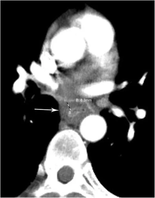
Özofajit; reflü, enfeksiyon, ilaç, radyasyon veya başka mukozal hasarlara bağlı özofagus inflamasyonudur. Özofajit değerlendirmesinde baryumlu özofagografi ve endoskopi BT'den daha duyarlıdır; acil başvuruda göğüs BT'si çekilmişse özofagus bulguları da değerlendirilebilir. Özofajit değerlendirmesinde baryumlu özofagografi ve endoskopi BT'den daha duyarlıdır. Fokal veya asimetrik özofagus duvar kalınlaşması maligniteyle ilişkili olabilir; özofagus perforasyonu mediastinal ya da akciğer apsesiyle komplike olabilir.
Faz ve pencere
- Venöz tanısal
- Özofagus duvarı ve çevresindeki mediasten değerlendirilir; perforasyon veya fistül kuşkusunda periözofageal ya da mediastinal hava ve sıvı, ekstraluminal kontrast veya fistül traktı aranabilir.
Önerilen pencereler G genişlik, M merkez; değerler Hounsfield birimi (HU)
- MediastenG 350 / M +50
- Yumuşak dokuG 400 / M +40
BT bulguları
- Duvar kalınlaşmasına rağmen korunmuş, düzgün dış kontur ve özofagus çevresindeki yağ planlarının büyük ölçüde seçilmesi
- Özofagus lümeninde sıvı veya hava-sıvı düzeyi; eşlik eden dismotilite ya da daralma varsa proksimal genişleme
- Özofagus perforasyonunda periözofageal veya mediastinal hava ya da sıvı görülebilir; inflamatuvar değişiklikler komşu mediastinal dokuları etkileyebilir.
- Özofagus perforasyonu kuşkusunda fokal duvar defekti veya kalınlaşması, ekstraluminal kontrast, periözofageal ya da mediastinal hava veya sıvı ve mediastinal apse bulguları aranabilir.
- Perforasyon kuşkusunda, uygun klinik ve protokolle oral suda çözünen kontrast kullanılarak ekstraluminal kontrast kaçağı araştırılabilir; kaçak görülmemesi perforasyonu tek başına dışlamaz.
Normalde
BT'de normal özofagus duvar kalınlığı anatomik konuma, distansiyon derecesine ve cinsiyete göre değişir; dış duvar sınırları ve komşu yapılar normalde seçilebilir olmalıdır. Özofagus duvar kalınlığı anatomik konuma, distansiyon derecesine ve cinsiyete göre değiştiğinden kıyaslamayı benzer düzey ve distansiyonda yap; normalde dış duvar sınırları seçilebilir olmalıdır.
Normal BT ile kıyasla
Aynı bölgenin, kaynak veri setinde patoloji içermediği belirtilen bir BT incelemesini kesit kesit kaydırın; organ sınırlarını açarak anatomiyi hasta görüntüsüyle karşılaştırın. Küçük rastlantısal bulgular tümüyle dışlanmamıştır.
Kaynak: TotalSegmentator v2.01 veri seti (Wasserthal ve ark., Radiology: Artificial Intelligence 2023), CC BY 4.0, vaka s1278; organ sınırları veri setinin otomatik segmentasyonundan, birleştirilerek sadeleştirildi. Görüntüler 2,5 mm kesit aralığına yeniden örneklendi.
Ayırıcı tanı
- Özofagus karsinomu
- çoğu kez fokal veya asimetrik kalınlaşma, düzensiz lümen daralması ve infiltratif sınır gösterir.
- Özofagus intramural hematomu
- duvar içinde kan birikimine bağlı konsantrik veya eksantrik duvar kalınlaşması ve yüksek atenüasyonlu kitle görülebilir.
- Özofagus dismotilitesi veya akalazya
- akalazyada alt özofagus sfinkterinin gevşemesi bozulur; kronik gıda ve sekresyon retansiyonu özofagusta belirgin genişlemeye yol açabilir.
- Radyasyon sonrası değişiklik
- radyasyon özofagus inflamasyonuna ve özofajite yol açabilir.
Tuzaklar
- Özofagus incelemelerin çoğunda dekomprese olduğundan duvar kalınlığını distansiyon derecesini dikkate alarak değerlendir; normal kalınlık anatomik konuma, distansiyona ve cinsiyete göre değişebilir.
- Özofajit değerlendirmesinde baryumlu özofagografi ve endoskopi BT'den daha duyarlıdır.
- Fokal asimetrik kalınlaşma veya dış kontur düzensizliğini yalnız özofajitle açıklama; tümör olasılığını ayrıca değerlendir.
Kendini dene
Acil özofagus patolojilerinde BT hangi yöntemlerle birlikte kullanıldığında yararlı bilgi sağlar?
Cevabı göster
Baryumlu özofagografi ve endoskopi. BT, acil özofagus patolojilerinde ilk başvurulan görüntüleme yöntemlerinden biri olabilir ve özofagografi ya da doğrudan görüntüleme ile birlikte kullanıldığında yararlı bilgi sağlar.
BT'de 13,5 mm'nin üzerindeki asimetrik özofagus duvar kalınlaşması hangi olasılık açısından anlamlı bulunmuştur?
Cevabı göster
Malignite. Çalışmada, BT'de 13,5 mm'yi aşan asimetrik özofagus duvar kalınlaşması malignite açısından anlamlı bulunmuştur; bu bulgu tek başına tanı koydurmaz.
Kaynaklar
Bu sayfadaki 29 cümle ve 2 quiz sorusu aşağıdaki kaynaklardan alıntılarla otomatik olarak karşılaştırıldı; 23 cümle bu karşılaştırmada düzeltildi (2026-10-09).
- Esophageal emergencies: another important cause of acute chest painpmc.ncbi.nlm.nih.gov
- ACR Appropriateness Criteria® Epigastric Pain.pubmed.ncbi.nlm.nih.gov
- Variants and Pitfalls in PET/CT Imaging of Gastrointestinal Cancers.pubmed.ncbi.nlm.nih.gov
- Cardiopulmonary and Gastrointestinal Manifestations of Eosinophil- associated Diseases and Idiopathic Hypereosinophilic Syndromes: Multimodality Imaging Approach.pubmed.ncbi.nlm.nih.gov
- Comprehensive update on select immune-mediated gastroenterocolitis syndromes: implications for diagnosis and management.pubmed.ncbi.nlm.nih.gov
- [Emphysematous gastritis secondary to gastric mucormycosis].pubmed.ncbi.nlm.nih.gov
- Radiation dose escalation with modified fractionation schedules for locally advanced NSCLC: A systematic review.pubmed.ncbi.nlm.nih.gov
- Role of endoscopy in GERD.pubmed.ncbi.nlm.nih.gov
- Identifying and distinguishing treatment effects and complications from malignancy at FDG PET/CT.pubmed.ncbi.nlm.nih.gov
- Esophageal Dysphagia in Children: State of the Art and Proposal for a Symptom-Based Diagnostic Approach.pubmed.ncbi.nlm.nih.gov
- Eosinophilic gastrointestinal diseases - Pathogenesis, diagnosis, and treatment.pubmed.ncbi.nlm.nih.gov
- Esophageal perforation in eosinophilic esophagitis: A systematic review on clinical presentation, management and outcomes.pubmed.ncbi.nlm.nih.gov
- Imaging of Drug-induced Complications in the Gastrointestinal System.pubmed.ncbi.nlm.nih.gov
- Gastrointestinal manifestations of leukemia.pubmed.ncbi.nlm.nih.gov
- Mediastinal Pancreatic Pseudocyst With Esophageal Compression Presenting as Acute Chest Pain.pubmed.ncbi.nlm.nih.gov
- Esophagitis Dissecans Superficialis: An Uncommon Endoscopic Finding.pubmed.ncbi.nlm.nih.gov
- The injured esophagus: imaging spectrum of esophageal emergencies.pubmed.ncbi.nlm.nih.gov
- Purulent Pericarditis and Tamponade From a Migrating Esophageal Foreign Body.pubmed.ncbi.nlm.nih.gov
- Retropharyngeal emphysema following adenoidectomy: a case report and review of literature.pubmed.ncbi.nlm.nih.gov
- Eosinophilic Cardiomyopathy in Eosinophilic Granulomatosis With Polyangiitis After Dupilumab Therapy.pubmed.ncbi.nlm.nih.gov
- Gastric perforation at the extremes of age: a comparative case report of NSAID-associated high-grade dysplasia versus methamphetamine-induced ischemic perforation.pubmed.ncbi.nlm.nih.gov
- Eosinophilic Enteritis Presenting with Recurrent Abdominal Pain and Bowel Obstruction in the Absence of Peripheral Eosinophilia: A Rare Case Report.pubmed.ncbi.nlm.nih.gov
- Dupilumab Alleviates Pulmonary Fibrosis by Interrupting Profibrotic Macrophage-Fibroblast Crosstalk and IL4R/JAK/STAT Signaling.pubmed.ncbi.nlm.nih.gov
- Case Report: Small bowel metastasis from esophageal squamous cell carcinoma: a case series and systematic review of clinical characteristics, tropism, and survival outcomes.pubmed.ncbi.nlm.nih.gov
- Stricturing Diseases of the Gastrointestinal Tract-Current Clinical Practice.pubmed.ncbi.nlm.nih.gov
- Verrucous Carcinoma of the Esophagus: A Deceiving Entity with an Uncertain Etiology - Case Report.pubmed.ncbi.nlm.nih.gov
- Severe Rectal Syphilis in the Setting of Profound HIV Immunosuppression: A Case Report Highlighting ERG/CD38 Immunophenotyping and a Review of the Literature.pubmed.ncbi.nlm.nih.gov
- Endoscopic Diagnosis of Eosinophilic Esophagitis: Basics and Recent Advances.pubmed.ncbi.nlm.nih.gov
- Rhinosinusitis: Evidence and experience - 2024.pubmed.ncbi.nlm.nih.gov
- Acute Esophageal Necrosis Secondary to Cardiogenic Shock: A Case Report and Review of the Literature.pubmed.ncbi.nlm.nih.gov
- Endoscopic Submucosal Dissection (ESD) of Upper Gastrointestinal Carcinomas: An Integrated Clinical and Pathological Perspective.pubmed.ncbi.nlm.nih.gov
- Computed Tomographic Findings and Clinical Manifestations of Acute Severe Hypersensitivity to a Non-Ionic Iodinated Contrast Agent in a Castrated Male Dog: A Case Report.pubmed.ncbi.nlm.nih.gov
- Boerhaave Syndrome Caused by Pancreatitis-Induced Gastric Outlet Obstruction: A Case Report.pubmed.ncbi.nlm.nih.gov
- Combined double-endoscopic surgery for a giant left-sided supradiaphragmatic esophageal diverticulum: a case report.pubmed.ncbi.nlm.nih.gov
- Hamman Syndrome: A Case Report of Interlobar Pneumothorax, Pneumopericardium, and Pneumorrhachis.pubmed.ncbi.nlm.nih.gov
- Streptococcal Esophagitis in an Immunocompetent Patient: A Rare Sequelae.pubmed.ncbi.nlm.nih.gov
- CT oral contrast and contemporary imaging pathways for suspected oesophageal perforation in the emergency department: a retrospective observational study.pubmed.ncbi.nlm.nih.gov
- Acute mediastinitis, mediastinal granuloma, and fibrosing mediastinitis: a narrative review.pubmed.ncbi.nlm.nih.gov
- Successful Multidisciplinary Management of Esophageal Perforation Complicated by Septic Shock: A Race Against Time.pubmed.ncbi.nlm.nih.gov
- Single-stage repair of congenital complete sternal agenesis with PFO-type interatrial communication and aortic arch variant complicated by an unexpected ascending aortic pseudoaneurysm: a case report.pubmed.ncbi.nlm.nih.gov
- Swallow contrast-enhanced ultrasonography detects a tiny cervical esophageal diverticulum missed on barium esophagography: a case report.pubmed.ncbi.nlm.nih.gov
- The Role of Multimodality Imaging in Atrial Fibrillation and Heart Failure: From Patient Selection to Procedural Ablation Guidance.pubmed.ncbi.nlm.nih.gov
- Endoscopic reduction of true gastrogastric intussusception in a dog.pubmed.ncbi.nlm.nih.gov
- Cervical Esophageal Characteristics in Smokers Versus Non-Smokers: An Ultrasonographic Comparative Analysis.pubmed.ncbi.nlm.nih.gov
- Case Report: Image-enhanced endoscopic characteristics of gastric amyloidosis with narrow-band imaging comparison.pubmed.ncbi.nlm.nih.gov
- Occult Esophageal Squamous Cell Carcinoma Presenting as Bone-Predominant Carcinoma of Unknown Primary: A Case Report of a p40-Negative Vertebral Metastasis.pubmed.ncbi.nlm.nih.gov
- Synchronous Metastatic Esophageal Squamous Cell Carcinoma and Acute Myeloid Leukemia: A Case Report.pubmed.ncbi.nlm.nih.gov
- Case Report: Synchronous Esophageal Squamous Cell Carcinoma and Gastric Cardia Adenocarcinoma with Hepatoid Differentiation After Neoadjuvant Chemoimmunotherapy.pubmed.ncbi.nlm.nih.gov
- Esophagogastric Variceal Bleeding in Cirrhotic Patients with Hepatocellular Carcinoma Receiving Systemic Therapy: A Retrospective Cohort Study.pubmed.ncbi.nlm.nih.gov
- Predictive value of high-risk esophageal varices in cirrhosis based on dual-energy CT combined with clinical and serologic features.pubmed.ncbi.nlm.nih.gov
- Findings of Endoscopic US and CT of Esophageal Disease.pubmed.ncbi.nlm.nih.gov
- Acute Traumatic Aortic Injury: What the Radiologist Needs to Know.pubmed.ncbi.nlm.nih.gov
- Imaging insights in veno-venous and veno-arterial extracorporeal membrane oxygenation (ECMO): CT protocols, underlying pathophysiology, and main complications.pubmed.ncbi.nlm.nih.gov
- Computed Tomography Imaging of Thoracic Aortic Surgery: Distinguishing Life-Saving Repairs from Life-Threatening Complications.pubmed.ncbi.nlm.nih.gov
- Can't Stomach It: A Dilated Tale of Achalasia.pubmed.ncbi.nlm.nih.gov
- Type II achalasia unmasked by EndoFLIP and high-resolution manometry in a child with eosinophilic esophagitis.pubmed.ncbi.nlm.nih.gov
- Intestinal Metaplasia of the Distal Esophagus in a Patient With Untreated Achalasia-An Unusual Case and Review of Literature.pubmed.ncbi.nlm.nih.gov
- Primary esophageal marginal zone lymphoma with respiratory symptoms: a case report and review of the literature.pubmed.ncbi.nlm.nih.gov
- Spontaneous Pneumothorax: A Review of Underlying Etiologies and Diagnostic Imaging Modalities.pubmed.ncbi.nlm.nih.gov
- Caught in a Cocoon: Operative Release of Primary Sclerosing Encapsulating Peritonitis With Endoscopic Management of Early Postoperative Duodenojejunal Intussusception.pubmed.ncbi.nlm.nih.gov
- Emphysematous conditions of the abdomen and pelvis: pearls and pitfalls.pubmed.ncbi.nlm.nih.gov
- A simple-to-use clinical nomogram to predict radiation esophagitis among esophageal cancer patients receiving radiotherapy: a retrospective cohort study.pubmed.ncbi.nlm.nih.gov
- Ileal malignant melanoma presenting with severe iron-deficiency anemia and active small-bowel bleeding: primary intestinal melanoma versus metastasis - a case report and narrative review.pubmed.ncbi.nlm.nih.gov
- High-Frequency Miniprobe Endoscopic Ultrasonography Across the Gastrointestinal Tract.pubmed.ncbi.nlm.nih.gov
- Mixed Neuroendocrine-Non-Neuroendocrine Neoplasm of the Esophagogastric Junction with Enteroblastic Differentiation and Morphologic-Immunophenotypic Discordance: A Case Report with Five-Year Disease-Free Survival.pubmed.ncbi.nlm.nih.gov
- Small Intestine Tumors: Diagnostic Role of Multiparametric Ultrasound.pubmed.ncbi.nlm.nih.gov
- German guidelines for the diagnosis and treatment of squamous-cell carcinoma and adenocarcinoma of the esophagus-version 4.0.pubmed.ncbi.nlm.nih.gov
- The comparison of wall thickness of esophagus and gastroesophageal junction using computed tomography with endoscopy and biopsy results.pubmed.ncbi.nlm.nih.gov
- Serious Adverse Events Following COVID-19 Vaccination: A Descriptive Case Series From a Rural Healthcare System.pubmed.ncbi.nlm.nih.gov
- Uncommon Causes of Interlobular Septal Thickening on CT Images and Their Distinguishing Features.pubmed.ncbi.nlm.nih.gov
- Imaging of abdominal and pelvic infections in the cancer patient.pubmed.ncbi.nlm.nih.gov
- Role of Transcatheter Arterial Embolization in Acute Refractory Non-variceal Upper Gastrointestinal Bleeding Not Controlled by Endoscopy: A Single-Center Experience and a Literature Review.pubmed.ncbi.nlm.nih.gov
- Successful multistage surgical management of Boerhaave syndrome: a case report and review of the literature.pubmed.ncbi.nlm.nih.gov
- EVT-Inspired Simplified Negative-Pressure Nasoesophageal Drainage for Cervical Abscess Secondary to Esophageal Perforation.pubmed.ncbi.nlm.nih.gov
- Pseudoachalasia as first manifestation of a diffusely infiltrative esophageal squamous cell carcinoma: A case report.pubmed.ncbi.nlm.nih.gov
- CA19-9-producing esophagogastric junction cancer with nodal recurrence outside the planned dissection field: a case report.pubmed.ncbi.nlm.nih.gov
- Diagnostic performance of whole-body diffusion-weighted imaging for staging esophageal cancer: a comparative study with PET-CT.pubmed.ncbi.nlm.nih.gov
- Anterior versus posterior peroral endoscopic myotomy: a prospective randomized controlled trial from a Thai tertiary center.pubmed.ncbi.nlm.nih.gov
- Massive Esophagogastric Variceal Bleeding from Chronic Portal Vein Thrombosis in a Non-Cirrhotic Young Man: A Resource-Limited Endoscopic Management Dilemma.pubmed.ncbi.nlm.nih.gov
- Pathophysiological management of gastric varices: From hemodynamics to targeted therapies.pubmed.ncbi.nlm.nih.gov
- Septic Pylephlebitis and Cavernous Transformation Presenting as Pyrexia of Unknown Origin: A Diagnostic Challenge.pubmed.ncbi.nlm.nih.gov
- Masked polycythemia vera presenting as non-cirrhotic portal hypertension with isolated gastric varices: a case report.pubmed.ncbi.nlm.nih.gov
- Metastatic Breast Cancer to the Esophagus Causing Pseudoachalasia.pubmed.ncbi.nlm.nih.gov
- Navigating Diagnostic Challenges in Pseudoachalasia: A Case Study of Esophagogastric Junction Adenocarcinoma.pubmed.ncbi.nlm.nih.gov
- Incidental esophageal and gastroesophageal junction uptake on ¹<sup>8</sup>F-FDG PET/CT: when is upper endoscopy warranted?pubmed.ncbi.nlm.nih.gov
- Orbital myositis last year and now Crohn's disease: A case report of a rare association.pubmed.ncbi.nlm.nih.gov
- Evaluation of Endoscopic Findings in Gastrointestinal Tract Wall Thickening Detected on Abdominal Radiological Imaging: A Two-Center Retrospective Descriptive Study.pubmed.ncbi.nlm.nih.gov
- Diagnosis and surgical management of an acquired cervical tracheoesophageal fistula in a Shih Tzu following foreign body removal.pubmed.ncbi.nlm.nih.gov
- Boerhaave Syndrome-Narrative Review.pubmed.ncbi.nlm.nih.gov
- Sodium glycididazole combined with definitive chemoradiotherapy for patients with esophageal squamous cell carcinoma: A multicenter randomized clinical trial.pubmed.ncbi.nlm.nih.gov
- From guidelines to radiology practice: navigating the 2023 ASCO guidelines for advanced gastroesophageal cancer and beyond.pubmed.ncbi.nlm.nih.gov
- Complications of Metabolic and Bariatric Surgery for the Gastroenterologist: A Comprehensive Review.pubmed.ncbi.nlm.nih.gov
- Esophageal peristalsis in health and disease: mechanistic insights.pubmed.ncbi.nlm.nih.gov
- Diagnosis, Treatment, and Follow-Up of Tracheo/Bronchomalacia in Children: The Italian Multicenter Experience.pubmed.ncbi.nlm.nih.gov
- Case Report: Endoscopic ultrasound in the diagnosis and endoscopic treatment of a delayed-presentation duodenal web in a toddler.pubmed.ncbi.nlm.nih.gov
- A Rare Case of Candida-Induced Acute Phlegmonous Esophagitis Complicated by Pericarditis and Myocarditis.pubmed.ncbi.nlm.nih.gov
- Management and Therapeutic Considerations of Hydrogen Peroxide-Induced Necrotizing Gastroenteritis: A Case Report.pubmed.ncbi.nlm.nih.gov
- Adverse Effects of Immune-Checkpoint Inhibitors: A Comprehensive Imaging-Oriented Review.pubmed.ncbi.nlm.nih.gov
- Non-neoplastic pathology at the crossroads between neck imaging and cardiothoracic imaging.pubmed.ncbi.nlm.nih.gov
- Esophageal Perforation and Acute Bacterial Mediastinitis: Other Causes of Chest Pain That Can Be Easily Missed.pubmed.ncbi.nlm.nih.gov
- Intramural hematoma of the esophagus.pubmed.ncbi.nlm.nih.gov
- Imaging the posterior mediastinum: a multimodality approach.pubmed.ncbi.nlm.nih.gov
- Esophageal emergencies: another important cause of acute chest pain.pubmed.ncbi.nlm.nih.gov
- Spontaneous bacteremia and spontaneous bacterial peritonitis share similar prognosis in patients with cirrhosis: a cohort study.pubmed.ncbi.nlm.nih.gov
- Patterns of injury and violence in Yaoundé Cameroon: an analysis of hospital data.pubmed.ncbi.nlm.nih.gov
- Involved-Field Irradiation Versus Elective Nodal Irradiation in Patients With Locally Advanced Esophageal Squamous Cell Carcinoma Treated With Neoadjuvant Chemoradiotherapy.pubmed.ncbi.nlm.nih.gov
- Fraction dose escalation of split-course concurrent chemoradiotherapy following induction chemo-immunotherapy in unresectable locally advanced oesophageal squamous carcinoma in China (GASTO-10102): a single-centre phase 1 study.pubmed.ncbi.nlm.nih.gov
- Stereotactic arrhythmia radioablation for ventricular tachycardia: Mechanisms, workflow, outcomes, and future directions.pubmed.ncbi.nlm.nih.gov
- Does laparoscopic surgery decrease the risk of atrial fibrillation after foregut surgery?pubmed.ncbi.nlm.nih.gov
Eğitim amaçlıdır; hasta başında tanı ve tedavi kararının yerine geçmez, tıbbi tavsiye değildir. Metinler yapay zekâ desteğiyle yazıldı ve otomatik bir adımda kaynak alıntılarıyla karşılaştırıldı; bu bir tıbbi doğrulama değildir ve içerik uzman radyolog incelemesinden geçmedi. Klinik kararlarda güncel kılavuzları, kurum protokollerini ve radyoloji raporunu esas alın.