Özet
Pulmoner nodüller solid ve subsolid olarak sınıflandırılır; tespit edilen nodüllerin %95'inden fazlası benign olup granülom veya intrapulmoner lenf nodudur. İnce kesit kontrastsız BT morfolojiyi, kalsifikasyonu, yağ içeriğini ve seri değişimi göstererek risk ayrımının temelini oluşturur. BT'de damar görüntüleri nedeniyle yalan pozitif saptamalar yaygındır ve benign lezyonların doğru tanısı gereksiz takibi önler.
Faz ve pencere
- Kontrastsız tanısal
- İnce kesit akciğer algoritmasında nodülün boyutu, kenar özellikleri, iç yoğunluk ve kalsifikasyonu değerlendirilir; çokfazlı kontrastlı BT benign ve malign nodüllerin ayırımında yüksek tanısal verimlilik sağlar.
Önerilen pencereler: Akciğer (G 1500 / M -600), Mediasten (G 350 / M 50).
BT bulguları
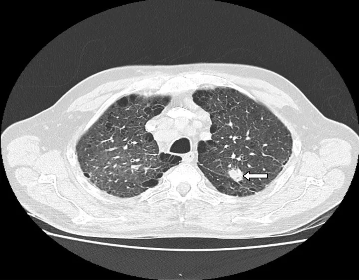
- Solid pulmoner nodüller, tipik olarak yuvarlak veya oval şekilli yumuşak doku odaklarıdır ve çevresindeki bronkovasküler yapılarla olan anatomik ilişkileri CT ile incelenir.
- Damar kesitinden ayırmak için ardışık ve koronal/sagittal kesitlerde devamlılığı izle.
- Nodül çapı, spikül (ışınımsı uzantı) varlığı, lobülasyon ve plevral çekinti gibi morfolojik özellikler malignite riskini artıran bağımsız prediktörlerdir.
- Santral, lameller, diffüz ya da konsantrik kalsifikasyon ve makroskopik yağ benign morfolojiyi destekler.
- Önceki ince kesit BT ile karşılaştırıldığında nodülün çap, hacim ve yoğunluktaki değişimler veya büyüme hızı (hacim katlanma süresi) izlenmelidir.
Ölçütler ve sınıflamalar
- Fleischner Society 2017
- Fleischner Society 2017 rehberi, tesadüfen saptanan solid ve subsolid nodüllerin yönetimine dair güncel önerileri sunar ve nodül boyutu ile morfolojisine dayalı takip protokolleri belirler.
- ACR Lung-RADS v2022
- Tarama BT’si bulguları için kategorik raporlama sistemidir; tesadüfi nodül rehberi değildir.
Normalde
Akciğerlerde oblik fissürler tam veya kısmi olabilir ve bu varyasyonlar BT'de normal anatomiyi etkiler; bronkovasküler demetlerin dallanma paterni nodül ayırımında önemlidir. Odağın damar dalıyla devam edip etmediğine ve normal parankim dokusunu örtüp örtmediğine bak.
Normal BT ile kıyasla
Aynı bölgenin, kaynak veri setinde patoloji içermediği belirtilen bir BT incelemesini kesit kesit kaydırın; organ sınırlarını açarak anatomiyi hasta görüntüsüyle karşılaştırın. Küçük rastlantısal bulgular tümüyle dışlanmamıştır.
Kaynak: TotalSegmentator v2.01 veri seti (Wasserthal ve ark., Radiology: Artificial Intelligence 2023), CC BY 4.0, vaka s1278; organ sınırları veri setinin otomatik segmentasyonundan, birleştirilerek sadeleştirildi. Görüntüler 2,5 mm kesit aralığına yeniden örneklendi.
Ayırıcı tanı
- Pulmoner damar kesiti
- multiplanar izde tübüler devamlılık gösterir.
- Perifissüral lenf nodu
- fissüre yakın düzgün oval, üçgen veya poligonal şekildedir.
- Granülom
- merkezi, diffüz veya konsantrik kalsifikasyon benign morfoloji lehinedir.
- Enfeksiyöz nodül
- tree-in-bud, sentrilobüler odak veya çevresel konsolidasyon eşlik edebilir.
Tuzaklar
- Damar komşuluğundaki nodül, multiplanar devamlılık izlenmeden damar kesiti veya parankimal nodül diye sınıflanabilir.
- Yüksek kesit kalınlığı ve nodülün kesit konumu, özellikle küçük nodüllerde ölçüm hatasına ve yalancı boyut/sıkılık değerlendirmesine neden olabilir.
- Lung-RADS sistemleri tarama BT'si bulguları için tasarlanırken, Fleischner Society rehberi sadece tesadüfen saptanan nodüllerin yönetimini kapsar; bu iki yaklaşım birbirinin yerine kullanılmamalıdır.
Kendini dene
İnce kesitte yuvarlak solid odak var. Koronal planda pulmoner arter dalı ile kesintisiz devam ediyor. En olası yorum?
Cevabı göster
Pulmoner damar kesiti. Multiplanar devamlılık damar kesitini gösterir; nodül parankim içinde bağımsız olmalıdır.
Tesadüfi solid pulmoner nodülün morfolojisi, kalsifikasyonu ve yağ içeriği en iyi hangi görüntüleme yöntemiyle değerlendirilir?
Cevabı göster
İnce kesit kontrastsız akciğer BT. İnce kesit kontrastsız akciğer penceresi BT, parankim yapısını, nodül kenarlarını ve iç kalsifikasyon/yağ varlığını en iyi gösteren standart yöntemdir.
Bu konunun yer aldığı turlar
Kaynaklar
Bu sayfadaki 33 cümle ve 2 quiz sorusu aşağıdaki kaynaklardan alıntılarla tek tek karşılaştırıldı; 11 cümle bu karşılaştırmada düzeltildi (2026-10-08).
- Evaluating the Patient With a Pulmonary Nodule: A Review.pubmed.ncbi.nlm.nih.gov
- Contribution of magnetic resonance imaging in lung cancer imaging.pubmed.ncbi.nlm.nih.gov
- Imaging of Solid Pulmonary Nodules.pubmed.ncbi.nlm.nih.gov
- Combined small cell lung carcinoma and giant cell carcinoma: a case report.pubmed.ncbi.nlm.nih.gov
- Multi-observer concordance and accuracy of the British Thoracic Society scale and other visual assessment qualitative criteria for solid pulmonary nodule assessment using FDG PET-CT.pubmed.ncbi.nlm.nih.gov
- The first case of gland inclusion in an intrapulmonary lymph node: a mimic of metastasis.pubmed.ncbi.nlm.nih.gov
- Solid pulmonary nodule risk assessment and decision analysis: comparison of four prediction models in 285 cases.pubmed.ncbi.nlm.nih.gov
- Multicenter external validation of two malignancy risk prediction models in patients undergoing 18F-FDG-PET for solitary pulmonary nodule evaluation.pubmed.ncbi.nlm.nih.gov
- Influencing factors of pneumothorax and parenchymal haemorrhage after CT-guided transthoracic needle biopsy: single-institution experience.pubmed.ncbi.nlm.nih.gov
- Primary solid lung cancerous nodules with different sizes: computed tomography features and their variations.pubmed.ncbi.nlm.nih.gov
- The effects of computed tomography with iterative reconstruction on solid pulmonary nodule volume quantification.pubmed.ncbi.nlm.nih.gov
- A New Outlook on the Ability to Accumulate an Iodine Contrast Agent in Solid Lung Tumors Based on Virtual Monochromatic Images in Dual Energy Computed Tomography (DECT): Analysis in Two Phases of Contrast Enhancement.pubmed.ncbi.nlm.nih.gov
- Pediatric pulmonary tuberculoma with a solid pulmonary nodule detected on chest computed tomography.pubmed.ncbi.nlm.nih.gov
- Vessel suppressed chest Computed Tomography for semi-automated volumetric measurements of solid pulmonary nodules.pubmed.ncbi.nlm.nih.gov
- Guidelines for Management of Incidental Pulmonary Nodules Detected on CT Images: From the Fleischner Society 2017.pubmed.ncbi.nlm.nih.gov
- Fleischner Society Guideline Recommendations for Incidentally Detected Pulmonary Nodules and the Probability of Lung Cancer.pubmed.ncbi.nlm.nih.gov
- [Not Available].pubmed.ncbi.nlm.nih.gov
- Small low-risk pulmonary nodules on chest digital radiography: can we predict whether the nodule is benign?pubmed.ncbi.nlm.nih.gov
- Pulmonary tuberculosis presenting as a hilar mass.pubmed.ncbi.nlm.nih.gov
- Pulmonary sclerosing pneumocytoma masquerading as lung metastasis: A case report.pubmed.ncbi.nlm.nih.gov
- Computer aided characterization of the solitary pulmonary nodule using volumetric and contrast enhancement features.pubmed.ncbi.nlm.nih.gov
- The incidental pulmonary nodule in a child. Part 2: Commentary and suggestions for clinical management, risk communication and prevention.pubmed.ncbi.nlm.nih.gov
- Efficiency of pulmonary nodule risk scoring systems in Turkish population.pubmed.ncbi.nlm.nih.gov
- Clinical Outcomes of Resected Pure Ground-Glass, Heterogeneous Ground-Glass, and Part-Solid Pulmonary Nodules.pubmed.ncbi.nlm.nih.gov
- Relationship between peripheral lung cancer and the surrounding bronchi, pulmonary arteries, pulmonary veins: a multidetector CT observation.pubmed.ncbi.nlm.nih.gov
- [Establishment of A Clinical Prediction Model of Solid Solitary Pulmonary Nodules].pubmed.ncbi.nlm.nih.gov
- Pulmonary nodules with explosive calcification were finally diagnosed as lung adenocarcinom: A case report.pubmed.ncbi.nlm.nih.gov
- [Pulmonary tumors].pubmed.ncbi.nlm.nih.gov
- Evaluation and management of pulmonary nodules: state-of-the-art and future perspectives.pubmed.ncbi.nlm.nih.gov
- Cytoreduction for colorectal metastases: liver, lung, peritoneum, lymph nodes, bone, brain. When does it palliate, prolong survival, and potentially cure?pubmed.ncbi.nlm.nih.gov
- MR Volumetry of Lung Nodules: A Pilot Study.pubmed.ncbi.nlm.nih.gov
- Impact on Prognosis of Stage I Non-Small Cell Lung Cancer Secondary to Delays in Diagnostic Workup.pubmed.ncbi.nlm.nih.gov
- Diagnostic Imaging: Appropriate and Safe Use.pubmed.ncbi.nlm.nih.gov
- Clinical spectrum of intrathoracic Castleman disease: a retrospective analysis of 48 cases in a single Chinese hospital.pubmed.ncbi.nlm.nih.gov
- Three-dimensionally printed navigational template: a promising guiding approach for lung biopsy.pubmed.ncbi.nlm.nih.gov
- Updated Fleischner Society Guidelines for Managing Incidental Pulmonary Nodules: Common Questions and Challenging Scenarios.pubmed.ncbi.nlm.nih.gov
- Management of Incidental Lung Nodules.pubmed.ncbi.nlm.nih.gov
- Management of incidental lung nodules <8 mm in diameter.pubmed.ncbi.nlm.nih.gov
- Lung-RADS v2022 Update.pubmed.ncbi.nlm.nih.gov
- ACR Lung-RADS v2022: Assessment Categories and Management Recommendations.pubmed.ncbi.nlm.nih.gov
- ACR Lung-RADS v2022: Assessment Categories and Management Recommendations.pubmed.ncbi.nlm.nih.gov
- Development and application of an electronic synoptic report for reporting and management of low-dose computed tomography lung cancer screening examination.pubmed.ncbi.nlm.nih.gov
- Typical CT Features of Intrapulmonary Lymph Nodes: A Review.pubmed.ncbi.nlm.nih.gov
- Pulmonary perifissural nodules on CT scans: rapid growth is not a predictor of malignancy.pubmed.ncbi.nlm.nih.gov
- Initial LDCT appearance of incident lung cancers in the ITALUNG trial.pubmed.ncbi.nlm.nih.gov
- The calcified lung nodule: What does it mean?pubmed.ncbi.nlm.nih.gov
- Multiple calcifying fibrous pseudotumors disseminated in the pleura.pubmed.ncbi.nlm.nih.gov
- Pulmonary Hyalinizing Granuloma: A Rare Cause of a Benign Lung Mass.pubmed.ncbi.nlm.nih.gov
- Clinical Differences between Community-Acquired Mycoplasma pneumoniae Pneumonia and COVID-19 Pneumonia.pubmed.ncbi.nlm.nih.gov
- Bronchiolar disorders: a clinical-radiological diagnostic algorithm.pubmed.ncbi.nlm.nih.gov
- Early identification of novel coronavirus (COVID-19) pneumonia using clinical and radiographic findings.pubmed.ncbi.nlm.nih.gov
- A mathematical simulation to assess variability in lung nodule size measurement associated with nodule-slice position.pubmed.ncbi.nlm.nih.gov
- CT features of peripheral pulmonary carcinoid tumors.pubmed.ncbi.nlm.nih.gov
- Pulmonary Nodules.pubmed.ncbi.nlm.nih.gov
- Probability of cancer in pulmonary nodules detected on first screening CT.pubmed.ncbi.nlm.nih.gov
- A narrative review on CT-based evaluation and prediction of lung nodule growth: current status and future directions.pubmed.ncbi.nlm.nih.gov
- CT-guided Preoperative Localization of Impalpable Pulmonary Nodules: Comparison Between Two Devices.pubmed.ncbi.nlm.nih.gov
- Diagnostic Performance of 3.0-Tesla Magnetic Resonance Imaging for Pulmonary Nodules and Masses: A Comparative Cross-Sectional Study With Multidetector Computed Tomography.pubmed.ncbi.nlm.nih.gov
- Metastatic Retroperitoneal Rhabdomyosarcoma in a Child: A Case Report Highlighting the Role of Imaging in Diagnosis, Staging, and Follow-Up.pubmed.ncbi.nlm.nih.gov
- Thoracolithiasis: a rare and frequently underrecognized entity - a retrospective single-center study and narrative literature review.pubmed.ncbi.nlm.nih.gov
- Lipoid Pneumonia: HRCT and MRI Spectrum, Diagnostic Pitfalls, and Imaging-Based Diagnostic Workflow.pubmed.ncbi.nlm.nih.gov
- Analysis of the clinicopathological characteristics of pulmonary sclerosing pneumocytoma (PSP).pubmed.ncbi.nlm.nih.gov
- A partial response to atezolizumab in alveolar soft part sarcoma with near-complete resolution of extensive pulmonary metastases: a case report.pubmed.ncbi.nlm.nih.gov
- Pulmonary cryptococcosis with or without immunocompromise: a retrospective comparative study of clinical and CT features and influential factors for lesion absorption.pubmed.ncbi.nlm.nih.gov
- Case Report: Pulmonary benign metastasizing leiomyoma with mild <sup>18</sup>F-FDG uptake occurring 30 years after hysterectomy.pubmed.ncbi.nlm.nih.gov
- Incidental Lung Lesions on the CT Component of Oncologic FDG PET/CT: A Pictorial Review of Interpretive Pitfalls and Diagnostic Clues.pubmed.ncbi.nlm.nih.gov
- MRI for Lung Cancer Management: Any Closer to Clinical Application?pubmed.ncbi.nlm.nih.gov
- Clinical and computed tomography (CT) features of nodular pulmonary histoplasmosis.pubmed.ncbi.nlm.nih.gov
- Imaging features of lung cancer with cystic airspaces: clinical utility, challenges, and perspectives.pubmed.ncbi.nlm.nih.gov
- Development of a Radiologic Nomogram to Predict Invasiveness in Pulmonary Pure Ground-Glass Opacities: Analysis of the GORDON Cohort.pubmed.ncbi.nlm.nih.gov
- Congenital pulmonary airway malformation mimicking lung cancer in adults: a pitfall in oncologic diagnosis.pubmed.ncbi.nlm.nih.gov
- Construction and validation of a prediction model for malignant pulmonary nodules based on imaging, demographic, and laboratory features.pubmed.ncbi.nlm.nih.gov
- Comparison of the efficacy of spiral CT combined with advanced multiplanar reconstruction in detecting the progression of pneumoconiosis.pubmed.ncbi.nlm.nih.gov
- Evaluation of benign and malignant pulmonary nodules based on clinical and contrast-enhanced computed tomography data: a multicenter study.pubmed.ncbi.nlm.nih.gov
- Serum tumor markers combined with HRCT for malignancy risk assessment of solitary pulmonary nodules: a retrospective study.pubmed.ncbi.nlm.nih.gov
- Application value of high-resolution CT imaging features combined with texture analysis in patients with solitary pulmonary nodules.pubmed.ncbi.nlm.nih.gov
- Clinical Relevance of Calcifications in Osteosarcoma Lung Metastases: Correlations Among Radiological Patterns, Histological Subtypes, and Chemotherapy Responses.pubmed.ncbi.nlm.nih.gov
- The role of radiological imaging in differentiating malignant and benign pulmonary nodules: a retrospective study.pubmed.ncbi.nlm.nih.gov
- Next-gen paediatric imaging: ultra-low dose chest photon-counting CT approaching chest radiograph doses.pubmed.ncbi.nlm.nih.gov
- Evaluating Korean Lung-RADS Category 2b: Diagnostic Performance and Interreader Agreement in Lung Cancer Screening.pubmed.ncbi.nlm.nih.gov
- Bridging Targeting Precision and Oncologic Safety: Localization Accuracy for Margin Adequacy in Cone-Beam Computed Tomography-Guided Pulmonary Nodule Resection.pubmed.ncbi.nlm.nih.gov
- Quantifying the Resolution Ceiling of Prototype-Based Few-Shot Segmentation on Sub-Centimetre Pulmonary Nodules: A Training-Free Geometric Analysis.pubmed.ncbi.nlm.nih.gov
- Unveiling Hidden Variations in Lung Fissures: A Cadaveric Perspective on Surgical and Diagnostic Challenges.pubmed.ncbi.nlm.nih.gov
- Thoracoscopic left apicoposterior segmentectomy for bronchiectasis with a displaced B¹⁺² bronchus: a case report.pubmed.ncbi.nlm.nih.gov
- Uniportal robot-assisted resection and reconstruction of the left bronchial secondary carina in a pediatric patient.pubmed.ncbi.nlm.nih.gov
- Arterial Supply to the Atrioventricular Node: Anatomical Variability and Clinical Implications.pubmed.ncbi.nlm.nih.gov
- Three-Dimensional Bronchovascular Modelling in Sublobar Pulmonary Resection: A Tool for Personalised Thoracic Surgery.pubmed.ncbi.nlm.nih.gov
- DeepBranchAI: a transferable 3D segmentation model for branching networks.pubmed.ncbi.nlm.nih.gov
- Are respiratory physicians adherent to guidelines when evaluating pulmonary nodules?pubmed.ncbi.nlm.nih.gov
- Coronary Calcium Screening and Incidental Lung Cancer Detection in Patients Ineligible for US Preventive Services Task Force Screening.pubmed.ncbi.nlm.nih.gov
- ACR Appropriateness Criteria® Indolent Lung Cancer.pubmed.ncbi.nlm.nih.gov
- Natural history and clinical characteristics of pulmonary subsolid nodules in pediatric patients: a cohort study.pubmed.ncbi.nlm.nih.gov
- Lung-RADS 0: How much is too much?pubmed.ncbi.nlm.nih.gov
- The CT colonography radiology and data system: history, updates, and future directions.pubmed.ncbi.nlm.nih.gov
- Lung Cancer Yield by Baseline Screening Lung CT Screening Reporting and Data System (Lung-RADS) Assignment and Patient Factors: The Population-Based Research to Optimize the Screening Process Lung Consortium.pubmed.ncbi.nlm.nih.gov
- Interobserver Variability in Lung-RADS Categorization Between Tertiary and Non-Tertiary Hospitals, and Its Implications for Patient Management.pubmed.ncbi.nlm.nih.gov
- Impact of COPD on Lung Cancer Screening Positivity in a Specialist-Managed Cohort From Brazil.pubmed.ncbi.nlm.nih.gov
- Incidental Breast Findings on Chest CT: Rates of Report and Follow-Up.pubmed.ncbi.nlm.nih.gov
- Exploring the key clinical and computed tomography features for distinguishing high-grade lung cancers from morphologically similar benign tumors: a two-center case-control study.pubmed.ncbi.nlm.nih.gov
- RADS ALPHABET: news and tips for young and general radiologists.pubmed.ncbi.nlm.nih.gov
- Lung cancer screening with low-dose CT: definition of positive, indeterminate, and negative screen results. A nodule management recommendation from the European Society of Thoracic Imaging.pubmed.ncbi.nlm.nih.gov
- The TSANZ Practical Guide for Clinicians in the Management of Screen- and Incidentally-Detected Nodules.pubmed.ncbi.nlm.nih.gov
- Prediction of visceral pleural invasion of clinical stage IA lung adenocarcinoma based on computed tomography features.pubmed.ncbi.nlm.nih.gov
- Antimicrobial prescribing guidelines for horses in Australia.pubmed.ncbi.nlm.nih.gov
- Unusual Presentation of Atrial Myxoma: A Case Report and Review of the Literature.pubmed.ncbi.nlm.nih.gov
- Acute Endobronchial Dissemination of a Tuberculoma Following EBUS-GS-Guided Transbronchial Biopsy: A Case Report.pubmed.ncbi.nlm.nih.gov
- Invasive pulmonary aspergillosis as a sentinel manifestation of good syndrome in a patient with thymoma.pubmed.ncbi.nlm.nih.gov
- Septic arthritis caused by Nocardia asiatica: a case report and narrative review.pubmed.ncbi.nlm.nih.gov
- Parameter-Efficient LoRA-GRL Adaptation for Cross-Center Classification of Benign and Malignant Lung Nodules on Heterogeneous Standard-Dose Chest CT: A Multi-Institutional Study from Palestine.pubmed.ncbi.nlm.nih.gov
- Diagnostic performance of commercial AI systems versus participating radiologists for pulmonary nodule detection in routine clinical practice.pubmed.ncbi.nlm.nih.gov
Eğitim amaçlıdır; hasta başında tanı ve tedavi kararının yerine geçmez. İçerik yapay zekâ destekli bir süreçle güncel literatüre göre cümle cümle kaynaklarla karşılaştırıldı, ancak uzman radyolog incelemesinden geçmedi. Klinik kararlarda güncel kılavuzları, kurum protokollerini ve radyoloji raporunu esas alın.