Özet
İntrakraniyal damarların değerlendirilmesinde MPR, CPR, MIP ve VR gibi yeniden yapılandırma yöntemleri kullanılabilir. BT anjiyografide yalancı oklüzyonun gerçek oklüzyon olarak yorumlanması, trombüs yerinin belirlenmesini ve tedavi planlamasını etkileyebilir.
Okuma sırası
- Petroz ICA. Petroz apeks düzeyinde karotid kanalı tanıyın ve opak ICA'yı kemik kanalının içinden kavernöz segmente doğru izlemeye başlayın.
- Karotid kanal çevresindeki artefakt. Lümen dar görünüyorsa aynı kesitteki koyu-açık çizgilenmenin yönünü kontrol edin; damar sınırını kesen bant, yoğun kafa tabanı kemiğiyle ilişkili olabilir.
- Petroz-kavernöz ICA geçişi. Ardışık kaynak kesitlerde ve eğrisel MPR'de petroz-kavernöz ICA geçişini yeniden izleyin; kesintinin başka düzlemde kaybolup kaybolmadığını gösterin.
- Karşı ICA ve vertebrobaziler bileşke. Aynı seviyedeki karşı ICA'yı ve vertebrobaziler bileşkeyi gözden geçirerek eşlik eden gerçek damar darlığı, oklüzyon veya diseksiyon bulgusunu arayın.
Faz ve pencere
- BTA tanısal
- BT anjiyografisinde yoğun kemik yapılarının neden olduğu ışın sertleşmesi ve çizgilenme artefaktları, damar yolu üzerindeki kontrast sütununu kesintiye uğratabilir ve yalancı darlık izlenimi verebilir. Vertebral arterin foramen magnuma girişi ve baziler arter başlangıcında posterior fossa artefaktı lümen sınırını bulanıklaştırabilir.
Önerilen pencereler: Anjiyo (G 600 / M 150), Kemik (G 1800 / M 400).
BT bulguları
- Artefakt kaynaklı lümen silinmesi ardışık aksiyel kesitlerde değişken görünür ve damar merkez hattı boyunca sabit bir kontur bozukluğu oluşturmayabilir.
- İntrakraniyal damar stenozunun değerlendirilmesinde MPR, CPR, MIP ve VR rekonstrüksiyonları kullanılabilir.
Normalde
Normal BTA'da petroz ICA, karotid kanal içinden kavernöz segmente düzenli konturla geçer; vertebral arterler foramen magnumda birleşip baziler arteri oluşturur.
Normal BT ile kıyasla
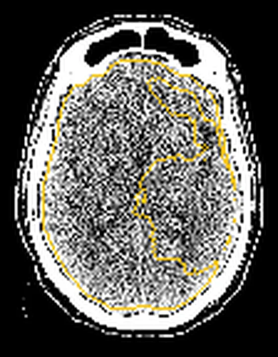
Aynı bölgenin, kaynak veri setinde patoloji içermediği belirtilen bir BT incelemesini kesit kesit kaydırın; organ sınırlarını açarak anatomiyi hasta görüntüsüyle karşılaştırın. Küçük rastlantısal bulgular tümüyle dışlanmamıştır.
Kaynak: TotalSegmentator v2.01 veri seti (Wasserthal ve ark., Radiology: Artificial Intelligence 2023), CC BY 4.0, vaka s0849; organ sınırları veri setinin otomatik segmentasyonundan, birleştirilerek sadeleştirildi. Görüntüler 2,5 mm kesit aralığına yeniden örneklendi.
Ayırıcı tanı
- Aterosklerotik stenoz
- damar duvarında anatomik olarak tutarlı eksantrik veya konsantrik daralma ve bazen kalsifiye plak görülür.
- Vazospazm
- birden fazla segmentte düzgün daralma ve klinik bağlama uygun yaygın damar kalibre değişikliği görülebilir.
- Yetersiz kontrastlanma, BTA'nın büyük damar oklüzyonunu değerlendirmedeki tanısal doğruluğunu azaltabilir.
Tuzaklar
- Petroz ICA'yı kemik penceresindeki çizgilenmeye bakarak ölçme; anjiyo penceresinde kaynak kesitleri ve eğrisel MPR'yi birlikte kullan.
- Karşı taraf damarın normal olması gerçek tek taraflı stenozu dışlamaz; karşılaştırmayı damar lümeninin kesitler boyunca sürekliliğiyle tamamla.
- Çift enerji kemik çıkarma sonrası görüntülerde kemik kalıntısı ve damar bütünlüğü ayrıca değerlendirilmelidir.
- Çift enerjili kemik çıkarma sonrası görüntülerde kemik kalıntılarını ve damar bütünlüğünü değerlendirin.
Kendini dene
64 kesitli BT anjiyografisinde diseksiyon veya damar oklüzyonunu taklit edebilen, klinik açıdan en önemli artefakt türü hangisidir?
Cevabı göster
Akım artefaktları. S66'da diseksiyon veya damar oklüzyonunu taklit edebilen, klinik açıdan en önemli artefaktların akım artefaktları olduğu belirtiliyor.
S66 çalışmasında 64 kesitli BT anjiyografide diseksiyon veya damar oklüzyonunu taklit eden akım artefaktları hastaların yüzde kaçında görüldü?
Cevabı göster
%14. S66'ya göre diseksiyon veya damar oklüzyonunu taklit eden akım artefaktları hastaların %14'ünde görüldü.
Kaynaklar
Bu sayfadaki 23 cümle ve 2 quiz sorusu aşağıdaki kaynaklardan alıntılarla otomatik olarak karşılaştırıldı; 8 cümle bu karşılaştırmada düzeltildi (2026-10-09).
- Differentiating True Occlusion from Pseudo-Occlusion: The Role of Extended Multiphase Computed Tomography Angiography Scan Range in Internal Carotid Artery Occlusionpmc.ncbi.nlm.nih.gov
- Multimodal imaging in neuro-ophthalmologypmc.ncbi.nlm.nih.gov
- Narrower jugular bulbs and their tributaries are associated with skull base venous hyperintensity on arterial spin-labeling MRIpmc.ncbi.nlm.nih.gov
- An Overview of Cone-Beam Computed Tomography and Dental Panoramic Radiography in Dentistry in the Communitypmc.ncbi.nlm.nih.gov
- Advanced Radiotherapy Technologies in Spine Tumors: What the Surgeon Needs to Knowpmc.ncbi.nlm.nih.gov
- Where Does the Signal Go? Technical Challenges of CT Perfusion in Lacunar and Infratentorial Stroke.pubmed.ncbi.nlm.nih.gov
- Dual-Energy CT in Head and Neck Imaging.pubmed.ncbi.nlm.nih.gov
- 128-slice acceletated-pitch dual energy CT angiography of the head and neck: comparison of different low contrast medium volumes.pubmed.ncbi.nlm.nih.gov
- Imaging and Management of Blunt Cerebrovascular Injury.pubmed.ncbi.nlm.nih.gov
- State of the art: noninvasive imaging and management of neurovascular trauma.pubmed.ncbi.nlm.nih.gov
- Acute Traumatic Aortic Injury: What the Radiologist Needs to Know.pubmed.ncbi.nlm.nih.gov
- Beyond Angiography: Cardiac CT for Planning Complex PCI in Calcified Coronary Lesions.pubmed.ncbi.nlm.nih.gov
- Coronary Computed Tomography Angiography to Guide Percutaneous Coronary Intervention: Expert Opinion from a SCAI/SCCT Roundtable.pubmed.ncbi.nlm.nih.gov
- Society for Cardiovascular Magnetic Resonance 2024 Cases of SCMR case series.pubmed.ncbi.nlm.nih.gov
- Aneurysms Arising from Persistent Arteries in the Vertebrobasilar Paramedian Longitudinal Axis: Two Case Reports.pubmed.ncbi.nlm.nih.gov
- Neuroangiography: Review of Anatomy, Periprocedural Management, Technique, and Tips.pubmed.ncbi.nlm.nih.gov
- Three-Dimensional Constructive Interference in Steady State (3D CISS) Imaging and Clinical Applications in Brain Pathology.pubmed.ncbi.nlm.nih.gov
- 2.5-mm articulated endoluminal forceps using a compliant mechanism.pubmed.ncbi.nlm.nih.gov
- Advances in human intracranial electroencephalography research, guidelines and good practices.pubmed.ncbi.nlm.nih.gov
- Commentary: an eye on PET quantification.pubmed.ncbi.nlm.nih.gov
- Coronary Computed Tomography Angiography for the Diagnosis and Revascularization Guidance of Coronary Bifurcation Lesions: A Contemporary Review.pubmed.ncbi.nlm.nih.gov
- Stroke or Seizure? Diagnostic Role of Neuroimaging in Acute Neurologic Mimics.pubmed.ncbi.nlm.nih.gov
- From intravascular imaging to adaptive vascular care: intelligent photonics and digital twins in panvascular disease.pubmed.ncbi.nlm.nih.gov
- Multimodal imaging in neuro-ophthalmology.pubmed.ncbi.nlm.nih.gov
- Artificial Intelligence for Computer-Aided Detection in Endovascular Interventions: Clinical Applications, Validation, and Translational Perspectives.pubmed.ncbi.nlm.nih.gov
- Treatment of carotid stenosis: an updated review.pubmed.ncbi.nlm.nih.gov
- Congenital Anomalies and Variations of the Carotid and Vertebral Arteries: A Case-Based Imaging Review.pubmed.ncbi.nlm.nih.gov
- An Atlas of Neonatal Neurovascular Imaging Anatomy as Depicted with Microvascular Imaging: The Intracranial Arteries.pubmed.ncbi.nlm.nih.gov
- A Practical Approach to Intracranial Dural Arteriovenous Fistulas: Pathogenesis, Classification and Management.pubmed.ncbi.nlm.nih.gov
- The role of contrast-enhanced transcranial color-coded real-time sonography combined with the common carotid artery compression test in preoperative evaluation of anterior communicating artery patency in patients with insufficient temporal bone windows.pubmed.ncbi.nlm.nih.gov
- Clinical analysis of 48 patients with carotid web diagnosed by digital subtraction angiography.pubmed.ncbi.nlm.nih.gov
- A systematic review on imaging and diagnostic modalities for the detection, evaluation, and surgical planning of upper and lower extremity lymphedema.pubmed.ncbi.nlm.nih.gov
- Vertebral Artery Pseudoaneurysm in a Single-Vessel Posterior Circulation With Chronic Contralateral Occlusion and Recent Methamphetamine Use.pubmed.ncbi.nlm.nih.gov
- Clinical implications of vessel wall imaging-State-of-the-art review.pubmed.ncbi.nlm.nih.gov
- A review of the pathogenic mechanism and clinical management progress of in-stent calcification restenosis.pubmed.ncbi.nlm.nih.gov
- Neuroimaging for neurovascular complications of traumatic brain injury.pubmed.ncbi.nlm.nih.gov
- Operative Procedures for Ultrasound Assessment of Extracranial Artery Disease: A Narrative Review by the Italian Society for Vascular Investigation (SIDV).pubmed.ncbi.nlm.nih.gov
- Posterior Circulation Stroke After Subclavian Artery Thrombosis Following Low-Voltage Electrical Injury in an Active Woman With Previously Unrecognized Prothrombotic Risk Factors: A Case Report.pubmed.ncbi.nlm.nih.gov
- Ruptured Brain Arteriovenous Malformation Coexisting With Reversible Cerebral Vasoconstriction Syndrome: A Case Report.pubmed.ncbi.nlm.nih.gov
- Malignant Hemispheric Stroke Secondary to Cavernous Internal Carotid Artery Stenosis in the Setting of Sepsis: A Case Report.pubmed.ncbi.nlm.nih.gov
- Radiation dose optimization in coronary CT angiography clinical application of absolute phase gating in patients with atrial fibrillation.pubmed.ncbi.nlm.nih.gov
- Cardiac CT in the Era of Precision Cardiology: From Calcium Scoring to Comprehensive Risk Profiling.pubmed.ncbi.nlm.nih.gov
- Flow diverter treatment for MCA M1 aneurysms: clinical outcomes and predictors of occlusion.pubmed.ncbi.nlm.nih.gov
- Compression Syndromes of the Stylohyoid Complex: Neural, Arterial, and Venous Phenotypes in a Dynamic Neurovascular Corridor.pubmed.ncbi.nlm.nih.gov
- Anatomic variations in basilar artery termination: a systematic review and meta-analysis with presentation of an illustrative case.pubmed.ncbi.nlm.nih.gov
- Intracranial vasculopathy with MR vessel wall imaging: a case series.pubmed.ncbi.nlm.nih.gov
- Understanding Photon-Counting CT: Physics, Detector Technology, and Image Reconstructions.pubmed.ncbi.nlm.nih.gov
- Clinical benefits and current challenges of photon-counting detector CT in vascular imaging.pubmed.ncbi.nlm.nih.gov
- Dual-energy computed tomography in children: Technique and clinical applications.pubmed.ncbi.nlm.nih.gov
- Long-term safety and effectiveness of the "OptEase" vena cava filter.pubmed.ncbi.nlm.nih.gov
- NIR Indocyanine-White Light Overlay Visualization for Neuro-Oto-Vascular Preservation During Anterior Transpetrosal Approaches: A Technical Note.pubmed.ncbi.nlm.nih.gov
- Endoscopic endonasal treatment of Meckel's cave epidermoid cysts: case series and systematic review.pubmed.ncbi.nlm.nih.gov
- Relationships of the vidian nerve and internal carotid artery: MRI and intraoperative surgical evaluation.pubmed.ncbi.nlm.nih.gov
- Plaque-Adapted Virtual Monoenergetic Image Selection for Carotid Plaque Visualization Using Photon-Counting CT.pubmed.ncbi.nlm.nih.gov
- Vessel-targeted compensation of deformable motion in interventional cone-beam CT.pubmed.ncbi.nlm.nih.gov
- Cardiac CT blooming artifacts: clinical significance, root causes and potential solutions.pubmed.ncbi.nlm.nih.gov
- Clinical importance of the persistent primitive hypoglossal artery in vascular lesions and endovascular treatment: a narrative review.pubmed.ncbi.nlm.nih.gov
- Middle cerebral artery dissection: pathophysiology, diagnosis, and therapeutic options.pubmed.ncbi.nlm.nih.gov
- Parent Artery Disease-Related Stroke: What Is the Impact on Endovascular Treatment? A Narrative Review.pubmed.ncbi.nlm.nih.gov
- Clinical and CT angiography characteristics of Chinese stroke patients with carotid web: a retrospective study.pubmed.ncbi.nlm.nih.gov
- Carotid Perivascular Adipose Tissue Density as a Marker of Large Artery Atherosclerotic Stroke in Patients Undergoing Mechanical Thrombectomy for Acute Middle Cerebral Artery Occlusion.pubmed.ncbi.nlm.nih.gov
- Exchange-free single-pass gateway balloon-assisted neuroform atlas stenting for symptomatic high-grade intracranial atherosclerotic stenosis: clinical and angiographic outcomes.pubmed.ncbi.nlm.nih.gov
- Narrower jugular bulbs and their tributaries are associated with skull base venous hyperintensity on arterial spin-labeling MRI.pubmed.ncbi.nlm.nih.gov
- Differentiating True Occlusion from Pseudo-Occlusion: The Role of Extended Multiphase Computed Tomography Angiography Scan Range in Internal Carotid Artery Occlusion.pubmed.ncbi.nlm.nih.gov
- Robust Segmentation of the Full Cerebral Vasculature in 4D CT of Suspected Stroke Patients.pubmed.ncbi.nlm.nih.gov
- Sixty-four-section multidetector CT angiography of carotid arteries: a systematic analysis of image quality and artifacts.pubmed.ncbi.nlm.nih.gov
- Multiparametric Abdominal Ultrasound as a Source of Imaging Biomarkers in Predictive and Personalized Medicine: A Narrative Review.pubmed.ncbi.nlm.nih.gov
- CT Angiography of the Upper Extremities: Review of Acute Arterial Entities.pubmed.ncbi.nlm.nih.gov
- Review of Clinical Applications of Dual-Energy CT in Patients after Endovascular Aortic Repair.pubmed.ncbi.nlm.nih.gov
- Integrative Approaches in Acute Ischemic Stroke: From Symptom Recognition to Future Innovations.pubmed.ncbi.nlm.nih.gov
- 2023 ACC/AHA/ACCP/HRS Guideline for the Diagnosis and Management of Atrial Fibrillation: A Report of the American College of Cardiology/American Heart Association Joint Committee on Clinical Practice Guidelines.pubmed.ncbi.nlm.nih.gov
- 2023 ACC/AHA/ACCP/HRS Guideline for the Diagnosis and Management of Atrial Fibrillation: A Report of the American College of Cardiology/American Heart Association Joint Committee on Clinical Practice Guidelines.pubmed.ncbi.nlm.nih.gov
- Insights into the pathophysiology of stroke-heart syndrome: a review of recent literature and clinical trials to illuminate promising therapeutic strategies.pubmed.ncbi.nlm.nih.gov
- From polygenic risk to digital twins: the future of personalised cardiovascular medicine.pubmed.ncbi.nlm.nih.gov
- 2023 Guidelines of the Taiwan Society of Cardiology on the Diagnosis and Management of Chronic Coronary Syndrome.pubmed.ncbi.nlm.nih.gov
- Navigated minimally invasive puncture of the trigeminal cistern in horses-a cadaveric study in preparation for a controlled rhizotomy.pubmed.ncbi.nlm.nih.gov
- Automated Vessel Occlusion Software in Acute Ischemic Stroke: Pearls and Pitfalls.pubmed.ncbi.nlm.nih.gov
- Improvement of Neurovascular Imaging Using Ultra-High-Resolution Computed Tomography Angiography.pubmed.ncbi.nlm.nih.gov
- Intracranial venous thrombosis imaging by anatomical location: an educational review with mimics and diagnostic pitfalls.pubmed.ncbi.nlm.nih.gov
- Effects of iterative metal artifact reduction techniques on diagnostic performance in patients with dental artifacts on carotid computed tomography angiography.pubmed.ncbi.nlm.nih.gov
- Spectral CT and Photon-counting Detector CT: Principles and Applications in Neuroradiology.pubmed.ncbi.nlm.nih.gov
- High-affinity alphavbeta3 integrin targeted optical probe as a new imaging biomarker for early atherosclerosis: initial studies in Watanabe rabbits.pubmed.ncbi.nlm.nih.gov
- Enhancing Prenatal Genetic Evaluation Through the Combination of Single-Gene Non-Invasive Prenatal Screening and Prenatal Imaging.pubmed.ncbi.nlm.nih.gov
- Role of Low-Field MRI in Acute Stroke.pubmed.ncbi.nlm.nih.gov
- Proceedings of the 15th International Congress on Circumpolar Health.pubmed.ncbi.nlm.nih.gov
- Monitoring cerebral oxygenation during balloon occlusion with multichannel NIRS.pubmed.ncbi.nlm.nih.gov
- Patterns of injury and violence in Yaoundé Cameroon: an analysis of hospital data.pubmed.ncbi.nlm.nih.gov
- Evolving cerebrovascular pathology in fibromuscular dysplasia leading to aneurysmal subarachnoid hemorrhage.pubmed.ncbi.nlm.nih.gov
- Congenital internal carotid artery hypoplasia in a symptomatic young adult: A case report.pubmed.ncbi.nlm.nih.gov
- Retrieval of an atherosclerotic plaque-vessel wall complex during mechanical thrombectomy: illustrative case.pubmed.ncbi.nlm.nih.gov
- Current Concepts in Imaging Diagnosis and Screening of Blunt Cerebrovascular Injuries.pubmed.ncbi.nlm.nih.gov
- From Bench to Bone: Translational Pathways of Nanomaterial-Based Therapies in Craniomaxillomandibulofacial and Cranial Vault Reconstruction.pubmed.ncbi.nlm.nih.gov
- A Dual-Bioresponsive and Programmable Microneedle Matrix as a Bioinspired Coupler for Orchestrating Diabetic Bone Regeneration.pubmed.ncbi.nlm.nih.gov
- Neuroimaging with photon-counting computed tomography: A review of clinical applications.pubmed.ncbi.nlm.nih.gov
- Novel Imaging-Based Biomarkers for Identifying Carotid Plaque Vulnerability.pubmed.ncbi.nlm.nih.gov
- Clinical applications of portable CT scanners.pubmed.ncbi.nlm.nih.gov
- Quality of extracranial carotid evaluation with 256-section CT.pubmed.ncbi.nlm.nih.gov
- The value of dual-energy computed tomography angiography-based virtual monoenergetic imaging for evaluations after cerebral aneurysm clipping.pubmed.ncbi.nlm.nih.gov
- Deep learning-based bone removal in head and neck computed tomography angiography: a comparative study with conventional algorithms.pubmed.ncbi.nlm.nih.gov
- Quantifying Carotid Stenosis: History, Current Applications, Limitations, and Potential: How Imaging Is Changing the Scenario.pubmed.ncbi.nlm.nih.gov
- Magnetic Resonance Imaging Techniques in Peripheral Arterial Disease.pubmed.ncbi.nlm.nih.gov
- Tissue clock-guided prediction and intervention of futile recanalization: towards precision therapy in mechanical thrombectomy.pubmed.ncbi.nlm.nih.gov
- Obesity-Driven Segment-Specific Cerebrovascular Remodeling: A 3D High-Resolution Vessel Wall Imaging Study.pubmed.ncbi.nlm.nih.gov
- Assessment of intracranial arterial stenosis with multidetector row CT angiography: a postprocessing techniques comparison.pubmed.ncbi.nlm.nih.gov
- Intracranial vascular stenosis and occlusive disease: evaluation with CT angiography, MR angiography, and digital subtraction angiography.pubmed.ncbi.nlm.nih.gov
- Initial magnetic resonance imaging/magnetic resonance angiography may miss intracranial artery dissection in young adults with stroke: the role of subsequent imaging.pubmed.ncbi.nlm.nih.gov
- Villaret's Syndrome Secondary to Internal Carotid Artery Dissection: A Case Report.pubmed.ncbi.nlm.nih.gov
- Diagnostic Evaluation and Surgical Management of Self-Inflicted Trans-Frontal Sinus Penetrating Brain Injury in a Patient with Schizophrenia.pubmed.ncbi.nlm.nih.gov
- Vessel Wall Magnetic Resonance Imaging in Cerebrovascular Diseases.pubmed.ncbi.nlm.nih.gov
- Blunt Cerebrovascular Injuries: Advances in Screening, Imaging, and Management Trends.pubmed.ncbi.nlm.nih.gov
- Chronic Left Gastric Artery and Acute Superior Mesenteric Artery Dissections During a Hypertensive Crisis.pubmed.ncbi.nlm.nih.gov
- Expert consensus on intracranial vessel wall MRI in cerebrovascular disease: Society for Magnetic Resonance Angiography recommendations.pubmed.ncbi.nlm.nih.gov
- Dual-Energy CT in Oncologic Imaging.pubmed.ncbi.nlm.nih.gov
- Role of whole body MRI in paraneoplastic/autoimmune syndromes: An MRPET study to standardize protocols, pattern interpretation, and establish yield of MRI.pubmed.ncbi.nlm.nih.gov
- RAPID CTA versus JLK LVO for large vessel occlusion detection: a pragmatic comparison of performance and common pitfalls.pubmed.ncbi.nlm.nih.gov
Eğitim amaçlıdır; hasta başında tanı ve tedavi kararının yerine geçmez, tıbbi tavsiye değildir. Metinler yapay zekâ desteğiyle yazıldı ve otomatik bir adımda kaynak alıntılarıyla karşılaştırıldı; bu bir tıbbi doğrulama değildir ve içerik uzman radyolog incelemesinden geçmedi. Klinik kararlarda güncel kılavuzları, kurum protokollerini ve radyoloji raporunu esas alın.