Özet
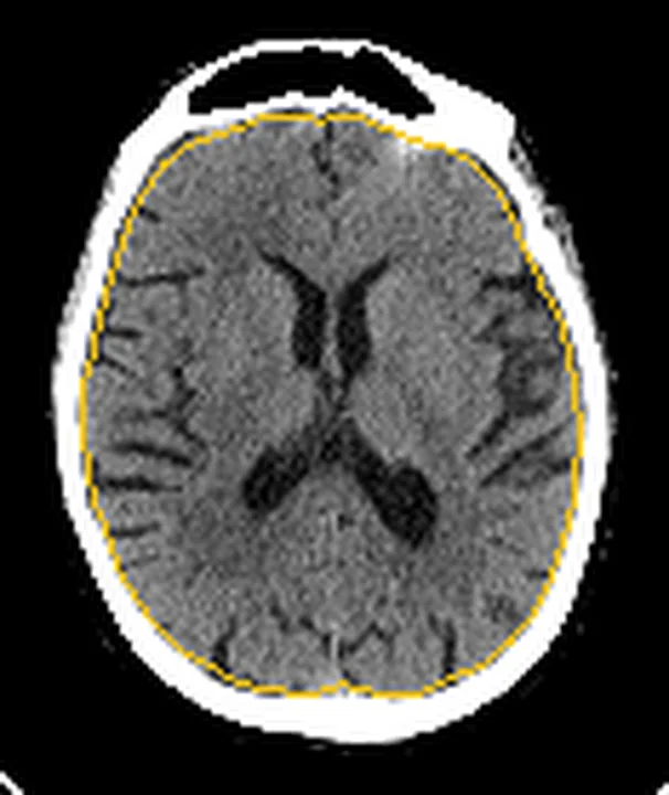
Lateral ventrikül boyutları ve Willis poligonu damar kalibrasyonu anatomik varyasyon gösterebilir; ventrikül asimetrisi tek başına normal varyant ya da hastalık tanısı koydurmaz. Kontrastsız BT ventrikül biçimini, septum pellucidumun konumunu ve komşu parankimi değerlendirmeye; BT anjiyografi (BTA) A1, P1 ve vertebral arter lümenlerini karşılaştırmaya yardımcı olur. Ayırımda ventrikül asimetrisine eşlik eden orta hat deviasyonu, kitle etkisi veya parankim bulguları ile arterlerin distal devamlılığı değerlendirilir. Normal anatomik varyantları patolojiyle karıştırmamak için görüntüleme bulguları birlikte yorumlanmalıdır.
Okuma sırası
- Frontal boynuzlar. Foramen Monro düzeyindeki aksiyel kesitte iki frontal boynuzun biçimini karşılaştır; gözlenen farkın tek ventrikülle sınırlı mı yoksa tüm ventriküler sistemde mi olduğunu belirle.
- Septum pellucidum. Septum pellucidumun orta hatta kalıp kalmadığını ve korpus kallozum ile kaudat başların konturunu incele; deviasyon varsa bası yönünü açıklayacak komşu lezyonu ara.
- Oksipital boynuzlar. Atrium ve oksipital boynuzlara ilerleyerek asimetrinin biçimsel devamlılığını, sulkus genişliğini ve periventriküler dansiteyi kontrol et.
- A1, P1 ve vertebral arterler. Son olarak BTA'da A1/P1 ve vertebral arterleri distal devamlılıklarıyla izle; kesilme, dolum defekti veya ilgili akut parankim bulgusu varsa varyant varsayımını bırak.
Faz ve pencere
- Kontrastsız tanısal
- Yüksek çözünürlüklü kesitsel görüntülemede ventrikül anatomisindeki biçim ve konum farklılıkları daha kolay saptanabilir; bu farklılıkların klinik anlamı ayrıca değerlendirilmelidir.
- BTA tanısal
- A1 ve P1 segmentleri, posterior komünikan arterler ve vertebral arterlerin opasifiye lümen çapı ile devamlılığı karşılaştırılır; hipoplastik segmentin distal bağlantısı ve karşı tarafın beslediği damar alanı aranır.
Önerilen pencereler: Beyin (G 80 / M 40), Anjiyo (G 600 / M 150).
BT bulguları
- İzole lateral ventrikül asimetrisinde boynuzlar farklı genişlikte olabilir; septum konumu, komşu parankimde hacim kaybı ve kitle etkisi ayrıca değerlendirilir.
- Oksipital boynuzların uzunluğu ve biçimi bireyler arasında farklılık gösterebilir; atrium ve oksipital boynuz konturunu seri kesitlerde izle.
- Foramen Monro stenozu veya obstrüksiyonu tek taraflı hidrosefaliye yol açabilir.
- A1 hipoplazisi veya aplazisinde karşı A1 ve anterior komünikan arter üzerinden iki A2 segmentine doluş görülebilir.
- Fetal tip PCA’da posterior komünikan arter belirgin, P1 hipoplastik veya aplastik olabilir; BTA’da P1’in ince ya da izlenmemesi tek başına akut oklüzyon tanısı koydurmaz.
- Dominant vertebral arter karşı tarafın hipoplazisine eşlik edebilir; küçük vertebral arterin PICA'ya sonlanması gelişimsel varyant lehinedir.
- Küçük kalibreli vertebral arterde gelişimsel hipoplazi ile kazanılmış stenozu yalnızca çapa bakarak ayırma; damar duvarını, plak/kalsifikasyonu ve tüm segmentin devamlılığını birlikte değerlendir.
- Ventrikül asimetrisi anatomik varyasyonlardan biri olabilir; normal varyasyonların patolojiyle karıştırılmaması gerekir.
Normalde
Bebeklerde asimetrik lateral ventriküller normal varyantlar arasında tanımlanmıştır. BTA’da sağ-sol A1 ve P1’i aynı düzeyde, vertebral arterleri baziler birleşime kadar izle; simetri zorunlu değildir, devamlı lümen ve uygun kollateral bağlantılar varyantı destekler.
Normal BT ile kıyasla
Aynı bölgenin, kaynak veri setinde patoloji içermediği belirtilen bir BT incelemesini kesit kesit kaydırın; organ sınırlarını açarak anatomiyi hasta görüntüsüyle karşılaştırın. Küçük rastlantısal bulgular tümüyle dışlanmamıştır.
Kaynak: TotalSegmentator v2.01 veri seti (Wasserthal ve ark., Radiology: Artificial Intelligence 2023), CC BY 4.0, vaka s0103; organ sınırları veri setinin otomatik segmentasyonundan, birleştirilerek sadeleştirildi. Görüntüler 2,5 mm kesit aralığına yeniden örneklendi.
Ayırıcı tanı
- Foramen Monro obstrüksiyonu
- ilgili lateral ventrikül genişler, septum pellucidum karşıya bombeleşebilir ve foramen komşuluğunda lezyon/koaptasyon bulunabilir.
- Ex-vaküo ventrikül genişlemesi
- parankimal hacim kaybına bağlı ventrikül genişlemesidir; sulkus ve fissür örüntüsünü birlikte değerlendir.
- Akut hidrosefali
- temporal boynuz genişlemesi ve diğer ventriküler genişleme bulguları görülebilir; periventriküler hipodensite transependimal BOS geçişini gösterebilir.
- Akut arter oklüzyonunda BTA'da ilgili arter lümeninde oklüzyon izlenebilir.
- Vertebral arter hipoplazisi ile kazanılmış aterosklerotik stenozun ayrımında damar duvarı ve plak bulguları değerlendirilir; hipoplaziye ateroskleroz da eşlik edebilir.
Tuzaklar
- Sadece bir kesitteki boynuz genişliğini hidrosefali sayma; aynı seviyeli seri kesitlerde ventrikülün biçimini, sulkusları ve septum konumunu kıyasla.
- Dar A1 veya P1'i otomatik oklüzyon olarak adlandırma; karşı taraftan doluşu, komünikan arter bağlantısını ve distal dalları izle.
- Küçük vertebral arteri tromboze kabul etmeden önce tüm V4 segmentini ve PICA'ya sonlanımı araştır; kontrastsız kalsifikasyon tek başına lümen açıklığını göstermez.
- Septum pellucidum deviasyonunu izole anatomik varyant sayma; komşu kitle, asimetrik ödem ve foraminal çıkışta tıkanıklık olup olmadığını kontrol et.
Kendini dene
Sol oksipital boynuz sağdan geniş; foramen Monro açık, diğer ventriküller normal, septum orta hatta, sulkuslar simetrik ve ekstraaksiyel kitle izlenmiyor. En uygun yorum hangisi?
Cevabı göster
İzole ventrikül varyantı. İzole ventrikül asimetrisi anatomik varyant olabilir; orta hattın korunması ve kitle etkisinin bildirilmemesi bu seçeneği destekler, ancak tek başına kesin tanı koydurmaz.
Önceki BTA’da da aynı görünüm var: sağ A1 tüm seyri boyunca düzgün kalibrasyonlu ve ince; sol A1 ile anterior komünikan arter opak, iki A2 de doluyor. Bulguyu en iyi ne açıklar?
Cevabı göster
A1 hipoplazisi. Uzun segment boyunca ince ancak opaklaşan sağ A1 ve karşı A1/anterior komünikan arter üzerinden iki A2’ye doluş gelişimsel hipoplaziyi destekler; ani kesilme veya distal doluş kaybı bildirilmemiştir.
Kaynaklar
Bu sayfadaki 36 cümle ve 2 quiz sorusu aşağıdaki kaynaklardan alıntılarla otomatik olarak karşılaştırıldı; 21 cümle bu karşılaştırmada düzeltildi (2026-10-09).
- The Lateral Ventricles: A Detailed Review of Anatomy, Development, and Anatomic Variationspubmed.ncbi.nlm.nih.gov
- Intracranial arterial variations: a comprehensive evaluation using CT angiographypubmed.ncbi.nlm.nih.gov
- Variants of cerebral arteries - anterior circulationpmc.ncbi.nlm.nih.gov
- Imaging Markers for Normal Pressure Hydrocephalus: An Overviewpubmed.ncbi.nlm.nih.gov
- Differences in Brain Morphology between Hydrocephalus Ex Vacuo and Idiopathic Normal Pressure Hydrocephaluspmc.ncbi.nlm.nih.gov
- Wall Imaging for Unilateral Intracranial Vertebral Artery Hypoplasia with Three-dimensional High-isotropic Resolution Magnetic Resonance Imagespmc.ncbi.nlm.nih.gov
- Morphology and Variations of the Posterior Cerebral Artery: A Literature Reviewpubmed.ncbi.nlm.nih.gov
- CT Imaging in Neurocritical Carepmc.ncbi.nlm.nih.gov
- Big Vents: Causes of Hydrocephaluspubs.rsna.org
- Diagnostic performance of single-phase CT angiography in detecting large vessel occlusion in ischemic stroke: A systematic reviewpubmed.ncbi.nlm.nih.gov
- Imaging of the septum pellucidum: normal, variants and pathologypubmed.ncbi.nlm.nih.gov
- Intracranial calcifications on CT: an updated reviewpmc.ncbi.nlm.nih.gov
- Functional Aplasia of the Contralateral A1 Segment Influences Clinical Outcome in Patients with Occlusion of the Distal Internal Carotid Arterypmc.ncbi.nlm.nih.gov
- Incidental vascular findings on brain magnetic resonance angiographypmc.ncbi.nlm.nih.gov
- Common and uncommon intracranial arterial anatomic variations in multi-detector computed tomography angiography (MDCTA). What radiologists should be aware ofpmc.ncbi.nlm.nih.gov
- Automated Midline Shift Detection in Head CT Using Localization and Symmetry Techniques Based on User-Selected Slice.pubmed.ncbi.nlm.nih.gov
- A unilateral open-lip schizencephaly in a neonate: A rare case report.pubmed.ncbi.nlm.nih.gov
- Brain Midline Shift Measurement and Its Automation: A Review of Techniques and Algorithms.pubmed.ncbi.nlm.nih.gov
- Paediatric cranial ultrasound: abnormalities of the brain in term neonates and young infants.pubmed.ncbi.nlm.nih.gov
- Cranial ultrasound for beginners.pubmed.ncbi.nlm.nih.gov
- Diagnostic Approach to Macrocephaly in Children.pubmed.ncbi.nlm.nih.gov
- Radiogenomics of congenital brain malformations: Linking embryology, genetics, and imaging.pubmed.ncbi.nlm.nih.gov
- A simple and quantitative ultrasonographic screening method for fetal Sylvian fissure abnormalities.pubmed.ncbi.nlm.nih.gov
- Aggravation of rainbow sign on hematoma membrane by arterial spin labeling predicts chronic subdural hematoma recurrence following middle meningeal artery embolization.pubmed.ncbi.nlm.nih.gov
- Treatment pitfalls in recurrent dolichoectatic basilar trunk aneurysm with flow diversion: illustrative case.pubmed.ncbi.nlm.nih.gov
- Proteomic Analysis of Mouse Cerebral Cortex Following Experimental Ischemic Stroke: Identifying Novel Biomarkers of Damage and Repair.pubmed.ncbi.nlm.nih.gov
- Decoding the Diverse Colours of Guillain-Barré Syndrome (GBS): A Case Report and Narrative Review of Atypical Miller Fisher-GBS Overlap Syndrome.pubmed.ncbi.nlm.nih.gov
- Venous-significant intracranial hemorrhage in neonatal HIE treated with therapeutic hypothermia: imaging phenotypes, diagnostic pitfalls, and prognostic implications.pubmed.ncbi.nlm.nih.gov
- A Misleading Cystic Brain Lesion: An Uncommon Presentation of Meningioma.pubmed.ncbi.nlm.nih.gov
- Case Report: Occlusion of the foramen of Monro treated with endoscopic septostomy and foraminotomy in a preterm neonate.pubmed.ncbi.nlm.nih.gov
- Prenatal diagnosis and postnatal management of congenital unilateral hydrocephalus for stenosis of the foramen of Monro.pubmed.ncbi.nlm.nih.gov
- Prenatal assessment of brain malformations on neuroimaging: an expert panel review.pubmed.ncbi.nlm.nih.gov
- CT Angiography of the Upper Extremities: Review of Acute Arterial Entities.pubmed.ncbi.nlm.nih.gov
- Spontaneous Coronary Artery Dissection Unveiled: Pathophysiology, Imaging, and Evolving Management Strategies.pubmed.ncbi.nlm.nih.gov
- Pulmonary arteries: imaging of pulmonary embolism and beyond.pubmed.ncbi.nlm.nih.gov
- Delayed M1 Dissection After Thrombectomy Treated With Rescue Stenting and Tirofiban: A Case Report.pubmed.ncbi.nlm.nih.gov
- Middle cerebral artery dissection: pathophysiology, diagnosis, and therapeutic options.pubmed.ncbi.nlm.nih.gov
- Prediction of collateral circulation grading and functional outcomes in acute ischemic stroke using FLAIR vascular hyperintensity combined with multimodal CT parameters.pubmed.ncbi.nlm.nih.gov
- Atherosclerosis-Like Spontaneous Middle Cerebral Artery Dissection.pubmed.ncbi.nlm.nih.gov
- The impact of thrombus surface morphology of the basilar artery on the successful rate of mechanical thrombectomy.pubmed.ncbi.nlm.nih.gov
- Clot Meniscus Sign: An Angiographic Clue for Choosing between Stent Retriever and Contact Aspiration in Acute Basilar Artery Occlusion.pubmed.ncbi.nlm.nih.gov
- Operative Procedures for Ultrasound Assessment of Extracranial Artery Disease: A Narrative Review by the Italian Society for Vascular Investigation (SIDV).pubmed.ncbi.nlm.nih.gov
- The genetic basis of hydrocephalus: genes, pathways, mechanisms, and global impact.pubmed.ncbi.nlm.nih.gov
- Endoscopic third ventriculostomy for diencephalosynapsis-related hydrocephalus in MPDZ gene syndrome: a case report and review of the literature.pubmed.ncbi.nlm.nih.gov
- 선천성 뇌 질환의 영상 소견: 임상 화보.pubmed.ncbi.nlm.nih.gov
- Acute aortic dissection presenting as crossed neurological symptoms: a case report.pubmed.ncbi.nlm.nih.gov
- Severe Neurovascular and Cardiovascular Manifestations of Takayasu Arteritis: A Case Series and Review of the Literature.pubmed.ncbi.nlm.nih.gov
- Management of Shamblin Type III Carotid Body Tumor With Preoperative Embolization, Surgical Resection, and Rescue Endovascular Stroke Treatment: A Case Report.pubmed.ncbi.nlm.nih.gov
- Delayed coil migration after endovascular treatment of a ruptured intracranial aneurysm managed by open microsurgical surgery: illustrative case.pubmed.ncbi.nlm.nih.gov
- External Carotid Artery Pseudoaneurysm: A Rare Cause of Ischemic Stroke via Internal Carotid Artery Compression-A Case Report.pubmed.ncbi.nlm.nih.gov
- Retrieval of an atherosclerotic plaque-vessel wall complex during mechanical thrombectomy: illustrative case.pubmed.ncbi.nlm.nih.gov
- A systematic approach to detection of epileptogenic foci on imaging: protocols, entities, search pattern, and reporting tools.pubmed.ncbi.nlm.nih.gov
- Structural neuroimaging markers of normal pressure hydrocephalus versus Alzheimer's dementia and Parkinson's disease, and hydrocephalus versus atrophy in chronic TBI-a narrative review.pubmed.ncbi.nlm.nih.gov
- Electroencephalographic Aspects in a Sheep with Coenurosis.pubmed.ncbi.nlm.nih.gov
- Biomechanical Mechanisms of Vocal Fold Nodule and Polyp Formation: A Review.pubmed.ncbi.nlm.nih.gov
- Multimodality Imaging of Pulmonary Regurgitation and Right Ventricular Remodeling in Adults with Repaired Tetralogy of Fallot: A Descriptive Single-Center Case Series.pubmed.ncbi.nlm.nih.gov
- Isolated absent pulmonary valve syndrome with intact ventricular septum in a child with CHARGE syndrome: a rare case report and review of the literature.pubmed.ncbi.nlm.nih.gov
- Prenatal Diagnosis of Autosomal Recessive Primary Microcephaly Type 2 Caused by Compound Heterozygous WDR62 Variants in a Family With Two Recurrent Cases.pubmed.ncbi.nlm.nih.gov
- Atypical presentation of Cavum Vergae.pubmed.ncbi.nlm.nih.gov
- Three-Dimensional Left Atrial Geometry in Atrial Fibrillation: Imaging Biomarkers, Substrate Phenotyping, and Ablation Outcome Prediction.pubmed.ncbi.nlm.nih.gov
- Fluoroless Workflow for Pulsed Field Ablation of Atrial Fibrillation Using the Sphere-9 and VARIPULSE Catheters.pubmed.ncbi.nlm.nih.gov
- Fetus-in-Fetu: A PRISMA-Informed Narrative Review With an Illustrative Prenatal Case.pubmed.ncbi.nlm.nih.gov
- Imaging More than Skin-Deep: Radiologic and Dermatologic Presentations of Systemic Disorders.pubmed.ncbi.nlm.nih.gov
- Multimodality imaging of neurodegenerative disorders with a focus on multiparametric magnetic resonance and molecular imaging.pubmed.ncbi.nlm.nih.gov
- Spontaneous atraumatic pediatric basal ganglia hemorrhage in the setting of COLGALT1-related collagenopathy: illustrative case.pubmed.ncbi.nlm.nih.gov
- Case Report: Intracranial hypertension occurring in association with subacute thyroiditis.pubmed.ncbi.nlm.nih.gov
- Dyke-Davidoff-Masson Syndrome Presenting With Recurrent Seizures and Left-Sided Motor Weakness: A Case Report.pubmed.ncbi.nlm.nih.gov
- Case Report: Giant acute hemorrhagic colloid cyst in the septum pellucidum-foramen of Monro region with obstructive hydrocephalus in an 8-month-old infant.pubmed.ncbi.nlm.nih.gov
- Myxoid glioneuronal tumor of the septum pellucidum with concurrent dual <i>PDGFRA</i> and <i>FGFR3</i> gene mutations: a case report and literature review.pubmed.ncbi.nlm.nih.gov
- Technical nuances of endoscopic transcortical transventricular fenestration for management of giant suprasellar arachnoid cysts in adults: illustrative cases.pubmed.ncbi.nlm.nih.gov
- Interstitial Lung Disease and Cardiotoxicity Associated with Trastuzumab Deruxtecan, Sacituzumab Govitecan, and Trastuzumab Emtansine: A Narrative Review.pubmed.ncbi.nlm.nih.gov
- From Modified Haller Index to a Novel Patented Anatomical Measurement Device: Engineering Development, Validation, and Clinical Applications of Non-Invasive Thoracic Morphometry.pubmed.ncbi.nlm.nih.gov
Eğitim amaçlıdır; hasta başında tanı ve tedavi kararının yerine geçmez, tıbbi tavsiye değildir. Metinler yapay zekâ desteğiyle yazıldı ve otomatik bir adımda kaynak alıntılarıyla karşılaştırıldı; bu bir tıbbi doğrulama değildir ve içerik uzman radyolog incelemesinden geçmedi. Klinik kararlarda güncel kılavuzları, kurum protokollerini ve radyoloji raporunu esas alın.