
Özet
Kontrastsız BT'de hiperdens arter işareti akut serebral arter trombüsünü gösterebilir; damar kalsifikasyonu bu işaretin tanınmasını güçleştirebilir. Çift enerjili BT’de VNCa ve renk kodlu kalsiyum üst üste bindirim görüntüleri, akut trombüsü damar duvarı kalsifikasyonundan ayırmaya yardımcı olabilir. M1 oklüzyonu olan hastalarda mCTA ile trombüsün gerçek uzunluğu ve yoğunluğu ile kollateral dolaşım değerlendirilebilir. Kalsifik plağı akut tıkanma saymak yalancı pozitif yorum doğurur; gerçek hiperdens damar işaretini kalsifikasyon diye geçmekse oklüzyonun erken fark edilmesini engelleyebilir.
Faz ve pencere
- Kontrastsız tanısal
- Nonkontrast BT'de hiperdens arter işareti (HAS) intralümeneral akut trombüsü gösterir ancak damar duvarı kalsifikasyonu bu tespiti zorlaştırır. Hematokrit artışı, Willis poligonu ve serebral venöz sinüslerde yaygın atenüasyon artışına yol açarak kontrastlı incelemeyi taklit edebilir. Nonkontrast BT'de yüksek CT numarası veren damar görünümü polikitemide de görülebilir ve ileri görüntülemeyi gerektirir.
- BTA
- Çokfazlı BT anjiyografide (mCTA) lümen içi dolam kusuru ve kollateral dolum derecesi, hiperdensidadenin trombotik oklüzyona neden olup olmadığını değerlendirmede kullanılır. Çiftenerjili BT ile elde edilen sanal kalsiyumsuz (VNCa) görüntüler, akut trombüsün damar duvarı kalsifikasyonundan ayrımını sağlar.
Önerilen pencereler G genişlik, M merkez; değerler Hounsfield birimi (HU)
- BeyinG 80 / M +40
- KemikG 1800 / M +400
- AnjiyoG 600 / M +150
BT bulguları
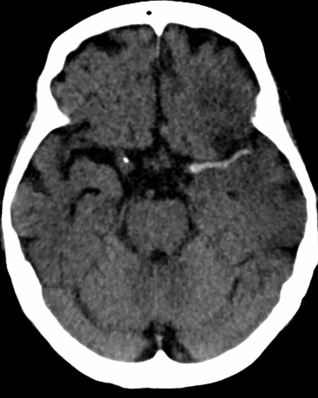
- Kontrastsız BT'de hiperdens arter işareti, serebral arter içindeki akut trombüsü gösterebilir.
- Polisitemide, Circle of Willis ve serebral venöz sinüslerde yaygın atenüasyon artışı görülebilir ve kontrastlı incelemeyi taklit edebilir.
- Damar duvarı kalsifikasyonu, hiperdens arter işaretinin tanınmasını güçleştirebilir; çift enerjili BT’de VNCa ve renk kodlu kalsiyum üst üste bindirim görüntüleri bu ayrımda yardımcı olabilir.
- VNCa ve renk kodlu kalsiyum üst üste bindirim görüntüleri, akut trombüsün damar duvarı kalsifikasyonundan ayrımına yardımcı olabilir.
- Yaygın intravasküler yoğunluk — arterler ve venler birlikte parlaksa hemokonsantrasyon olasılığı; izole tek damar trombüsü paterni değildir.
- Kalsifiye serebral emboliler, serebral arterlerin distal segmentlerinde yuvarlak, yüksek dansiteli kalsifik odaklar olarak görülebilir; çift enerjili BT karakterizasyona yardımcı olabilir.
- Hiperdens arter işareti, büyük damar oklüzyonunun bulunduğu bölgeye karşılık gelebilir.
Normalde
Nonkontrast BT'de hiperdens arter işareti (HAS), intrakraniyal arterlerde akut trombüs varlığını gösteren önemli bir görüntüleme bulgusudur. Çiftenerjili BT sanal kalsiyumsuz (VNCa) görüntüleri, akut trombüsün damar duvarı kalsifikasyonundan ayrımını sağlayarak doğru tanıya yöneltir.
Normal BT ile kıyasla
Aynı bölgenin, kaynak veri setinde patoloji içermediği belirtilen bir BT incelemesini kesit kesit kaydırın; organ sınırlarını açarak anatomiyi hasta görüntüsüyle karşılaştırın. Küçük rastlantısal bulgular tümüyle dışlanmamıştır.
Kaynak: TotalSegmentator v2.01 veri seti (Wasserthal ve ark., Radiology: Artificial Intelligence 2023), CC BY 4.0, vaka s0103; organ sınırları veri setinin otomatik segmentasyonundan, birleştirilerek sadeleştirildi. Görüntüler 2,5 mm kesit aralığına yeniden örneklendi.
Ayırıcı tanı
- Akut intraluminal trombüs, kontrastsız BT'de hiperdens arter işaretiyle görülebilir ve bu işaret büyük damar oklüzyonunu düşündürebilir.
- İntrakraniyal aterosklerotik darlığa bağlı büyük damar oklüzyonunun işlem öncesi tanısı güç olabilir.
- Yüksek hematokrit
- tek damar yerine çok sayıda arter ve venin benzer biçimde yoğun görünmesine yol açabilir; hemogram korelasyonu gerekir.
Tuzaklar
- MCA duvar kalsifikasyonu hiperdens arter işaretinin tanınmasını güçleştirebilir; çift enerjili BT'de VNCa ve kalsiyum üst üste bindirim görüntüleri trombüsü kalsifikasyondan ayırmaya yardımcı olabilir.
- Spontan kalsifiye serebral emboliler (SCCE), BT'de tanı konulsa sıkça yanlış tanınır veya tamamen kaçırılır ve bu durum katastrofik sonuçlara yol açabilir.
- Bir M1 oklüzyonu kohortunda, daha kısa gerçek trombüs uzunluğu ve daha yüksek trombüs yoğunluğu iyi klinik sonuçla ilişkili bulunmuş; gerçek uzunluk için çalışmanın iyi sonuç öngören kesim noktası 18,7 mm olarak bildirilmiştir.
- Hiperdens damar işaretinin yokluğunu oklüzyon yokluğu gibi yorumlama; işaretin duyarlılığı sınırlıdır ve BTA damar lümenini doğrudan değerlendirir.
- Hiperdens arter işaretinin görülmemesi oklüzyonu dışlamaz; bir kohortta işaretin yokluğu dirençli trombektomiyle ilişkili bulunmuştur.
- Hiperdens arter işaretinin görülmemesi büyük damar oklüzyonunu dışlamaz; bir kohortta bu işaretin yokluğu dirençli trombektomiyle ilişkili bulunmuştur.
Kendini dene
Kontrastsız BT'de hiperdens arter işareti, MRA'da saptanan büyük damar oklüzyonuyla aynı bölgeye karşılık geliyor. En olası bulgu nedir?
Cevabı göster
Akut intraluminal trombüs. Hiperdens arter işareti, kontrastsız BT'de serebral arter içindeki akut trombüsü düşündürür. Çift enerjili BT'de VNCa ve renk kodlu kalsiyum üst üste bindirim görüntüleri, akut trombüsün damar duvarı kalsifikasyonundan ayrılmasına yardımcı olabilir.
Kontrastsız BT'de Circle of Willis ve serebral venöz sinüslerde yaygın artmış atenüasyon görülüyor. Hangi taklitçi daha olasıdır?
Cevabı göster
Yüksek hematokrit. Polisitemide Willis poligonundaki arterlerde ve serebral venöz sinüslerde yaygın atenüasyon artışı görülebilir. Bu yaygın damar görünümü, kontrastsız BT'de kontrastlı incelemeyi taklit edebilir.
Kaynaklar
Bu sayfadaki 40 cümle ve 2 quiz sorusu aşağıdaki kaynaklardan alıntılarla otomatik olarak karşılaştırıldı; 33 cümle bu karşılaştırmada düzeltildi (2026-10-09).
- Noncontrast CT-based habitat analysis for differentiating hemorrhagic transformation from contrast extravasation after endovascular therapy of acute ischemic stroke.pubmed.ncbi.nlm.nih.gov
- Effects of Computed Tomography Hyperdense Middle Cerebral Artery Sign on the Efficacy of Endovascular Therapy in Acute Ischemic Stroke With Large Infarcts: A Subgroup Analysis of the ANGEL-ASPECT Trial.pubmed.ncbi.nlm.nih.gov
- Polycythemia, a radiologic mimic in plain CT head: A case report.pubmed.ncbi.nlm.nih.gov
- Vasculitis syndromes: large vessel vasculitides and ANCA-associated vasculitides from a neuroradiologist's perspective.pubmed.ncbi.nlm.nih.gov
- Diagnostic imaging and emerging image-guided neurointervention in brain tumors: a narrative review.pubmed.ncbi.nlm.nih.gov
- Endovascular Treatment Strategies for Acute Intracranial Atherosclerotic Stenosis-Related Large Vessel Occlusion.pubmed.ncbi.nlm.nih.gov
- A Near-Fatal Leak: Acute Cardiac Tamponade From Contrast Extravasation via a Thrombosed Port-a-Cath.pubmed.ncbi.nlm.nih.gov
- Localized renal artery vasculopathy: avoiding misdiagnosis between inflammatory arteritis and isolated renal artery dissection.pubmed.ncbi.nlm.nih.gov
- Mechanical Thrombectomy for Large-Vessel Occlusion after Fontan Procedure in an Adult.pubmed.ncbi.nlm.nih.gov
- Spontaneous calcified cerebral emboli: a comprehensive review and proposed diagnostic criteria.pubmed.ncbi.nlm.nih.gov
- When Cancer Clots: An Extensive Radiologic Analysis of Cancer-Associated Thromboembolism.pubmed.ncbi.nlm.nih.gov
- Artificial Intelligence in Acute Ischemic Stroke Subtypes According to Toast Classification: A Comprehensive Narrative Review.pubmed.ncbi.nlm.nih.gov
- Large-particle aerosol exposure to the Bangladesh or Malaysia strain of Nipah virus results in markedly divergent disease presentation in African Green Monkeys.pubmed.ncbi.nlm.nih.gov
- Cerebral Venous Sinus Thrombosis Mimicking Tumor Hemorrhage: Successful Anticoagulation in a Resource-Limited Setting.pubmed.ncbi.nlm.nih.gov
- Bilateral Vein of Trolard Thrombosis Presenting with Seizure and Minimal Deficits: A Rare Case Report.pubmed.ncbi.nlm.nih.gov
- Sharp recanalization of extensive multilevel central venous occlusions in a young patient with familial Mediterranean fever: A case report.pubmed.ncbi.nlm.nih.gov
- Using Multimodal Imaging to Correlate Link Between Intracranial Aneurysms and Acute Ischemic Stroke.pubmed.ncbi.nlm.nih.gov
- Otogenic Sigmoid Sinus Thrombosis in a 34-Year-Old Woman: A Rare Intracranial Complication.pubmed.ncbi.nlm.nih.gov
- Redefining HIV-associated large-vessel vasculopathy in the antiretroviral therapy era: Multi-detector CT imaging findings and their correlation with intervention and patient outcomes.pubmed.ncbi.nlm.nih.gov
- Carotid Web as a Treatable Etiology of Recurrent Cryptogenic Ischemic Stroke Highlighting Neurointervention: A Case Report and Literature Review.pubmed.ncbi.nlm.nih.gov
- Diagnostic performance of prehospital large-vessel occlusion detection scales in patients with suspected acute stroke: a retrospective validation study. The Akershus Study of Ischemic Stroke and Thrombolysis.pubmed.ncbi.nlm.nih.gov
- Emergency microsurgical trapping, decompression, and revascularization of a middle cerebral artery dissecting aneurysm using a minipterional approach in a pediatric patient: illustrative case.pubmed.ncbi.nlm.nih.gov
- Heart Meets Brain: Insights into Neurocardiac Pathophysiology.pubmed.ncbi.nlm.nih.gov
- Refractory Thrombectomy: Incidence and Related Factors in a Third-Level Stroke Treatment Center.pubmed.ncbi.nlm.nih.gov
- Blind spots in brain imaging: a pictorial essay.pubmed.ncbi.nlm.nih.gov
- Chronic Subdural Hematoma: Pathophysiology, Diagnosis, and the Emerging Role of Middle Meningeal Artery Embolization.pubmed.ncbi.nlm.nih.gov
- Multimodal imaging in neuro-ophthalmology.pubmed.ncbi.nlm.nih.gov
- The Train-Line Pattern on Duplex Ultrasound Helps Differentiate Intramural Thrombus from Atheromatous Plaque in Common Carotid Artery Dissection.pubmed.ncbi.nlm.nih.gov
- Operative Procedures for Ultrasound Assessment of Extracranial Artery Disease: A Narrative Review by the Italian Society for Vascular Investigation (SIDV).pubmed.ncbi.nlm.nih.gov
- Congenital and acquired anomalies of the basilar artery: A pictorial essay.pubmed.ncbi.nlm.nih.gov
- Fully Automated Thrombus Segmentation on CT Images of Patients with Acute Ischemic Stroke.pubmed.ncbi.nlm.nih.gov
- Aggressive Vaccine-Induced Immune Thrombocytopenia and Thrombosis in a Young Woman with a Past Mild SARS-CoV-2 Infection.pubmed.ncbi.nlm.nih.gov
- Beyond Ischemia: The Rare Occurrence of Hemorrhagic Strokes in Pediatric Sickle Cell Anemia.pubmed.ncbi.nlm.nih.gov
- Imaging Evaluation of Acute Traumatic Brain Injury.pubmed.ncbi.nlm.nih.gov
- Individualized treatment for pediatric intracranial aneurysms in China: case series and literature review.pubmed.ncbi.nlm.nih.gov
- Retroperitoneal hematoma secondary to rupture of uterine artery pseudoaneurysm after cesarean section, managed by selective embolization: A case report.pubmed.ncbi.nlm.nih.gov
- Computed tomography perfusion-defined ischemic core predicts functional outcome after basilar artery thrombectomy.pubmed.ncbi.nlm.nih.gov
- Traumatic basilar artery incarceration following longitudinal clival fracture: illustrative case.pubmed.ncbi.nlm.nih.gov
- Anatomical variability of the lingual artery: a comprehensive narrative review with clinical and surgical applications.pubmed.ncbi.nlm.nih.gov
- Inflammation of central nervous system fiber tracts is active in patients over six months post-intracerebral hemorrhage.pubmed.ncbi.nlm.nih.gov
- Morphological and Imaging Characterization of the Eye and Adnexa of the Lesser Anteater (Tamandua tetradactyla).pubmed.ncbi.nlm.nih.gov
- Middle cerebral artery dissection: pathophysiology, diagnosis, and therapeutic options.pubmed.ncbi.nlm.nih.gov
- Non-Thrombotic Filling Defects in Cerebral Veins and Sinuses: When Normal Structures Mimic a Disease.pubmed.ncbi.nlm.nih.gov
- Imaging of Cerebral Venous Thrombosis.pubmed.ncbi.nlm.nih.gov
- Clinical implications of vessel wall imaging-State-of-the-art review.pubmed.ncbi.nlm.nih.gov
- Mechanical stress from the hyoid bone as a potential etiology of bony-related ischemic stroke secondary to carotid artery dissection: A case report and systematic review.pubmed.ncbi.nlm.nih.gov
- Profound Vision Loss With Minimal Retinal Findings: A Case of Microvascular Ischemia Unraveled by Multimodal Imaging.pubmed.ncbi.nlm.nih.gov
- Contrast-Induced Encephalopathy Due to Contrast Extravasation From a Chronic Post-hemorrhagic Encephalomalacic Region: Prior Hemorrhage as a Potential Risk Factor.pubmed.ncbi.nlm.nih.gov
- Triple-modality management of complex septated chronic subdural hematoma: a preliminary technical note on feasibility and safety.pubmed.ncbi.nlm.nih.gov
- Contrast extravasation on Dyna-CT as a predictor of malignant brain edema after mechanical thrombectomy for acute anterior circulation large vessel occlusion.pubmed.ncbi.nlm.nih.gov
- The Evolving Role and Use of Echocardiography in the Evaluation of Cardiac Source of Embolism.pubmed.ncbi.nlm.nih.gov
- Multi-modal CT scanning in the evaluation of cerebrovascular disease patients.pubmed.ncbi.nlm.nih.gov
- Neurovascular disorders in children: an updated practical guide.pubmed.ncbi.nlm.nih.gov
- Cerebral venous thrombosis: diagnosis dilemma.pubmed.ncbi.nlm.nih.gov
- Intracranial venous thrombosis imaging by anatomical location: an educational review with mimics and diagnostic pitfalls.pubmed.ncbi.nlm.nih.gov
- The Importance of Predicting Bowel Necrosis in Acute Mesenteric Ischemia: Narrative Review.pubmed.ncbi.nlm.nih.gov
- Update on cerebrovascular manifestations of COVID-19.pubmed.ncbi.nlm.nih.gov
- Increasing arterial compliance by laser modification of fibro-calcific plaques.pubmed.ncbi.nlm.nih.gov
- Clinical usability of 3D gradient-echo-based ultrashort echo time imaging: Is it enough to facilitate diagnostic decision in real-world practice?pubmed.ncbi.nlm.nih.gov
- Imaging of penetrating vascular trauma of the body and extremities secondary to ballistic and stab wounds.pubmed.ncbi.nlm.nih.gov
- Sequential Flow-Based Assessment of a Suspected Inferior Pancreaticoduodenal Artery-Derived Pathway Entering the Right Hepatic Artery during Pancreaticoduodenectomy: A Case Report.pubmed.ncbi.nlm.nih.gov
- Occult ascending pharyngeal artery pseudoaneurysm masked by CTA artifact after penetrating facial trauma: a case report.pubmed.ncbi.nlm.nih.gov
- Endovascular Revascularization for Chronic Vertebral Artery Occlusion with Poor Collateral Circulation and a Large Perfusion Abnormality Identified by Automated Perfusion Imaging: A Case Report.pubmed.ncbi.nlm.nih.gov
- Infarcts and ischemia in the abdomen: an imaging perspective with an emphasis on cross-sectional imaging findings.pubmed.ncbi.nlm.nih.gov
- Proceedings of the 15th International Congress on Circumpolar Health.pubmed.ncbi.nlm.nih.gov
- Neuroimaging for neurovascular complications of traumatic brain injury.pubmed.ncbi.nlm.nih.gov
- Developmental venous anomaly coexisting with arteriovenous malformation: a case report.pubmed.ncbi.nlm.nih.gov
- Parent Artery Disease-Related Stroke: What Is the Impact on Endovascular Treatment? A Narrative Review.pubmed.ncbi.nlm.nih.gov
- A Review of the Current Literature on Cerebral Aneurysms.pubmed.ncbi.nlm.nih.gov
- Ischemic posterior circulation stroke: a review of anatomy, clinical presentations, diagnosis, and current management.pubmed.ncbi.nlm.nih.gov
- Gallbladder cancer revisited: the evolving role of a radiologist.pubmed.ncbi.nlm.nih.gov
- Bladder multilithiasis across different clinical scenarios: Multimodality imaging findings, differential diagnosis, and complication assessment in 4 patients.pubmed.ncbi.nlm.nih.gov
- High-volume mucinous ascites secondary to low-grade mucinous neoplasm arising in a mature cystic teratoma: a case report.pubmed.ncbi.nlm.nih.gov
- Laparoscopic partial gastrectomy for gastric calcifying fibrous tumor: a case report and literature review.pubmed.ncbi.nlm.nih.gov
- Spontaneous bacteremia and spontaneous bacterial peritonitis share similar prognosis in patients with cirrhosis: a cohort study.pubmed.ncbi.nlm.nih.gov
- Analysis of the baseline multiphase computed tomographic angiography findings to predict clinical outcomes in patients with middle cerebral artery M1 occlusion treated with mechanical thrombectomy.pubmed.ncbi.nlm.nih.gov
- Incidental aneurysm rupture during endovascular thrombectomy for acute ischaemic stroke.pubmed.ncbi.nlm.nih.gov
- MDCT Imaging of Non-Traumatic Thoracic Aortic Emergencies and Its Impact on Diagnosis and Management-A Reappraisal.pubmed.ncbi.nlm.nih.gov
- Giant serpentine aneurysm: Imaging features including potential diagnostic utility of arterial spin labeling.pubmed.ncbi.nlm.nih.gov
- Craniocervical arterial dissections with cerebral infarction: a five-case series with diagnostic application of vessel wall MRI.pubmed.ncbi.nlm.nih.gov
- Multimodal Imaging Reveals Labbé Vein Thrombosis Mimicking Subdural Hematoma: A Diagnostic Pitfall in Emergency Neuroimaging.pubmed.ncbi.nlm.nih.gov
- Carotid web: an underrecognized cause of ischemic stroke in a young patient.pubmed.ncbi.nlm.nih.gov
- A rare case report of pulmonary artery sarcoma diagnosed and managed based on multimodal imaging.pubmed.ncbi.nlm.nih.gov
- Acute aortic syndromes and aortic emergencies.pubmed.ncbi.nlm.nih.gov
- Embolization in Pediatric Patients: A Comprehensive Review of Indications, Procedures, and Clinical Outcomes.pubmed.ncbi.nlm.nih.gov
- Delayed evacuation of bilateral chronic subdural hematoma with herniation-induced bilateral posterior cerebral artery infarction: illustrative case.pubmed.ncbi.nlm.nih.gov
- Mechanical thrombectomy for tandem internal carotid artery-middle cerebral artery occlusion in a patient with a persistent hypoglossal artery: illustrative case.pubmed.ncbi.nlm.nih.gov
- A bioactive magnesium alloy scaffold integrated with BMSCs-Loaded 3D microspheres synergistically promotes femoral head osteonecrosis repair by improving the osteogenic-angiogenic microenvironment.pubmed.ncbi.nlm.nih.gov
- Laser Interstitial Thermal Therapy for High-Grade Gliomas: Current Evidence, Clinical Applications and Emerging Role of Artificial Intelligence.pubmed.ncbi.nlm.nih.gov
- Parry-Romberg Syndrome: Radioclinical Dissociation in a Paucisymptomatic Form and a Proposed Diagnostic Framework.pubmed.ncbi.nlm.nih.gov
- Spectral CT of the abdomen: Where are we now?pubmed.ncbi.nlm.nih.gov
- Pediatric Stroke Imaging.pubmed.ncbi.nlm.nih.gov
- Application of deep learning reconstruction combined with time-resolved post-processing method to improve image quality in CTA derived from low-dose cerebral CT perfusion data.pubmed.ncbi.nlm.nih.gov
- Imaging of Skull Base Tumors.pubmed.ncbi.nlm.nih.gov
- Clinical and CT imaging features of different types of thoracic complex lymphatic anomaly.pubmed.ncbi.nlm.nih.gov
- Rhinosinusitis: Evidence and experience - 2024.pubmed.ncbi.nlm.nih.gov
- Rare Diseases of the Orbit.pubmed.ncbi.nlm.nih.gov
- Painless Acute Type A Aortic Dissection Masquerading as an Acute Neurological Event: A Case Report.pubmed.ncbi.nlm.nih.gov
- Imaging of the aortic root on high-pitch non-gated and ECG-gated CT: awareness is the key!pubmed.ncbi.nlm.nih.gov
- Case Report: Sellar Ependymomas: A Clinic-Pathological Study and Literature Review.pubmed.ncbi.nlm.nih.gov
- Photon-counting CT characterization of carotid perivascular adipose tissue: a layer-by-layer quantitative analysis. A preliminary analysis in an asymptomatic population.pubmed.ncbi.nlm.nih.gov
- From Magnetic Moment to Magnetic Particle Imaging: A Comprehensive Review on MPI Technology, Tracer Design and Biological Applications.pubmed.ncbi.nlm.nih.gov
- Carotid artery dissection linked to intermittent apnoeic swimming: A case-control study.pubmed.ncbi.nlm.nih.gov
- Dual-Energy Computed Tomography Virtual Noncalcium Imaging of Intracranial Arteries in Acute Ischemic Stroke: Differentiation Between Acute Thrombus and Calcification.pubmed.ncbi.nlm.nih.gov
- Transient ischemic attack due to multiple spontaneous calcified embolus of the cerebral arteries on a calcified mitral and aortic stenosis.pubmed.ncbi.nlm.nih.gov
- Magnetic resonance imaging of the orbit, Part 2: Characterization of orbital pathologies.pubmed.ncbi.nlm.nih.gov
- Multimodality imaging in the detection and management of coronary and peripheral arterial disease in patients with cancer receiving cardiotoxic antineoplastic treatments: A clinical consensus statement of the ESC Council of Cardio-Oncology and the European Association of Cardiovascular Imaging (EACVI) of the ESC.pubmed.ncbi.nlm.nih.gov
- Tissue infarction secondary to brucellosis: a systematic review of case reports and case series.pubmed.ncbi.nlm.nih.gov
- Dual-Energy Heart CT: Beyond Better Angiography-Review.pubmed.ncbi.nlm.nih.gov
- Gastrointestinal stromal tumors: a comprehensive radiological review.pubmed.ncbi.nlm.nih.gov
Eğitim amaçlıdır; hasta başında tanı ve tedavi kararının yerine geçmez, tıbbi tavsiye değildir. Metinler yapay zekâ desteğiyle yazıldı ve otomatik bir adımda kaynak alıntılarıyla karşılaştırıldı; bu bir tıbbi doğrulama değildir ve içerik uzman radyolog incelemesinden geçmedi. Klinik kararlarda güncel kılavuzları, kurum protokollerini ve radyoloji raporunu esas alın.