Özet
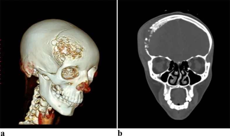
Kalvaryal osteomiyelit, sinüzit, saçlı deri enfeksiyonu, travma veya kraniyotomi sonrası gelişebilen kemik enfeksiyonudur; frontal kemik tutulumu subperiostal apse ve kafa içi yayılımla komplike olabilir. Kontrastsız ince kesit BT, kalvaryumda litik lezyonlar, dış tabula ve diploeyi tutan düzensiz kortikal erozyonlar ile epidural kalınlaşma veya koleksiyon bulgularını gösterir. Erken kemik iliği enfeksiyonu BT’de silik kalabilir ve MR ile daha iyi değerlendirilir. Kloaka, subperiostal apse veya epidural/subdural yayılımın gözden kaçması hastalığın kapsamını ve aciliyetini olduğundan düşük gösterebilir.
Faz ve pencere
- Kontrastsız tanısal
- Kemik penceresinde diploe ve dış tabulayı tutan osteolitik lezyonlar görülebilir; erken kemik iliği tutulumu BT’de belirgin olmayabilir ve MR ile daha iyi değerlendirilir. Kontrastsız BT osteolitik kemik lezyonlarını gösterebilir; kontrastlı görüntüler eşlik eden yumuşak doku veya epidural koleksiyonun kontrastlanmasını ortaya koyabilir.
- Kemik algoritması tanısal
- İnce kesit BT’de kalvaryal osteolitik lezyonların yayılımı ve eşlik eden skleroz değerlendirilebilir; MR kemik iliği ve yumuşak doku tutulumunu daha iyi gösterir.
Önerilen pencereler: Kemik (G 1800 / M 400), Yumuşak doku (G 400 / M 40).
BT bulguları
- Diploik tutulum — dış tabula ve diploe'de düzensiz litik lezyonlar, kemik yoğunluğunun yer yer kaybolması ve normal trabeküler yapının bozulması izlenir.
- Kortikal erozyon — iç ya da dış tabulada düzensiz incelme, fokal kesinti ve destrüksiyon görülebilir.
- Periostit enfeksiyona eşlik edebilir ve osteolitik lezyonlarla birlikte görülebilir.
- Reaktif skleroz — kronikleşen hastalıkta diploe ve kortekste kalınlaşma/yüksek kemik yoğunluğu gelişebilir.
- Kronik osteomiyelitte nekrotik doku parçaları ayrılarak sekestr oluşturabilir.
- Skalp veya subperiostal koleksiyon — dış tabula üzerinde sıvı birikimi ve komşu cilt altı dokuda şişlik frontal enfeksiyonu destekler.
- Epidural yayılım — iç tabula defekti boyunca koleksiyon veya ekstraaksiyel yumuşak doku izlenebilir; MR intrakraniyal yumuşak dokuyu daha iyi karakterize eder.
- Komşu sinüs hastalığı — frontal sinüzite bağlı doğrudan yayılım frontal kemik osteomiyelitine yol açabilir; sinüs opasifikasyonu tek başına kemik enfeksiyonunu göstermez.
- Pott puffy tümör, frontal kemik osteomiyeliti ile üzerindeki subperiostal apsenin birlikteliğidir; intrakraniyal komplikasyonlar arasında epidural apse, subdural ampiyem, serebral apse ve menenjit bulunabilir.
Normalde
Kalvaryal kemik defekti, lizis ve skleroz BT'de; lezyonun yumuşak doku bileşenleri ve çevre dokularla ilişkisi MR'da değerlendirilir. Kalvaryal kemik defekti, lizis ve skleroz BT'de; lezyonun yumuşak doku bileşenleri ve çevre dokularla ilişkisi MR'da değerlendirilir.
Ayırıcı tanı
- Kalvaryal metastaz veya primer tümörler
- fokal litik veya sklerotik kemik destrüksiyonu oluşturabilir; enfeksiyonda ise çevresel yumuşak doku reaksiyonu ve subperiostal apse bulguları baskındır.
- Eozinofilik granülom
- Langerhans hücreli histiyositozun bir varyantıdır ve kalvaryal litik lezyonların ayırıcı tanısında yer alır.
- İntraosseöz venöz malformasyonda BT'de dilate venöz kanallara eşlik eden karakteristik iç kemik spikülleri görülebilir.
- Kraniyotomi sonrası geç dönemde de kalvaryal osteomiyelit gelişebilir; başlangıçta belirtisiz seyreden olgular bildirilmiştir.
Tuzaklar
- Erken osteomiyelitte belirgin kortikal değişiklik henüz oluşmamış olabilir; negatif BT, kemik iliği enfeksiyonunu dışlamaz ve klinik şüphe sürüyorsa MR gerekir.
- Kontrastsız seride koleksiyonun sınırı ve çevresel inflamasyon daha belirsiz olabilir; sinüs ve yumuşak doku yayılımını kontrastlı seriyle değerlendirin.
- Frontal sinüs enfeksiyonu doğrudan yayılımla frontal kemiği tutabilir; kalvaryal osteomiyelit şüphesinde kemik ve eşlik eden yumuşak doku yayılımı görüntülemeyle değerlendirilmelidir.
- Kraniyotomi sonrası kemik kenarı düzensizliği enfeksiyonla karışabilir; ameliyat alanının beklenen konturunu, kortikal değişikliklerin ilerleyişini ve komşu koleksiyonu birlikte okuyun.
- İntrakraniyal uzanım kuşkusunda MR, kemik iliğindeki inflamatuvar değişiklikleri yüksek duyarlılıkla değerlendirir ve kalvaryal lezyonlarda komşu dura tutulumunun yayılımını göstermeye yardımcı olur.
Kendini dene
Frontal sinüzitin komplikasyonu olan Pott puffy tümör hangi bulgularla tanımlanır?
Cevabı göster
Frontal kemik osteomiyeliti ve subperiostal apse. Pott puffy tümör, frontal sinüzit bağlamında frontal kemik osteomiyeliti ve subperiostal apseyle tanımlanır.
İlk BT erken osteomiyeliti göstermemiş, ancak kemik iliği tutulumu veya intrakraniyal yayılım kuşkusu sürüyor. Hangi inceleme daha duyarlıdır?
Cevabı göster
MR. MR, kemik iliğindeki inflamatuvar değişiklikleri ve komşu yumuşak doku tutulumunu değerlendirmede duyarlıdır; osteomiyelit saptamada tercih edilen görüntüleme yöntemidir.
Kaynaklar
Bu sayfadaki 37 cümle ve 2 quiz sorusu aşağıdaki kaynaklardan alıntılarla otomatik olarak karşılaştırıldı; 21 cümle bu karşılaştırmada düzeltildi (2026-10-09).
- A Rare Case of Orbital Cellulitis with Progressive Calvarial Osteomyelitispmc.ncbi.nlm.nih.gov
- Calvarial osteomyelitis in secondary syphilis: evaluation by MRI and CT, including cinematic renderingpmc.ncbi.nlm.nih.gov
- A novel mouse calvarial defect model of osteomyelitis induced by Staphylococcus aureus (ATCC 6538)pmc.ncbi.nlm.nih.gov
- A Rare Clinical Presentation of Multifocal Tuberculous Osteomyelitis of the Right Parietal and Left Calcaneal Bones in an 11‐Year‐Old Male: A Case Reportpmc.ncbi.nlm.nih.gov
- Salvage reconstruction of an irradiated and infected scalp–calvarial defect following recurrent atypical meningioma: recipient-vessel strategy and multidisciplinary decision-makingpmc.ncbi.nlm.nih.gov
- Neuromelioidosis: A Single-Center Experience with Emphasis on Imaging.pubmed.ncbi.nlm.nih.gov
- Immediate single-stage reconstruction of complex frontofaciobasal injuries: part I.pubmed.ncbi.nlm.nih.gov
- A Rare Case of Orbital Cellulitis with Progressive Calvarial Osteomyelitis.pubmed.ncbi.nlm.nih.gov
- Varicella-Zoster Virus Infection and Osteomyelitis of the Skull.pubmed.ncbi.nlm.nih.gov
- Multifocal pure tubercular osteomyelitis: An unusual presentation in childhood.pubmed.ncbi.nlm.nih.gov
- Propionibacterium acnes osteomyelitis occurring 23 years after craniotomy: case report and review of literature.pubmed.ncbi.nlm.nih.gov
- Calvarial osteomyelitis and intracranial extension post-Mohs micrographic surgery.pubmed.ncbi.nlm.nih.gov
- Calvarial osteomyelitis in secondary syphilis: evaluation by MRI and CT, including cinematic rendering.pubmed.ncbi.nlm.nih.gov
- Human botfly (Dermatobia hominis) larva in a child's scalp mimicking osteomyelitis.pubmed.ncbi.nlm.nih.gov
- Flavonoids of Herba Epimedii Enhances Bone Repair in a Rabbit Model of Chronic Osteomyelitis During Post-infection Treatment and Stimulates Osteoblast Proliferation in Vitro.pubmed.ncbi.nlm.nih.gov
- Calvarial tubercular osteomyelitic abscess in an elderly.pubmed.ncbi.nlm.nih.gov
- Calvarial lytic lesions in neurosyphilis with ocular involvement.pubmed.ncbi.nlm.nih.gov
- An Algorithm for Reconstruction of Electrical Injuries of the Scalp.pubmed.ncbi.nlm.nih.gov
- Osteomyelitis Secondary to Infected Cephalhematoma in a Female Neonate: A Case Report and Clinical Insights.pubmed.ncbi.nlm.nih.gov
- Pott's Puffy Tumour - Rare and Forgotten, yet Relevant and Life-Threatening: Case Report.pubmed.ncbi.nlm.nih.gov
- Review of imaging modalities and radiological findings of calvarial lesions.pubmed.ncbi.nlm.nih.gov
- A Unique Case of Frontotemporal Dermoid Cyst Presenting as Orbital Cellulitis.pubmed.ncbi.nlm.nih.gov
- Imaging of Skull Base Tumors.pubmed.ncbi.nlm.nih.gov
- Massive calvarial melioidosis abscess following minor trauma in rural areas of Thailand.pubmed.ncbi.nlm.nih.gov
- An Update on Adipose-Derived Stem Cells for Regenerative Medicine: Where Challenge Meets Opportunity.pubmed.ncbi.nlm.nih.gov
- Imaging in tuberculosis.pubmed.ncbi.nlm.nih.gov
- High-affinity alphavbeta3 integrin targeted optical probe as a new imaging biomarker for early atherosclerosis: initial studies in Watanabe rabbits.pubmed.ncbi.nlm.nih.gov
- Management of Calvarial Osteoradionecrosis After Treatment of Cutaneous Malignancy: A Systematic Review.pubmed.ncbi.nlm.nih.gov
- Current and emerging antibacterial strategies for periprosthetic joint infection: An overview.pubmed.ncbi.nlm.nih.gov
- Therapeutic Potential of Exosomes in Bone Infection: Mechanisms of Action, Engineering Strategies, and Translational Applications.pubmed.ncbi.nlm.nih.gov
- From Bench to Bone: Translational Pathways of Nanomaterial-Based Therapies in Craniomaxillomandibulofacial and Cranial Vault Reconstruction.pubmed.ncbi.nlm.nih.gov
- Stromal Defense Against Cancer: An Unprecedented Mechanism Limiting Cancer Invasion Into the Bone.pubmed.ncbi.nlm.nih.gov
- Non-odontogenic tumors of the facial bones in children and adolescents: role of multiparametric imaging.pubmed.ncbi.nlm.nih.gov
- Acute Soft Head Syndrome: A Case Report in Kuwait and a Clinical Framework for Management.pubmed.ncbi.nlm.nih.gov
- Acute soft head syndrome in sickle cell disease: A rare and under-recognized complication with diagnostic challenges.pubmed.ncbi.nlm.nih.gov
- Solitary plasmacytoma of the skull compressing the superior sagittal sinus: Case report and literature review.pubmed.ncbi.nlm.nih.gov
- Metastasis in Adult Brain Tumors.pubmed.ncbi.nlm.nih.gov
- The solitary skull mass: An atypical presentation of anaplastic large cell lymphoma.pubmed.ncbi.nlm.nih.gov
- Incidental finding of Langerhans cell histiocytosis of temporoparietal bone - A case report.pubmed.ncbi.nlm.nih.gov
- Intraosseous Dermoid Presenting as an Expansile Lytic Lesion.pubmed.ncbi.nlm.nih.gov
- First report of <i>Rhinocladiella mackenziei</i> osteomyelitis with CNS dissemination in a pediatric CGD patient post-HSCT: A case report and literature review.pubmed.ncbi.nlm.nih.gov
- Conference Proceedings: Select Abstracts Presented at the 2025 Advocate Aurora Scientific Day.pubmed.ncbi.nlm.nih.gov
- A Rare Clinical Presentation of Multifocal Tuberculous Osteomyelitis of the Right Parietal and Left Calcaneal Bones in an 11-Year-Old Male: A Case Report.pubmed.ncbi.nlm.nih.gov
- Chitosan and Chitin-Derived Biomaterials in Orthopedics: A Structured Narrative Review of Polymer Design, Quantitative Performance, and Clinical Translation.pubmed.ncbi.nlm.nih.gov
- MXene Chemistries for Biomedicine: On-Body, In-Body, and Beyond.pubmed.ncbi.nlm.nih.gov
- Skull base inverted papilloma: a comprehensive review.pubmed.ncbi.nlm.nih.gov
- A systematic approach in the diagnosis of paediatric skull lesions: what radiologists need to know.pubmed.ncbi.nlm.nih.gov
- Unique Imaging Findings of a Calvarial Ossifying Fibroma Successfully Managed With Surgery and Definitive Intent Radiotherapy.pubmed.ncbi.nlm.nih.gov
- Computed Tomographic Grading of Ear Diseases in Guinea Pigs (Cavia porcellus).pubmed.ncbi.nlm.nih.gov
- Pott's Puffy Tumor: A Case Report of a Rare Complication of Frontal Sinusitis.pubmed.ncbi.nlm.nih.gov
- A novel mouse calvarial defect model of osteomyelitis induced by Staphylococcus aureus (ATCC 6538).pubmed.ncbi.nlm.nih.gov
- The headband that held in the brain: illustrative case.pubmed.ncbi.nlm.nih.gov
- Calvarial Langerhans cell histiocytosis in an Adult: Typical imaging findings in an atypical age group.pubmed.ncbi.nlm.nih.gov
- Growing intradiploic epidermoid cyst presenting as a diagnostic challenge in the setting of Langerhans cell histiocytosis: A case report.pubmed.ncbi.nlm.nih.gov
- Imaging for Pediatric Head and Neck Emergencies: Critical Insights.pubmed.ncbi.nlm.nih.gov
- Pott's Puffy Tumor Following Frontal Craniotomy: A Rare Delayed Complication in a Diabetic Elderly Woman.pubmed.ncbi.nlm.nih.gov
- Pott's Puffy Tumor: A Rare Complication of Frontal Sinusitis.pubmed.ncbi.nlm.nih.gov
- Pott's puffy tumour based on <i>Aggregatibacter aphrophilus</i> in an adult patient with bilateral cheilognathopalatoschisis.pubmed.ncbi.nlm.nih.gov
- Pott puffy tumor: Rare complication of sinusitis in a toddler.pubmed.ncbi.nlm.nih.gov
- Periostitis Ossificans in a Litter of Belgian Malinois Puppies.pubmed.ncbi.nlm.nih.gov
- Case Report: Idiopathic calvarial hyperostosis and concurrent nasal discharge in a 22-week-old Staffordshire bull terrier.pubmed.ncbi.nlm.nih.gov
- Dysosteosclerosis: Clinical and Radiological Evolution Reflecting Genetic Heterogeneity.pubmed.ncbi.nlm.nih.gov
- Imaging of skull vault tumors in adults.pubmed.ncbi.nlm.nih.gov
- IDNet: A Diffusion Model-Enhanced Framework for Accurate Cranio-Maxillofacial Bone Defect Repair.pubmed.ncbi.nlm.nih.gov
- Current concepts of craniofacial fibrous dysplasia: pathophysiology and treatment.pubmed.ncbi.nlm.nih.gov
- Safety and Efficacy of Recombinant Human Bone Morphogenetic Protein-2 (rhBMP-2) in Craniofacial Surgery.pubmed.ncbi.nlm.nih.gov
- Insights into the pathogenesis and differential diagnosis of clival lesions in an individual from a 16th-century-CE mass grave at Mohács (Southwestern Hungary).pubmed.ncbi.nlm.nih.gov
- Radiological and Surgical Findings of Patients with Intradiploic Mass Lesion: Case Series and Systematic Review.pubmed.ncbi.nlm.nih.gov
- The CT and MRI features of benign calvarium and skull base osteoblastoma.pubmed.ncbi.nlm.nih.gov
- Incorporation of anterior iliac crest or calvarial bone grafts in reconstructed atrophied maxillae: A randomized clinical trial with histomorphometric and micro-CT analyses.pubmed.ncbi.nlm.nih.gov
- Calvarial Bone Defects Heal Better in Short-Term Ovariectomized Rats Compared to Healthy Rats.pubmed.ncbi.nlm.nih.gov
- Subchondral Bone Regeneration of the Porcine Knee Joint Using α-TCP Biocement Enriched with Manuka Honey-Qualitative Pilot Study.pubmed.ncbi.nlm.nih.gov
- Global but not myeloid lineage-directed Girk3 deletion increases bone mass in female mice.pubmed.ncbi.nlm.nih.gov
- Intraosseous Venous Malformations of the Head and Neck.pubmed.ncbi.nlm.nih.gov
- Radiological review of skull lesions.pubmed.ncbi.nlm.nih.gov
- A unique case of multiple calvarial hemangiomas with one large symplastic hemangioma.pubmed.ncbi.nlm.nih.gov
- Evaluation of the time-dependent osteogenic activity of glycerol incorporated magnesium oxide nanoparticles in induced calvarial defects.pubmed.ncbi.nlm.nih.gov
- Focal venous hypertension as a pathophysiologic mechanism for tissue hypertrophy, port-wine stains, the Sturge-Weber syndrome, and related disorders: proof of concept with novel hypothesis for underlying etiological cause (an American Ophthalmological Society thesis).pubmed.ncbi.nlm.nih.gov
- Intentional Self-Inflicted Transcranial Screwdriver Injury: A Case Report.pubmed.ncbi.nlm.nih.gov
- Role of the "In-Window" Craniectomy in the Management of Serious Head Trauma: 12 Case Series, Technical Note, Literature Review, and Comparison With Other Surgical Managements.pubmed.ncbi.nlm.nih.gov
- Beyond the bone flap: integrating biomarkers, imaging, and emerging technologies in the surgical continuum of acute subdural hematoma (ASDH).pubmed.ncbi.nlm.nih.gov
- Operative diagnosis of a meningioma-like intraosseous skull lesion revealing multiple myeloma: Case report and literature review.pubmed.ncbi.nlm.nih.gov
- Case report: Primary intraosseous meningioma: a radiological study of two cases confirmed pathologically.pubmed.ncbi.nlm.nih.gov
- Occipital Bone Angiomatosis and Chronic Subdural Hematoma Causing Severe Cerebellar and Brainstem Compression and Obstructive Hydrocephalus in a Young Adult Cat.pubmed.ncbi.nlm.nih.gov
- Fully Automated AI-Based Digital Workflow for Mirroring of Healthy and Defective Craniofacial Models.pubmed.ncbi.nlm.nih.gov
- Use of magnetic source imaging to assess recovery after severe traumatic brain injury-an MEG pilot study.pubmed.ncbi.nlm.nih.gov
- Secondary Reconstruction of the Zygomaticomaxillary Complex.pubmed.ncbi.nlm.nih.gov
- Case Report: Intracranial epidermoid cyst accompanied by bone hyperplasia forming a bone encapsulation.pubmed.ncbi.nlm.nih.gov
- So the Bone Flap Hit the Floor, Now What? An In Vitro Comparison of Cadaveric Bone Flap Decontamination Procedures.pubmed.ncbi.nlm.nih.gov
- Clival and paraclival pathologies: imaging features and differential diagnosis.pubmed.ncbi.nlm.nih.gov
- A practical overview of CT and MRI features of developmental, inflammatory, and neoplastic lesions of the sphenoid body and clivus.pubmed.ncbi.nlm.nih.gov
- Pott's puffy tumor: An unusual complication of rhino-orbito-cerebral mucormycosis.pubmed.ncbi.nlm.nih.gov
- Pott's Puffy Tumor in an Inflammatory Bowel Disease Patient on Anti-TNF Therapy.pubmed.ncbi.nlm.nih.gov
- Not quite Pott's puffy tumour.pubmed.ncbi.nlm.nih.gov
- Long-term evaluation of patient-specific carbon fiber implants for calvarial defects.pubmed.ncbi.nlm.nih.gov
- Surgical Technique for Repair of Tegmen Dehiscence with Encephalocele: A Retrospective Study.pubmed.ncbi.nlm.nih.gov
- Identifying risk factors for osteoradionecrosis of the jaws: a systematic review and meta-analysis.pubmed.ncbi.nlm.nih.gov
- Calvarial Syphilitic Osteomyelitis as a Rare Manifestation of Secondary Syphilis in an Immunocompetent Patient: A Rare Diagnostic Challenge.pubmed.ncbi.nlm.nih.gov
- Bilateral intradiploic dermoid cysts with unilateral rupture causing hearing loss: illustrative case.pubmed.ncbi.nlm.nih.gov
- Physicochemical, microstructural, and histological evaluation of hexagonal boron nitride-reinforced mineral trioxide aggregate in experimental rat tibial bone defects.pubmed.ncbi.nlm.nih.gov
- Bioactive Scaffolds as a Promising Alternative for Enhancing Critical-Size Bone Defect Regeneration in the Craniomaxillofacial Region.pubmed.ncbi.nlm.nih.gov
- What happens to brain outside the thermal ablation zones? An assessment of needle-based therapeutic ultrasound in survival swine.pubmed.ncbi.nlm.nih.gov
- Repair of Occipital Bone Defects in Neurofibromatosis Type 1 by Means of CAD/CAM Prefabricated Titanium Plates.pubmed.ncbi.nlm.nih.gov
- Calvarial melorheostosis: an extremely rare case and diagnostic challenge.pubmed.ncbi.nlm.nih.gov
- Diagnostic Performance of ⁶⁸Ga-FAPI PET/CT, ¹⁸F-FDG PET/CT, and Breast MRI for Assessing Tumor Extent and Axillary Metastasis in Breast Cancer: A Histopathology-Referenced Retrospective Study.pubmed.ncbi.nlm.nih.gov
- Stem Cells From Human Exfoliated Deciduous Teeth as a Potent Cell Therapy for Radiation-Induced Osteonecrosis Model.pubmed.ncbi.nlm.nih.gov
Eğitim amaçlıdır; hasta başında tanı ve tedavi kararının yerine geçmez, tıbbi tavsiye değildir. Metinler yapay zekâ desteğiyle yazıldı ve otomatik bir adımda kaynak alıntılarıyla karşılaştırıldı; bu bir tıbbi doğrulama değildir ve içerik uzman radyolog incelemesinden geçmedi. Klinik kararlarda güncel kılavuzları, kurum protokollerini ve radyoloji raporunu esas alın.