Özet
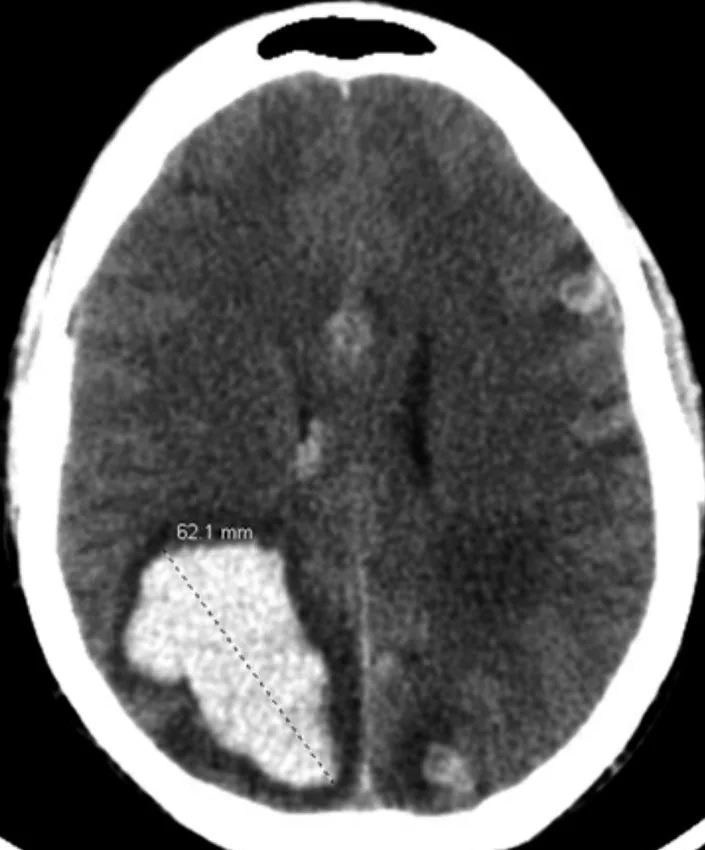
Ani nörolojik kötüleşme veya yeni nöbetle başvuran, bilinen kanseri olan ya da tümör riski taşıyan hastada intraparenkimal kanama bazen altta yatan lezyonun ilk bulgusudur. Kontrastsız BT kanamanın yerini, kitle etkisini ve ventrikül yayılımını hızlı gösterir; kontrastlı BT'de hematom içinde veya kenarında solid nodüler kontrastlanma tümör şüphesini artırır. Hematom akut dönemde lezyon dokusunu örtebilir, bu nedenle şüpheli olguda MRI daha duyarlı karakterizasyon sağlar. Kanamanın yalnız primer damar hastalığına bağlanması tümör tanısını geciktirebilir ve ek lezyonları atlatabilir.
Faz ve pencere
- Kontrastsız tanısal
- Akut kan çoğunlukla hiperdens hematom olarak izlenir; hematom çevresindeki vazojenik ödem, beklenen kitle hacmine göre yaygın olabilir ve sınırlar düzensizleşebilir. Aynı görüntüde küçük nodüler solid bileşen kan tarafından örtülebilir.
- Kontrastlı tanısal
- Kontrast sonrası kesitlerde hematomun kenarında veya içinde nodüler veya solid iyot tutulumu aranır; bu bulgular metastaz şüphesini artırır. Akut kanama dokuyu maskeleyebileceği için negatif kontrastlı BT tümörü dışlamaz.
Önerilen pencereler: Beyin (G 80 / M 40).
BT bulguları
- Orantısız vazojenik ödem — hematom çevresindeki beyaz cevher hipodensitesi, kanama boyutuna göre geniş bir alana yayılır.
- Akut kanamalı metastazlar beyine göre hiperdens görünür; kontrast sonrası nodüler veya halka şeklinde tutulum ve lezyon boyutundan bağımsız olarak belirgin vazojenik ödem beklenir.
- Nodüler kontrastlanma — hematom içinde ya da hemen kenarında damar dışı solid kontrastlanan odak.
- Çoklu hemorajik odak — özellikle gri-beyaz cevher bileşkesindeki ayrı lezyonlar metastaz olasılığını yükseltir.
- Melanom, renal hücreli, tiroid ve koryokarsinom öyküsü şüpheyi artırır; akciğer ve meme metastazları en sık kanayan tümörlerdir.
- Kitle etkisi — ventrikül basısı, orta hat itilmesi veya herniasyon bulguları eşlik edebilir.
Normalde
Kontrastsız BT tek başına metastaz taraması için yeterli değildir; ancak bulgular tanıda ipucu verebilir ve kontrastlı değerlendirmeye yönlendirir. Perihematomal vazojenik ödem ile kontrast alan lezyon boyutu arasındaki oran değerlendirilmeli; metastazlarda bu oran yüksek deregli gliomalara göre belirgin şekilde artmıştır.
Normal BT ile kıyasla
Aynı bölgenin, kaynak veri setinde patoloji içermediği belirtilen bir BT incelemesini kesit kesit kaydırın; organ sınırlarını açarak anatomiyi hasta görüntüsüyle karşılaştırın. Küçük rastlantısal bulgular tümüyle dışlanmamıştır.
Kaynak: TotalSegmentator v2.01 veri seti (Wasserthal ve ark., Radiology: Artificial Intelligence 2023), CC BY 4.0, vaka s0103; organ sınırları veri setinin otomatik segmentasyonundan, birleştirilerek sadeleştirildi. Görüntüler 2,5 mm kesit aralığına yeniden örneklendi.
Ayırıcı tanı
- Hipertansif primer hematom
- bazal ganglion kanaması, hipertansif intrakraniyal kanama ile uyumlu olabilir.
- Metastatik lezyonlar genellikle gri-beyaz cevher bileşkesinde ve büyük arter vasküler bölgelerin sınır zonlarında yerleşim gösterir.
- Bilinmeyen malignitede yeni tanı alan tek odaklu enhansan lezyonlarda doğru tanı koymada proton MR spektroskopisi ve dinamik kontrast perfüzyon gibi ileri MR teknikleri yardımcı olur.
- MRI'da karakteristik hemosiderin halkası ile heterojen lezyon olarak izlenir; kanama yapma eğilimindedir.
- Hemorajik arteriyel infarkt
- iskemik infarktı hemorajik dönüşüm izleyebilir.
Tuzaklar
- İyotlu kontrast tutulumu metastaz tespitinde kritik olup; metastazlar kontrastlı BT'de halka, nodüler veya solid enhansan paternler gösterebilir.
- Kanamalı metastazların hepsi tek bir primer tümör tipine özgü değildir; çoklu lezyon ve klinik öykü tanıyı destekler ama BT'de kesin histoloji vermez.
- Akut hematom tümörü örtebilir; çevresel ödem ve düzensiz kontur şüphe uyandırıyorsa negatif ilk kontrastlı incelemeyi dışlama kabul etmeyin.
Kendini dene
Bilinen kanserli hastada lobar hematom çevresinde geniş ödem ve kenarda kontrastlanan nodül var. En olası açıklama nedir?
Cevabı göster
Kanamalı metastaz. Perihematomal geniş vazojenik ödem ile kalıcı nodüler kontrastlanma altta yatan tümörü destekler. Hipertansif hematom tipik derin yerleşimlidir; kavernomda MRI hemosiderin halkası yardımcıdır; arteriyel enfarkt damar alanına uyan hipodens iskemi gösterir.
Kontrastlı görüntülemede, akut hematomun kenarında veya içinde nodüler veya halka şeklinde iyot tutulumu izlenirse altta yatan metastatik lezyon şüphesi artar.
Cevabı göster
Metastaz. İyotlu kontrast tutulumu metastaz tespitinde kritik olup; metastazlar kontrastlı BT'de halka, nodüler veya solid enhansan paternler gösterebilir.
Kaynaklar
Bu sayfadaki 36 cümle ve 2 quiz sorusu aşağıdaki kaynaklardan alıntılarla tek tek karşılaştırıldı; 12 cümle bu karşılaştırmada düzeltildi (2026-10-09).
- Imaging of brain metastasespmc.ncbi.nlm.nih.gov
- Delayed intracranial lesions following cardiac myxoma resection in a patient with concurrent cerebral embolism: a diagnostic challenge in the absence of pathological evidencepmc.ncbi.nlm.nih.gov
- Giant neglected cutaneous squamous cell carcinoma of the scalp with full-thickness calvarial erosion and early fatal outcome: CT findings and treatment-planning implicationspmc.ncbi.nlm.nih.gov
- Metastatic gestational trophoblastic neoplasia presenting initially as intraparenchymal and subdural hematoma: a case reportpmc.ncbi.nlm.nih.gov
- Tumor-to-tumor metastasis of small cell carcinoma of presumed pulmonary origin to a sphenoid ridge meningioma: A diagnostic pitfall in preoperative systemic evaluationpmc.ncbi.nlm.nih.gov
- Paranasal sinus cancer.pubmed.ncbi.nlm.nih.gov
- Circulating Liquid Biopsy Biomarkers in Glioblastoma: Advances and Challenges.pubmed.ncbi.nlm.nih.gov
- Imaging diagnosis of brain metastasis.pubmed.ncbi.nlm.nih.gov
- [Brain Calcification].pubmed.ncbi.nlm.nih.gov
- P-glycoprotein Mediates Ceritinib Resistance in Anaplastic Lymphoma Kinase-rearranged Non-small Cell Lung Cancer.pubmed.ncbi.nlm.nih.gov
- Cerebrospinal fluid tumor DNA for liquid biopsy in glioma patients' management: Close to the clinic?pubmed.ncbi.nlm.nih.gov
- GDF-15 in solid vs non-solid treatment-naïve malignancies.pubmed.ncbi.nlm.nih.gov
- Subdural Hematoma Mimickers: A Systematic Review.pubmed.ncbi.nlm.nih.gov
- Central neurogenic hyperventilation secondary to suspected metastatic renal cell carcinoma.pubmed.ncbi.nlm.nih.gov
- Brain metastasis-related microRNAs in patients with advanced breast cancer.pubmed.ncbi.nlm.nih.gov
- Focused Ultrasound Treatment, Present and Future.pubmed.ncbi.nlm.nih.gov
- "You Always Worry": Scan-Related Anxiety Among Patients With Metastatic Lung Cancer.pubmed.ncbi.nlm.nih.gov
- BSREM for Brain Metastasis Detection with 18F-FDG-PET/CT in Lung Cancer Patients.pubmed.ncbi.nlm.nih.gov
- Case Report: Craniotomy and radiotherapy for multiple hemorrhagic cerebral lesions from resected cardiac myxoma.pubmed.ncbi.nlm.nih.gov
- Case Report: Clinical and magnetic resonance imaging findings in intracranial hemangiosarcoma in a cat.pubmed.ncbi.nlm.nih.gov
- Delayed intracranial lesions following cardiac myxoma resection in a patient with concurrent cerebral embolism: a diagnostic challenge in the absence of pathological evidence.pubmed.ncbi.nlm.nih.gov
- Case Report: Cerebrospinal fluid cytology as a diagnostic clue in hemorrhagic glioblastoma mimicking hypertensive intracranial hemorrhage.pubmed.ncbi.nlm.nih.gov
- Chronic expanding intracerebral hematoma: A case report.pubmed.ncbi.nlm.nih.gov
- Middle meningeal artery embolization of meningioma en plaque misdiagnosed as subdural hematoma: illustrative cases.pubmed.ncbi.nlm.nih.gov
- Machine learning in neuroradiology: Recent developments and applications.pubmed.ncbi.nlm.nih.gov
- Health system learning enables generalist neuroimaging models.pubmed.ncbi.nlm.nih.gov
- Coats disease: A narrative review of clinical spectrum, imaging features, and contemporary management.pubmed.ncbi.nlm.nih.gov
- Emergency MRI in acute dizziness and headache: current insights and future directions.pubmed.ncbi.nlm.nih.gov
- Case Report: Primary epithelioid sarcoma of orbit: first case in Asia and literature review.pubmed.ncbi.nlm.nih.gov
- Minimally invasive port stent surgery: Re-purposing a balloon-expandable covered vascular stent for minimally invasive resection of deep cerebral masses: Report of 4 first-in-human stent port cases.pubmed.ncbi.nlm.nih.gov
- Intracranial venous thrombosis imaging by anatomical location: an educational review with mimics and diagnostic pitfalls.pubmed.ncbi.nlm.nih.gov
- Advances in Stereotactic Hematoma Evacuation for Hypertensive Intracerebral Hemorrhage and the Value of Multimodal Monitoring.pubmed.ncbi.nlm.nih.gov
- Postoperative Pituitary MRI Findings in Acromegaly: A Pictorial Review.pubmed.ncbi.nlm.nih.gov
- Subcutaneous extraskeletal osteosarcoma of the gluteal region: a case report from a resource-limited setting.pubmed.ncbi.nlm.nih.gov
- Conventional ultrasound combined with contrast-enhanced ultrasound for differentiating benign and malignant thyroid nodules.pubmed.ncbi.nlm.nih.gov
- Herpes simplex virus encephalitis following acute brain injury: illustrative cases.pubmed.ncbi.nlm.nih.gov
- Hemorrhagic presentation of a DICER-1-mutant primary intracranial sarcoma with negative MR spectroscopy: illustrative case.pubmed.ncbi.nlm.nih.gov
- Mesenchymal Nonmeningothelial Tumors of the CNS: Evolving Molecular Landscape and Implications for Neuroradiologists.pubmed.ncbi.nlm.nih.gov
- Seizures, Edema, Thrombosis, and Hemorrhages: An Update Review on the Medical Management of Gliomas.pubmed.ncbi.nlm.nih.gov
- Brain metastases: An update on the multi-disciplinary approach of clinical management.pubmed.ncbi.nlm.nih.gov
- Surgical management of plexiform neurofibromas: An imaging-based perioperative framework.pubmed.ncbi.nlm.nih.gov
- Microsurgical Reconstruction of the Ear and Temporal Region: Structural and Functional Considerations Including Hearing Rehabilitation-A Narrative Review.pubmed.ncbi.nlm.nih.gov
- A Rare Instance of Adult Medulloblastoma Presenting Imaging Heterogeneity Correlating With Molecular Subtypes and Anatomical Locations.pubmed.ncbi.nlm.nih.gov
- Magnetic resonance imaging of primary central nervous system neoplasms of dogs and cats: Basic concepts for diagnostic pathologists.pubmed.ncbi.nlm.nih.gov
- Pituicytoma in the third ventricle suggesting a third ventricular floor origin: illustrative case.pubmed.ncbi.nlm.nih.gov
- Metastatic gestational trophoblastic neoplasia presenting initially as intraparenchymal and subdural hematoma: a case report.pubmed.ncbi.nlm.nih.gov
- Severe Recurrent Hemorrhage of an Oncogenic Vertebrobasilar Junction Aneurysm Following Stent-Assisted Coil Embolization in Metastatic Testicular Choriocarcinoma.pubmed.ncbi.nlm.nih.gov
- Primary cerebellar angiosarcoma: A rare case of posterior fossa vascular tumor with hemorrhagic presentation and favorable surgical outcome.pubmed.ncbi.nlm.nih.gov
- Synchronous Duodenal and Pancreatic Metastases From Clear Cell Renal Cell Carcinoma 18 Years After Nephrectomy: A Case Report.pubmed.ncbi.nlm.nih.gov
- Ileal malignant melanoma presenting with severe iron-deficiency anemia and active small-bowel bleeding: primary intestinal melanoma versus metastasis - a case report and narrative review.pubmed.ncbi.nlm.nih.gov
- Dural-based melanoma metastasis mimicking a posterior fossa meningioma: A case report and review of the literature.pubmed.ncbi.nlm.nih.gov
- Intraventricular giant cavernous malformation mimicking central neurocytoma characterized by digital subtraction angiography: illustrative case.pubmed.ncbi.nlm.nih.gov
- Glymphatic System Dysfunction in Epilepsy: Clinical and Translational Perspectives.pubmed.ncbi.nlm.nih.gov
- Synaptic micromechanics and brain softening as a mechanobiological hypothesis for Alzheimer's disease.pubmed.ncbi.nlm.nih.gov
- The Glymphatic System in α-Synucleinopathies.pubmed.ncbi.nlm.nih.gov
- Endoscopic versus catheter-based minimally invasive evacuation of spontaneous supratentorial intracerebral hemorrhage: a systematic review and meta-analysis.pubmed.ncbi.nlm.nih.gov
- Isolated Hemichorea-Hemiballismus as the Presenting Manifestation of Thalamic Intracerebral Hemorrhage: A Long-Term Treatment Course With Tetrabenazine and Aripiprazole.pubmed.ncbi.nlm.nih.gov
- The Anticoagulation Dilemma: Concurrent Traumatic Brain Hemorrhage and Cerebral Venous Thrombosis in a Blast Injury Patient.pubmed.ncbi.nlm.nih.gov
- Hemorrhagic posterior reversible encephalopathy syndrome with pontine infarction in early-onset eclampsia: An atypical MRI spectrum.pubmed.ncbi.nlm.nih.gov
- Microsurgical obliteration of a high-flow intraorbital arteriovenous malformation fed by the ophthalmic artery: Illustrative case.pubmed.ncbi.nlm.nih.gov
- Co-occurring scalp arteriovenous malformation and dural arteriovenous fistula with diploic vein involvement: illustrative case.pubmed.ncbi.nlm.nih.gov
- Unique Presentation of an Arteriovenous Malformation (AVM) in the Hand: A Case Report.pubmed.ncbi.nlm.nih.gov
- Spontaneous hemorrhage of cavernous malformation in the region of the cochlear nucleus: A case report.pubmed.ncbi.nlm.nih.gov
- Cavernous venous malformation of the frontal sinus: CT and MRI imaging findings.pubmed.ncbi.nlm.nih.gov
- Radiation-induced cavernous malformation in the brainstem after Gamma Knife radiosurgery for vestibular schwannoma: A case report and literature review.pubmed.ncbi.nlm.nih.gov
- Association of 24-Hour Computed Tomography Infarct Density on Functional Outcomes in Stroke: Secondary Analysis From the AcT Trial.pubmed.ncbi.nlm.nih.gov
- Redefining the Cerebral Ischemic Core-Penumbra-Oligemia Continuum.pubmed.ncbi.nlm.nih.gov
- A Cardiac Time Bomb: Stroke as the Initial Presentation of a Large Atrial Myxoma.pubmed.ncbi.nlm.nih.gov
- Liver Imaging Reporting and Data System (LI-RADS): A Comprehensive Review.pubmed.ncbi.nlm.nih.gov
- Understanding Photon-Counting CT: Physics, Detector Technology, and Image Reconstructions.pubmed.ncbi.nlm.nih.gov
- O-RADS MRI risk stratification system: pearls and pitfalls.pubmed.ncbi.nlm.nih.gov
- Retroperitoneal paraganglioma mimicking a solid pseudopapillary neoplasm of the pancreatic tail and confirmed by percutaneous biopsy: a case report.pubmed.ncbi.nlm.nih.gov
- Paraneoplastic neurologic syndrome in advanced lung cancer with mixed adenocarcinoma and squamous features: a diagnostic challenge-a case report.pubmed.ncbi.nlm.nih.gov
- Hyperparathyroidism revealed by brown tumors of the jaws: a retrospective case series.pubmed.ncbi.nlm.nih.gov
- Duodenal Obstruction and Grade III Acute Cholangitis Following Pancreaticoduodenal Artery Aneurysm Rupture During Conservative Management of Acute Type B Aortic Dissection: A Case Report.pubmed.ncbi.nlm.nih.gov
- Comparing Handcrafted Radiomics Versus Latent Deep Learning Features of Admission Head CT for Hemorrhagic Stroke Outcome Prediction.pubmed.ncbi.nlm.nih.gov
- Supratentorial Brain Lesions in Unconscious Patients Map to a Common Human Brain Circuit.pubmed.ncbi.nlm.nih.gov
- Brain Endothelial Glycocalyx as a Blood-Facing Translational Interface in Alzheimer's Disease: Beyond "Leaky" Barriers Toward Repair-First Stratification.pubmed.ncbi.nlm.nih.gov
- Radiogenomics of congenital brain malformations: Linking embryology, genetics, and imaging.pubmed.ncbi.nlm.nih.gov
- Cerebral microbleeds: Causes, clinical relevance, and imaging approach - A narrative review.pubmed.ncbi.nlm.nih.gov
- Remote diffusion-weighted imaging lesions in spontaneous intracerebral hemorrhage: research progress and new perspectives.pubmed.ncbi.nlm.nih.gov
- Neutrophil extracellular traps and microglia/macrophages interactions in stroke: from thromboinflammation to immunotherapy.pubmed.ncbi.nlm.nih.gov
- Neuron-Derived Sema3B Facilitates Microglial Hematoma Clearance After Intracerebral Hemorrhage.pubmed.ncbi.nlm.nih.gov
- Primary splenic angiosarcoma presenting with multiple bone destruction as the initial manifestation: a case report and narrative review.pubmed.ncbi.nlm.nih.gov
- Early detection of a new pseudoaneurysm from an alternative feeder after emergent embolization for ruptured giant renal angiomyolipoma: A case report.pubmed.ncbi.nlm.nih.gov
- Diagnostic Pitfall in Cardiac Angiosarcoma: Initial Misdiagnosis as Masson Tumor Due to Sampling of Necrotic Tissue.pubmed.ncbi.nlm.nih.gov
- Extensive intraventricular hemorrhage, complicated by dengue-induced coagulopathy, initially misdiagnosed as gastroenteritis in a toddler: A case report.pubmed.ncbi.nlm.nih.gov
- Time Is Key: Early Diagnosis of Post-Transplant Lymphoproliferative Disorder Presenting as Primary CNS Diffuse Large B-Cell Lymphoma.pubmed.ncbi.nlm.nih.gov
- A Diagnostic Challenge: From Suspected Ischemic Stroke to Confirmed Herpetic Encephalitis.pubmed.ncbi.nlm.nih.gov
- Spectrum of Neuroimaging Findings in CNS Lymphoproliferative Disorders.pubmed.ncbi.nlm.nih.gov
- Evaluation of glial tumors: correlation between magnetic resonance imaging and histopathological analysis.pubmed.ncbi.nlm.nih.gov
- Inflammatory disorders that affect the cerebral small vessels.pubmed.ncbi.nlm.nih.gov
- Cerebrovascular Manifestations of Neurosarcoidosis - An Overview.pubmed.ncbi.nlm.nih.gov
- Magnetic Resonance Imaging Features of Hepatic Hydatid Disease: A Pictorial Review with Emphasis on Atypical Presentations and Differential Diagnosis.pubmed.ncbi.nlm.nih.gov
- Cardiac Angiosarcoma Presenting as Large Pericardial Effusion With Effusive-Constrictive Pericarditis.pubmed.ncbi.nlm.nih.gov
- Anatomy-Guided Microsurgical Resection of a Dominant Frontal Lobe Tumor Without Intraoperative Adjuncts: A Case Report from a Resource-Limited Context.pubmed.ncbi.nlm.nih.gov
- Spinal lesions: a comprehensive radiologic overview.pubmed.ncbi.nlm.nih.gov
- AOSNP-ADAPTR resource level-based recommendations on practical diagnostic strategies for medulloblastomas and other embryonal tumors.pubmed.ncbi.nlm.nih.gov
- Percutaneous Cryoablation Versus Microwave Ablation for Hepatocellular Carcinoma: Comparative Study of Safety and Oncologic Outcomes.pubmed.ncbi.nlm.nih.gov
- A hybrid approach via video-assisted thoracoscopic surgery and limited sternal incision for resection of invasive thymoma with superior vena cava prosthetic replacement: a case report.pubmed.ncbi.nlm.nih.gov
- Delayed multiple hemorrhagic brain metastases from atrial myxoma: a case report and literature review.pubmed.ncbi.nlm.nih.gov
- Dual-energy CT in the evaluation of intracerebral hemorrhage of unknown origin: differentiation between tumor bleeding and pure hemorrhage.pubmed.ncbi.nlm.nih.gov
- Pathologic findings and causes of death of stranded cetaceans in the Canary Islands (2006-2012).pubmed.ncbi.nlm.nih.gov
- Efficacy and safety of online adaptive magnetic resonance-guided fractionated stereotactic radiotherapy for brain metastases in non-small cell lung cancer (GASTO-1075): a single-arm, phase 2 trial.pubmed.ncbi.nlm.nih.gov
- Special Issue: EACR 2026 Congress: Innovative Cancer Science, 8-11 June 2026.pubmed.ncbi.nlm.nih.gov
- Paratubal Leiomyoma Mimicking Ovarian Malignancy: A Case Report and Literature Review.pubmed.ncbi.nlm.nih.gov
- Endometrial Cancer: A Daily Clinical Practice Guide for Radiologists.pubmed.ncbi.nlm.nih.gov
- Uncommon Nasal Mass Presentation: A Radiological Case Series.pubmed.ncbi.nlm.nih.gov
- Population-based Brain Templates for Ultra-Low-Field MRI.pubmed.ncbi.nlm.nih.gov
- Anatomically Precise Microsurgical Resection of a Posterior Fossa Cerebellar Metastasis in an Elderly Patient with Preservation of Venous Outflow, Dentate Nucleus, and Cerebrospinal Fluid Pathways.pubmed.ncbi.nlm.nih.gov
- Toward reliable computer-aided brain tumor diagnosis: a contrast-enhanced deep learning approach with hybrid KNN classification.pubmed.ncbi.nlm.nih.gov
- Applications of Advanced Imaging for Radiotherapy Planning and Response Assessment in the Central Nervous System.pubmed.ncbi.nlm.nih.gov
- Pictorial Review on Imaging Findings in Cerebral CTP in Patients with Acute Stroke and Its Mimics: A Primer for General Radiologists.pubmed.ncbi.nlm.nih.gov
- Cerebral Superficial Siderosis : Etiology, Neuroradiological Features and Clinical Findings.pubmed.ncbi.nlm.nih.gov
- Simultaneous subarachnoid hemorrhage and cerebral infarction due to aneurysm rupture and occlusion of the parent terminal artery: A cause of angiogram-negative non-perimesencephalic subarachnoid hemorrhage.pubmed.ncbi.nlm.nih.gov
Eğitim amaçlıdır; hasta başında tanı ve tedavi kararının yerine geçmez. İçerik yapay zekâ destekli bir süreçle güncel literatüre göre cümle cümle kaynaklarla karşılaştırıldı, ancak uzman radyolog incelemesinden geçmedi. Klinik kararlarda güncel kılavuzları, kurum protokollerini ve radyoloji raporunu esas alın.