Özet
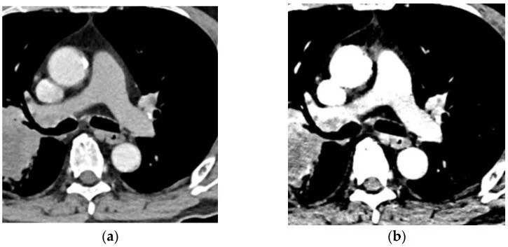
İntravenöz kontrastlı BT'de hedef damarın yeterince opaklaşmaması, akut pulmoner emboli veya arteriyel tıkanma gibi acil tanıların güvenilir biçimde dışlanmasını engelleyebilir. Yetersiz opaklaşma, kontrast enjeksiyon tekniği veya ekipman sorunları ve hastaya bağlı etmenlerle gelişebilir; derin inspirasyon da pulmoner arterlerde yetersiz opaklaşma riskini artırabilir. Pulmoner trunkusta yetersiz opaklaşma bulunan tanısal olmayan incelemede PE gözden kaçabilir; bu nedenle inceleme PE’yi güvenle dışlamaz.
Faz ve pencere
- BTA tanısal
- Yeterli kontrast opaklaşması tanısal kalite için kritik olup hasta ağırlığı, kardiyak debi, tarama süresi ve kontrast verme protokolüne bağlıdır.
- Arteriyel tanısal
- Kontrastın pulmoner arterlere ulaşma süresini belirlemek için bolus takibi veya zamanlama bolusu kullanılabilir; bu yöntemler homojen opaklaşma ve tanısal görüntü kalitesi sağlar.
Önerilen pencereler G genişlik, M merkez; değerler Hounsfield birimi (HU)
- AnjiyoG 600 / M +150
- MediastenG 350 / M +50
BT bulguları
- Pulmoner arter opaklaşması hasta ağırlığı, kardiyak debi, tarama süresi ve kontrast verme protokolüne bağlıdır; bolus takibi veya zamanlama bolusu, kontrastın pulmoner arterlere ulaşma süresini belirlemek ve homojen opaklaşma sağlamak için kullanılabilir.
- Bolus zamanlaması pulmoner arter opaklaşmasını etkileyebilir; bolus takibi, kontrastın pulmoner arterlere ulaşma süresini belirlemek için kullanılabilir.
- Derin inspirasyon, pulmoner arterlerde yetersiz opaklaşma riskini artırabilir.
- EKG eşzamanlaması olmadan yapılan DECT pulmoner anjiyografilerinde, pulmoner damarlarda çizgi artefaktları ve kontrast karışımına bağlı artefaktlar görülebilir.
- Enjeksiyon tekniği veya ekipman sorunları, tanısal olmayan CTA'ya yol açabilir.
Ölçütler ve sınıflamalar
- Pulmoner arter opaklaşması için literatürde tanımlanan yaklaşık nicel alt eşik
- yaklaşık 200–250 HU
Normalde
DECT-PA ve DECT-R protokollerinde pulmoner arterlerde (segmental dallara kadar) ve aortada yeterli kontrast yoğunluğu gösterilmiştir. Yeterli kontrast opaklaşması tanısal kalite için kritik olup hasta ağırlığı, kardiyak debi, tarama süresi ve kontrast verme protokolüne bağlıdır.
Normal BT ile kıyasla
Aynı bölgenin, kaynak veri setinde patoloji içermediği belirtilen bir BT incelemesini kesit kesit kaydırın; organ sınırlarını açarak anatomiyi hasta görüntüsüyle karşılaştırın. Küçük rastlantısal bulgular tümüyle dışlanmamıştır.
Kaynak: TotalSegmentator v2.01 veri seti (Wasserthal ve ark., Radiology: Artificial Intelligence 2023), CC BY 4.0, vaka s1278; organ sınırları veri setinin otomatik segmentasyonundan, birleştirilerek sadeleştirildi. Görüntüler 2,5 mm kesit aralığına yeniden örneklendi.
Ayırıcı tanı
- EKG eşzamanlaması olmadan yapılan DECT pulmoner anjiyografilerinde çizgi ve kontrast karışım artefaktları damar içi ürat birikimi izlenimi verebilir.
- Pulmoner arter dolum defektlerinin en sık nedeni tromboembolidir; ancak bu bulgu her zaman özgül olmadığından ayırıcı tanı gerekir.
- Kronik tromboembolik pulmoner hipertansiyon, pulmoner arterlerde organize tromboembolik lezyonlarla karakterizedir.
Tuzaklar
- Çekim öncesinde solunum manevrasını prova edin; derin inspirasyondan kaçınmak pulmoner arterlerde yetersiz opaklaşma riskini azaltmaya yardımcı olabilir.
- Bir CTPA çalışmasında ana pulmoner arterde 250 HU, teknik yeterliliği değerlendirmek için eşik olarak kullanılmıştır.
- Pulmoner trunkusta yetersiz opaklaşma bulunan tanısal olmayan incelemede PE gözden kaçabilir; bu nedenle inceleme PE'yi güvenle dışlamaz.
Kendini dene
BTA’da pulmoner trunkusta yetersiz kontrast opaklaşması nedeniyle inceleme tanısal değil. Bu teknik sınırlılıkta incelemede pulmoner emboli gözden kaçabilir mi?
Cevabı göster
İnceleme PE’yi güvenle dışlamaz; teknik sınırlılık belirtilmelidir. Pulmoner trunkusta kontrast opaklaşmasının yetersiz olduğu tanısal olmayan incelemelerde insidental PE başlangıç raporunda gözden kaçabilir. Pulmoner trunkusta kontrast opaklaşması yetersiz olan tanısal olmayan incelemelerde incidental pulmoner emboli başlangıç raporunda gözden kaçabilir.
EKG ile eşzamanize edilmemiş DECT pulmoner anjiyografilerinde en sık görülen iki damar içi artefakt türü nedir?
Cevabı göster
Çizgi artefaktı ve kontrast karışımına bağlı artefakt. Çalışmada gut hastalarında çizgi artefaktı %56, kontrast karışımına bağlı artefakt %51; kontrol grubunda ise sırasıyla %57 ve %65 oranında saptanmıştır.
İlgili konular
Kaynaklar
Bu sayfadaki 30 cümle ve 2 quiz sorusu aşağıdaki kaynaklardan alıntılarla otomatik olarak karşılaştırıldı; 24 cümle bu karşılaştırmada düzeltildi (2026-10-09).
- Looking beyond the thrombus: essentials of pulmonary artery imaging on CTpmc.ncbi.nlm.nih.gov
- Dual energy CT in clinical routine: how it works and how it adds value.pubmed.ncbi.nlm.nih.gov
- Suboptimal contrast opacification of dynamic head and neck MR angiography due to venous stasis and reflux: technical considerations for optimization.pubmed.ncbi.nlm.nih.gov
- Added value of an artificial intelligence algorithm in reducing the number of missed incidental acute pulmonary embolism in routine portal venous phase chest CT.pubmed.ncbi.nlm.nih.gov
- Early experience with a split-bolus single-pass CT protocol in paediatric trauma.pubmed.ncbi.nlm.nih.gov
- Multi-energy CT and iodinated contrast.pubmed.ncbi.nlm.nih.gov
- Improving Contrast Enhancement in Pulmonary CTA: The value of breathing maneuvers.pubmed.ncbi.nlm.nih.gov
- Stress echocardiography for the diagnosis of coronary artery disease: an evidence-based analysis.pubmed.ncbi.nlm.nih.gov
- Comparison of early- and late-phase CT angiography findings in brain death.pubmed.ncbi.nlm.nih.gov
- Dual energy CT angiography for lower extremity trauma: comparison with conventional CT.pubmed.ncbi.nlm.nih.gov
- Stress echocardiography with contrast for the diagnosis of coronary artery disease: an evidence-based analysis.pubmed.ncbi.nlm.nih.gov
- Low contrast volume dual-energy CT of the chest: Quantitative and qualitative assessment.pubmed.ncbi.nlm.nih.gov
- Improved diagnostic accuracy when combining computed tomography angiography and corrected coronary opacification in patients with coronary stents.pubmed.ncbi.nlm.nih.gov
- Effect of region-of-interest placement in bolus tracking cerebral computed tomography angiography.pubmed.ncbi.nlm.nih.gov
- Computed tomography pulmonary angiography using a 20% reduction in contrast medium dose delivered in a multiphasic injection.pubmed.ncbi.nlm.nih.gov
- Artificial intelligence in computed tomography imaging for pulmonary embolism: a narrative review from computed tomography pulmonary angiography to non-contrast computed tomography.pubmed.ncbi.nlm.nih.gov
- Deep Learning-Based Iodine Contrast Augmentation for Suboptimally Enhanced CT Pulmonary Angiography: Implications for Pulmonary Embolism Diagnosis.pubmed.ncbi.nlm.nih.gov
- Commercial artificial intelligence-assisted performance and interpretation time of first-on-call radiology residents using computed tomography pulmonary angiography to detect pulmonary embolism: a multireader, multicenter study.pubmed.ncbi.nlm.nih.gov
- Patient-specific timing acquisition for coronary CT angiography: A retrospective patient validation study.pubmed.ncbi.nlm.nih.gov
- Update on Contrast-Induced Nephropathy: Recent Developments in Its Prevention, Early Diagnosis, and Therapy.pubmed.ncbi.nlm.nih.gov
- Emerging Techniques and Treatment Outcomes in Pulmonary Arteriovenous Malformations Embolisation: A Narrative Review.pubmed.ncbi.nlm.nih.gov
- Efficiency and Optimization of Iodinated Contrast Agents in Cerebral Aneurysm CT Angiography: A Systematic Review.pubmed.ncbi.nlm.nih.gov
- Diagnosing lung cancer invading the pulmonary artery using ultra-low-dose CT pulmonary angiography: a feasibility study.pubmed.ncbi.nlm.nih.gov
- Epicardial Fat Tissue Volume Within 1 mm of the Left Atrium Combined With Left Atrial Appendage Volume is Associated With Thrombus in Nonvalvular Atrial Fibrillation: A CCTA-Based Risk Stratification Model.pubmed.ncbi.nlm.nih.gov
- Acute Traumatic Aortic Injury: What the Radiologist Needs to Know.pubmed.ncbi.nlm.nih.gov
- Contrast opacification on thoracic CT angiography: challenges and solutions.pubmed.ncbi.nlm.nih.gov
- Single-Bolus Sequential Triple-Rule-Out CT Angiography: Image Quality and Radiation Dose on Wide-Area Detector and Dual-Source CT.pubmed.ncbi.nlm.nih.gov
- Four-Dimensional (4D) Volume-Based Bolus-Tracking Monitoring for Post-endovascular Aneurysm Repair CT: Feasibility of Combined Hemodynamic and Morphological Imaging.pubmed.ncbi.nlm.nih.gov
- Sequential Helical-Axial-Helical Triple-Rule-Out CT Angiography: Technical Feasibility and Territory-Specific Image Quality in the Emergency Department.pubmed.ncbi.nlm.nih.gov
- Contrast-Enhanced CT of the Coelom in a Female Bald Eagle (<i>Haliaeetus leucocephalus</i>): Development of a Preliminary CT-Based Anatomical Segmentation Workflow.pubmed.ncbi.nlm.nih.gov
- Giant left main coronary artery aneurysm with fistulous drainage to the right atrium: a rare case report and surgical management approach.pubmed.ncbi.nlm.nih.gov
- Confounder-Matched Deep Learning on Cardiac CT for the Diagnosis of Tetralogy of Fallot: A Proof of Concept.pubmed.ncbi.nlm.nih.gov
- An indolent course of undifferentiated pleomorphic cardiac sarcoma mimicking myxoma: A case report.pubmed.ncbi.nlm.nih.gov
- Meningococcal Meningitis Complicated by Cerebral Venous Sinus Thrombosis Presenting as Pseudo-Subarachnoid Hemorrhage in a Toddler: A Case Report.pubmed.ncbi.nlm.nih.gov
- The Initial Diagnostic Approach to Adult Congenital Heart Disease: A Practical Imaging Pathway for General Cardiologists and Cardiac Imagers.pubmed.ncbi.nlm.nih.gov
- A case report of iatrogenic coronarocameral fistula after surgical aortic valve replacement: multimodality diagnosis and intravascular ultrasound-guided left main intervention.pubmed.ncbi.nlm.nih.gov
- Cardiovascular Flow Imaging.pubmed.ncbi.nlm.nih.gov
- Analysis of Cardiac Computed Tomography: Investigating the Relationship Between Coronary Microvascular Dysfunction and Left Heart Remodeling in Patients With Myocardial Ischemia Due to Non-Obstructive Coronary Artery Disease.pubmed.ncbi.nlm.nih.gov
- Intracoronary physiological indices for coronary microvascular dysfunction: from concept to clinical application.pubmed.ncbi.nlm.nih.gov
- Application of 50-keV virtual monochromatic imaging combined with a personalized contrast agent protocol in computed tomography pulmonary angiography: a randomized controlled trial.pubmed.ncbi.nlm.nih.gov
- Frequently Encountered Artifacts in the Application of Dual-Energy Computed Tomography to Cardiovascular Imaging for Urate Crystals in Gout: A Matched-Control Study.pubmed.ncbi.nlm.nih.gov
- Management of pneumonia by the emergency radiologist.pubmed.ncbi.nlm.nih.gov
- When Cancer Clots: An Extensive Radiologic Analysis of Cancer-Associated Thromboembolism.pubmed.ncbi.nlm.nih.gov
- Intravenous leiomyomatosis of the uterus: A case report and literature review.pubmed.ncbi.nlm.nih.gov
- Deep learning-assisted 3D CT angiography for supply-vessel localization in inflammatory peripheral pulmonary artery pseudoaneurysms.pubmed.ncbi.nlm.nih.gov
- Advances in the imaging of pulmonary hypertension.pubmed.ncbi.nlm.nih.gov
- Arterial and biliary complications after transarterial chemoembolization for hepatocellular carcinoma.pubmed.ncbi.nlm.nih.gov
- High-pitch free-breathing triple rule-out CT angiography for acute chest pain: a prospective sequential two-cohort comparison of radiation dose and diagnostic image quality.pubmed.ncbi.nlm.nih.gov
- Optimizing coronary CT angiography in patients with delayed contrast arrival using a free-breathing, high-threshold bolus-tracking protocol.pubmed.ncbi.nlm.nih.gov
- Photon-counting CT iodine maps for late iodine enhancement: a retrospective feasibility study of image quality and reader confidence.pubmed.ncbi.nlm.nih.gov
- Patient-Specific Timing for Pulmonary CT Angiography: Human Validation of a Physiologically Derived Post-Trigger Delay Model.pubmed.ncbi.nlm.nih.gov
- Optimal approach to performing and reporting computed tomography angiography for suspected acute pulmonary embolism: a clinical consensus statement of the ESC Working Group on Pulmonary Circulation & Right Ventricular Function, the Fleischner Society, the Association for Acute CardioVascular Care (ACVC) and the European Association of Cardiovascular Imaging (EACVI) of the ESC, endorsed by European Respiratory Society (ERS), Asian Society of Thoracic Radiology (ASTR), European Society of Thoracic Imaging (ESTI), and Society of Thoracic Radiology (STR).pubmed.ncbi.nlm.nih.gov
- Artificial Intelligence in Cardiology Examination: A Comparative Study of Physicians, ChatGPT-4, and ChatGPT-5.pubmed.ncbi.nlm.nih.gov
- Adult-onset idiopathic peripheral pulmonary artery stenosis.pubmed.ncbi.nlm.nih.gov
- High-pressure-resistant totally implantable venous power ports for improving image quality in contrast-enhanced computed tomography.pubmed.ncbi.nlm.nih.gov
- Weight-based flow rate triphasic contrast injection protocol for pediatric cardiac computed tomography.pubmed.ncbi.nlm.nih.gov
- Preconditioned endothelialized grafts reduce stenosis and improve vascular remodeling in a rat pulmonary artery model.pubmed.ncbi.nlm.nih.gov
- Intracoronary imaging demonstrating vasculitis as a rare cause of myocardial infarction: a case report.pubmed.ncbi.nlm.nih.gov
- Multimodal imaging in neuro-ophthalmology.pubmed.ncbi.nlm.nih.gov
- Performance of Cardiac MRI in Pediatric and Adult Patients with Fontan Circulation.pubmed.ncbi.nlm.nih.gov
- Assessment of pulmonary arterial enhancement on CT pulmonary angiography using a leg vein for contrast media administration.pubmed.ncbi.nlm.nih.gov
- Accuracy of electrical impedance tomography to detect perfusion defects in pulmonary embolism.pubmed.ncbi.nlm.nih.gov
- Diagnoses of pulmonary embolism from non-contrast 4DCT using image processing-derived quantitative perfusion scores.pubmed.ncbi.nlm.nih.gov
- Artificial Intelligence in Pulmonary Hypertension: Current State and Future Prospects.pubmed.ncbi.nlm.nih.gov
- Emerging quantitative CCTA imaging biomarkers for cardiovascular risk stratification: a narrative review.pubmed.ncbi.nlm.nih.gov
- Coronary-bronchial artery fistula arising from the sinoatrial nodal artery in a patient with bronchiectasis: a multimodality imaging case report.pubmed.ncbi.nlm.nih.gov
- Case Report: Intra-procedural fatal cerebral lipiodol embolism without angiographically visible shunting during right inferior phrenic artery TACE.pubmed.ncbi.nlm.nih.gov
- Type III endoleaks following endovascular aortic repair: a case series.pubmed.ncbi.nlm.nih.gov
- Case Report: Leiomyosarcoma of the inferior vena cava presenting as a right atrial mass: the value of multimodality imaging.pubmed.ncbi.nlm.nih.gov
- Diagnosis and Management of Patients with Patent Foramen Ovale: Current Evidence and Future Perspective.pubmed.ncbi.nlm.nih.gov
- Quantitative CT Pulmonary Angiography and Echo Cardiography Analysis for Enhanced Cardiovascular Assessment of Right Ventricular Failure in Pulmonary Hypertension.pubmed.ncbi.nlm.nih.gov
- Imaging in chronic thromboembolic pulmonary hypertension: review of the current literature.pubmed.ncbi.nlm.nih.gov
- Genicular Artery Embolization for Osteoarthritis Related Knee Pain: A Systematic Review and Qualitative Analysis of Clinical Outcomes.pubmed.ncbi.nlm.nih.gov
- Long-term safety and effectiveness of the "OptEase" vena cava filter.pubmed.ncbi.nlm.nih.gov
- Multimodality Imaging for Left Atrial Appendage Occlusion Devices.pubmed.ncbi.nlm.nih.gov
- Deep Learning and Cardiovascular Diseases: An Updated Narrative Review.pubmed.ncbi.nlm.nih.gov
- Obesity and Cardiovascular Disease: An Obesity Medicine Association (OMA) Clinical Practice Statement (CPS) 2026.pubmed.ncbi.nlm.nih.gov
- Protocol for Post-Mortem Micro-CT Imaging of Coronary Arteries in Low-Mass Neonatal Puppy Hearts Using Barium-Based Contrast.pubmed.ncbi.nlm.nih.gov
- Optimizing CT pulmonary angiography with patient-adaptive triggering-a novel approach for a "one-stop-shop" evaluation of pulmonary and aortic vasculature.pubmed.ncbi.nlm.nih.gov
- Spontaneous Extrapleural Hematoma in a Hemodialysis Patient Receiving Anticoagulation: A Rare Cause of Acute Chest Pain.pubmed.ncbi.nlm.nih.gov
- Optimized Contrast-Enhanced Micro-Computed Tomography for Simultaneous Imaging of the Ductus Arteriosus and Pulmonary Arteries During the Perinatal Transition.pubmed.ncbi.nlm.nih.gov
- Delayed innominate vein perforation and massive hemothorax following left-sided PICC placement: A case report.pubmed.ncbi.nlm.nih.gov
- Diagnostic and management dilemmas in pulmonary hypertension.pubmed.ncbi.nlm.nih.gov
- Valvular Heart Disease and Heart Failure in the Post-COVID-19 Era: A Narrative Review of Mechanisms, Diagnosis, Differential Assessment, and Clinical Outcomes.pubmed.ncbi.nlm.nih.gov
- Current Perspectives on Balloon Pulmonary Angioplasty for Chronic Thromboembolic Pulmonary Hypertension.pubmed.ncbi.nlm.nih.gov
- Assessing Venous Congestion in Heart Failure: A Review of Splanchnic, Cardiac, and Pulmonary Ultrasound: Part 2: Contrast-Enhanced Ultrasound and Shear Wave.pubmed.ncbi.nlm.nih.gov
- Advancing MRI with magnetic nanoparticles: a comprehensive review of translational research and clinical trials.pubmed.ncbi.nlm.nih.gov
- Blood Pressure and Penumbral Sustenance in Stroke from Large Vessel Occlusion.pubmed.ncbi.nlm.nih.gov
- Tricuspid regurgitation: standardized stepwise work-up from referral to expert heart valve centre.pubmed.ncbi.nlm.nih.gov
- Development of Ultrasound Microbubbles and Phase Shift Microbubbles in Theranostics.pubmed.ncbi.nlm.nih.gov
- Advancements in 3D Transoesophageal Echocardiography (TOE) and Computed Tomography (CT) for Stroke Prevention in Left Atrial Appendage Occlusion Interventions.pubmed.ncbi.nlm.nih.gov
- MR perfusion imaging in acute ischemic stroke.pubmed.ncbi.nlm.nih.gov
- High-affinity alphavbeta3 integrin targeted optical probe as a new imaging biomarker for early atherosclerosis: initial studies in Watanabe rabbits.pubmed.ncbi.nlm.nih.gov
- The filling defect of pulmonary artery, an imaging finding what we should know.pubmed.ncbi.nlm.nih.gov
- Patterns of injury and violence in Yaoundé Cameroon: an analysis of hospital data.pubmed.ncbi.nlm.nih.gov
- Multimodality diagnostic imaging and role of interventional radiology in pancreas transplantation.pubmed.ncbi.nlm.nih.gov
- Evaluation of Living Liver Donors Using Dual-Contrast-Agent Magnetic Resonance Imaging (MRI) and Cholangiopancreatography (MRCP)-Technical Development.pubmed.ncbi.nlm.nih.gov
- Poor Distal Left Atrial Appendage Opacification on Early-Phase Cardiac CTA and Association With Arterial Thromboembolism.pubmed.ncbi.nlm.nih.gov
- Novel Computed Tomography Angiography Parameter Is Associated with Low Cardiac Index in Patients with Chronic Thromboembolic Pulmonary Hypertension: A Retrospective Analysis.pubmed.ncbi.nlm.nih.gov
- Endoscopic Management of Post-Bariatric Surgery Complications: Diagnostic Work-Up and Innovative Approaches for Leak, Fistula, and Stricture Management.pubmed.ncbi.nlm.nih.gov
- Chronic Thromboembolic Pulmonary Hypertension: A Narrative Review.pubmed.ncbi.nlm.nih.gov
- Spectral CT-derived peripheral iodine concentration in acute pulmonary embolism: association with subsegmental emboli and pulmonary infarction.pubmed.ncbi.nlm.nih.gov
- Incidental pulmonary embolism in patients with cancer: prevalence, underdiagnosis and evaluation of an AI algorithm for automatic detection of pulmonary embolism.pubmed.ncbi.nlm.nih.gov
- Spontaneous bacteremia and spontaneous bacterial peritonitis share similar prognosis in patients with cirrhosis: a cohort study.pubmed.ncbi.nlm.nih.gov
- Urgent and emergent pediatric cardiovascular imaging.pubmed.ncbi.nlm.nih.gov
- Superselective bronchial artery embolization guided by bronchial artery CT angiography for pediatric hemoptysis and pulmonary hemorrhage: a retrospective single-center study.pubmed.ncbi.nlm.nih.gov
- Endovascular management of iliac vessel injury complicating lumbar disc surgery: a case report and review of literature.pubmed.ncbi.nlm.nih.gov
- Radiologic Interventions for Gastrointestinal Bleeding.pubmed.ncbi.nlm.nih.gov
- Homocystinuria presenting with cerebral venous thrombosis: a case report highlighting progressive thrombosis.pubmed.ncbi.nlm.nih.gov
- Pulmonary valve endocarditis in a postpartum patient: a case report of unconventional risk.pubmed.ncbi.nlm.nih.gov
- Clinical benefits and current challenges of photon-counting detector CT in vascular imaging.pubmed.ncbi.nlm.nih.gov
Eğitim amaçlıdır; hasta başında tanı ve tedavi kararının yerine geçmez, tıbbi tavsiye değildir. Metinler yapay zekâ desteğiyle yazıldı ve otomatik bir adımda kaynak alıntılarıyla karşılaştırıldı; bu bir tıbbi doğrulama değildir ve içerik uzman radyolog incelemesinden geçmedi. Klinik kararlarda güncel kılavuzları, kurum protokollerini ve radyoloji raporunu esas alın.