
Özet
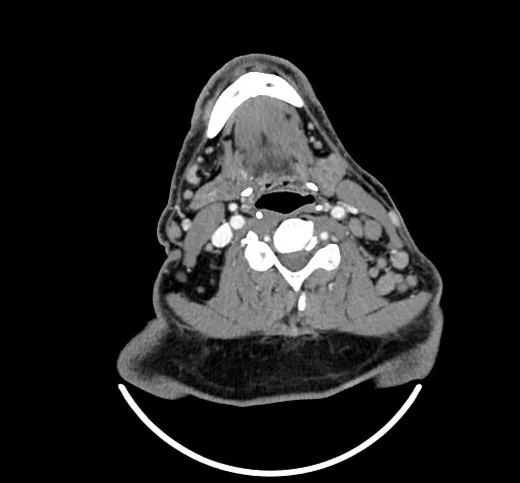
Servikal lenfoma, Hodgkin veya non-Hodgkin lenfomanın boyun nodal zincirlerini ya da Waldeyer halkasını tuttuğu hastalarda görülür; tutulan lenf nodları boyut olarak büyümüş veya sayıca çok olabilir. Kontrastlı boyun BT’si nodların sayısını, boyutunu, komşu boşluklarla ilişkisini ve toraks girişine uzanımı haritalar; FDG tutan lenfoma alt tiplerinde sistemik evreleme için PET/BT genellikle daha belirleyicidir. Waldeyer halkasında submukozal, düzgün sınırlı kitle ile nekroz göstermeyen çoklu nodların birlikte bulunması lenfomayı düşündürür, ancak BT tek başına histolojik tanı koydurmaz. Hastalığın komşu istasyonlara veya ekstranodal dokulara uzanımını atlamak evre ve tümör yükü değerlendirmesini eksik bırakabilir.
Faz ve pencere
- Portal tanısal
- İntravenöz kontrast sonrası servikal nodların yumuşak doku dansitesi ve çoğunlukla homojen kontrastlanması; Waldeyer halkasında eşlik eden submukozal yumuşak doku kalınlaşması ve nodal zincirlerin kraniokaudal dağılımı değerlendirilir. Nekroz bulunmaması lenfomayı destekleyebilir, fakat tek başına tanısal değildir.
Önerilen pencereler: Yumuşak doku (G 400 / M 40).
BT bulguları
- Birden fazla servikal istasyonda, tek tek ya da birleşik yumuşak doku yoğunluklu nodlar görülmesi lenfatik dağılımı düşündürür.
- Lenfomatöz nodlar sıklıkla homojen görünür; belirgin santral nekroz olmaması metastatik skuamöz hücreli noddan ayrımda yardımcıdır, ancak istisnalar vardır.
- Birbirine bitişik nodların konglomere kitle oluşturması damarları ve komşu kasları yer değiştirebilir; damar lümenleri korunabilir.
- Palatin tonsiller, nazofaringeal tonsil veya lingual tonsilde düzgün sınırlı, submukozal yumuşak doku kitlesi görülebilir.
- Servikal lenf nodları tutulumuyla birlikte görülen çok bölgeli Waldeyer halkası tutulumu, tek odaklı mukozal kitlelerden farklı bir dağılım örüntüsü oluşturur.
- İnce kesitlerde nod çevresi yağ planının, sternokleidomastoid kasın ve karotis kılıfının seçilebilir kalması belirgin invaziv uzanım olmadığını gösterir.
- Alt boyun kesitlerinde supraklaviküler ve üst mediastinal nodal zincir devamlılığı, hastalık haritasının servikal sınırın ötesine uzanıp uzanmadığını gösterir.
Ölçütler ve sınıflamalar
- Lugano sınıflaması (değiştirilmiş Ann Arbor evreleri)
- Evre I: tek lenfatik bölge/bölge grubu veya nodal hastalık olmadan tek ekstralenfatik odak. Evre II: diyaframın aynı tarafındaki en az iki nodal bölge; sınırlı ve bitişik ekstralenfatik uzanım varsa IIE olarak belirtilir. Evre III: diyaframın iki tarafında nodal bölgeler (dalak tutulumu eşlik edebilir). Evre IV: bir veya daha fazla ekstralenfatik organda yaygın ya da nodlardan ayrı tutulum. FDG-tutan lenfomalarda PET/BT evrelemeyi destekler; boyun BT’si tek başına tüm vücut evresi vermez.
Normalde
Normal kontrastlı boyunda Waldeyer halkası kabaca simetriktir; servikal zincirlerde küçük, oval ve homojen nodlar ayrı izlenir. Servikal lenf nodları genel olarak kısa aks ölçüsü 10 mm'nin üzerindeyse anormal kabul edilir; ancak lenfomada normal boyutlu nodlar hastalanabilir ve reaktif büyüme yanıltıcı bulgu verebilir. Karotis arter ile palatin tonsil arasındaki SUVmax farkı ve oranı gibi PET parametreleri, fizyolojik ve patolojik tutulumun ayrımında yardımcı olur.
Normal BT ile kıyasla
Aynı bölgenin, kaynak veri setinde patoloji içermediği belirtilen bir BT incelemesini kesit kesit kaydırın; organ sınırlarını açarak anatomiyi hasta görüntüsüyle karşılaştırın. Küçük rastlantısal bulgular tümüyle dışlanmamıştır.
Kaynak: TotalSegmentator v2.01 veri seti (Wasserthal ve ark., Radiology: Artificial Intelligence 2023), CC BY 4.0, vaka s0622; organ sınırları veri setinin otomatik segmentasyonundan, birleştirilerek sadeleştirildi. Görüntüler 2,5 mm kesit aralığına yeniden örneklendi.
Ayırıcı tanı
- Metastatik skuamöz hücreli karsinom
- özellikle kistik veya nekrotik nodlar, eşlik eden mukozal primer ve düzensiz kapsül dışı sınırlar aratır.
- Reaktif lenfadenopati
- çoğu kez enfeksiyon odağına komşu dağılım gösterir; çevre yağ planları korunur ve eşzamanlı çok istasyonlu konglomere kitle beklenmez.
- Tüberküloz lenfadeniti
- merkezinde düşük atenüasyon, çevresinde kontrastlanan nod duvarı ve birbirine yapışık nodlar oluşturabilir.
- Castleman hastalığı
- genellikle sınırlı nodal bölgede belirgin kontrastlanan kitle biçimindedir; yaygın homojen servikal nodal zincir örüntüsü tipik değildir.
- Waldeyer halkası skuamöz hücreli karsinomu dahil diğer baş-boyun maligniteleriyle ayrım için kontrastlı BT tek başına yetersiz kalabilir; fizyolojik tonsiller tutulum ve metabolik aktivite ayrımı amacıyla PET/BT tamamlanmalıdır.
Tuzaklar
- Lenfomaların BT'de genellikle homojen kontrastlanma gösterdiği ve nekroz ile kalsifikasyon içermediği gözlenir; bu özellik nekrozlu metastatik skuamöz hücreli kanserden ayrımda yardımcıdır ancak tek başına tanı koydurucu değildir.
- Tonsillerde fizyolojik lenfoid doku ve hafif asimetri sık görülebilir; kitleyi tonsil kriptleri, yüzey konturu ve karşı taraf hacmiyle kıyasla.
- Birbirine yaslanan nodlar tek kitle gibi görünebilir; ardışık aksiyel kesitlerde aradaki yağ çizgilerini ve farklı nodal istasyonlara devamı izle.
- Kontrastlı BT’de normal boyutlu nodlarda lenfoma bulunabileceği için yalnız kısa aks ölçüsüne dayanarak hastalığı dışlama; dağılımı ve eşlik eden bulguları değerlendir.
Kendini dene
Palatin tonsilde düzgün sınırlı submukozal kitleye birkaç servikal nodal istasyonda homojen, nekrozsuz nodlar eşlik ediyor; belirgin ülseratif mukozal primer yok. En olası tanı nedir?
Cevabı göster
Servikal lenfoma. Waldeyer halkasında düzgün sınırlı submukozal kitleyle birlikte birden çok servikal istasyonda homojen nodal büyüme lenfomayı destekler. Belirgin ülseratif mukozal primerin olmaması skuamöz hücreli kanser olasılığını azaltır; nekroz olmaması lenfomayı kanıtlamaz ve nekroz görülen lenfoma dışlanmaz.
Nekrotik üst juguler nodlara eşlik eden ülseratif tonsil kitlesi hangi tanıyı daha çok düşündürür?
Cevabı göster
Tonsil skuamöz hücreli kanseri. Ülseratif, asimetrik mukozal tonsil odağı ve nekrotik bölgesel nodlar skuamöz hücreli kanser lehinedir. Lenfomada Waldeyer halkası kitlesi daha çok submukozal ve düzgün sınırlı, eşlik eden nodlar sıklıkla nekrozsuzdur; Castleman genellikle sınırlı ve belirgin kontrastlanan nodal kitle yapar; reaktif nodlar ülseratif primer kitleyi açıklamaz.
İlgili konular
Kaynaklar
Bu sayfadaki 44 cümle ve 2 quiz sorusu aşağıdaki kaynaklardan alıntılarla tek tek karşılaştırıldı; 7 cümle bu karşılaştırmada düzeltildi (2026-10-08).
- PET-CT Imaging of Lymphomapmc.ncbi.nlm.nih.gov
- Perfusion CT of head and neck cancer.pubmed.ncbi.nlm.nih.gov
- Primary non-Hodgkin's lymphoma masquerading as cervical cancer.pubmed.ncbi.nlm.nih.gov
- A rare presentation of lymphoma of the cervix with cross-sectional imaging correlation.pubmed.ncbi.nlm.nih.gov
- Multiple schwannomas of the facial nerve mimicking cervical lymphoma: a case report.pubmed.ncbi.nlm.nih.gov
- A rare lymphoma in a patient with amyopathic dermatomyositis.pubmed.ncbi.nlm.nih.gov
- Primary B-cell lymphoma of the uterine cervix: Presentation in Pap-test slide and cervical biopsy.pubmed.ncbi.nlm.nih.gov
- Ultrasound-Based Radiomics Can Classify the Etiology of Cervical Lymphadenopathy: A Multi-Center Retrospective Study.pubmed.ncbi.nlm.nih.gov
- Primary cervical lymphoma: a rare presentation to a genitourinary medicine clinic.pubmed.ncbi.nlm.nih.gov
- Advanced Doppler imaging and trucut biopsy of a late-stage cervical lymphoma.pubmed.ncbi.nlm.nih.gov
- Primary Uterine Cervical Lymphoma Manifesting as Menolipsis Staged and Followed Up by FDG PET/CT.pubmed.ncbi.nlm.nih.gov
- Primary diffuse large B-Cell lymphoma of the uterine cervix with severe lower urinary tract Symptoms: A rare case report and review of the literature.pubmed.ncbi.nlm.nih.gov
- Dose and image quality of high-pitch dual source computed tomography for the evaluation of cervical lymph node status - comparison to regular 128-slice single source computed tomography.pubmed.ncbi.nlm.nih.gov
- [Alleviation of carotid sinus syncope and removal of cardiac pacing after regression of cervical malignant lymphoma].pubmed.ncbi.nlm.nih.gov
- Perfluorobutane CEUS for early-stage cervical lymphoma: diagnostic value of the postvascular phase starfield sign.pubmed.ncbi.nlm.nih.gov
- CXCR4 PET imaging of mantle cell lymphoma using [<sup>68</sup>Ga]Pentixafor: comparison with [<sup>18</sup>F]FDG-PET.pubmed.ncbi.nlm.nih.gov
- Whole-body diffusion-weighted imaging for staging malignant lymphoma in children.pubmed.ncbi.nlm.nih.gov
- Comorbidities associated with HIV and antiretroviral therapy (clinical sciences): a workshop report.pubmed.ncbi.nlm.nih.gov
- Nasopharyngeal classical Hodgkin lymphoma: a case report.pubmed.ncbi.nlm.nih.gov
- Primary synovial sarcoma of the thyroid mimicking medullary thyroid cancer.pubmed.ncbi.nlm.nih.gov
- Nasopharyngeal carcinoma masquerading as a primary sinonasal mass: a case report.pubmed.ncbi.nlm.nih.gov
- Non-Squamous Cell Carcinoma of the Larynx: A State-of-the-Art Review.pubmed.ncbi.nlm.nih.gov
- Duodenal imaging on the spotlight: from A to Z.pubmed.ncbi.nlm.nih.gov
- Imaging characteristics of primary laryngeal lymphoma.pubmed.ncbi.nlm.nih.gov
- Diffuse Large B-Cell Lymphoma Presenting as an Infiltrative Masticator Space Lesion Mimicking Inflammatory Disease: A Case Report.pubmed.ncbi.nlm.nih.gov
- Malignant lymph nodes of the head and neck: imaging pearls, pitfalls, and practical approach to reporting in the era of extranodal extension.pubmed.ncbi.nlm.nih.gov
- Diagnostic work-up in obstructive and inflammatory salivary gland disorders.pubmed.ncbi.nlm.nih.gov
- Application of conventional and contrast-enhanced ultrasound in diagnosing cervical tuberculous lymphadenitis.pubmed.ncbi.nlm.nih.gov
- Multifocal osseous and cutaneous Rosai-Dorfman-Destombes disease presenting as an aggressive intramedullary femoral lesion.pubmed.ncbi.nlm.nih.gov
- Primary Lymphomas of the Female Genital Tract: Recognizing the Rare Mimicker of Gynecologic Malignancy.pubmed.ncbi.nlm.nih.gov
- A collision tumor of buccal squamous cell carcinoma and diffuse large B-cell lymphoma: a case report and literature review.pubmed.ncbi.nlm.nih.gov
- Seeing Angiogenesis: Multiparametric Ultrasound as a Non-Invasive Window into the Vascularization Status of Cervical Lymph Nodes in Head and Neck Squamous Cell Carcinoma.pubmed.ncbi.nlm.nih.gov
- Dermatopathic Lymphadenopathy with <i>Talaromyces marneffei</i> Infection Mediated by Anti-Interferon γ Autoantibodies: A Case Report with a Literature Review.pubmed.ncbi.nlm.nih.gov
- Multiparametric MRI Assessment of Cervical Lymphadenopathy: Combined Diagnostic Performance of Morphological Features and Apparent Diffusion Coefficient.pubmed.ncbi.nlm.nih.gov
- Lymph Node Assessment with Multiparametric Ultrasound: Normal Values, Morphologic Patterns, and Diagnostic Algorithms.pubmed.ncbi.nlm.nih.gov
- Unilateral and Reversible Hypoglossal Nerve Palsy in Infectious Mononucleosis Syndromes: Two Rare Cases from Our Clinic.pubmed.ncbi.nlm.nih.gov
- Pathology of the carotid space.pubmed.ncbi.nlm.nih.gov
- Mucosal immune response in biology, disease prevention and treatment.pubmed.ncbi.nlm.nih.gov
- Excision of a Giant Bilobed Second Branchial Cleft Cyst: A Case Report.pubmed.ncbi.nlm.nih.gov
- Parapharyngeal Schwannoma: Limitations Regarding Diagnosis and Surgical Intervention-A Case Report and Focused Literature Review.pubmed.ncbi.nlm.nih.gov
- Construction of a system for head and neck tumor traceless resection with non-inflatable transaxillary total endoscopic surgery.pubmed.ncbi.nlm.nih.gov
- The Efficiency of Allotransplant in a Case of Acute Biphenotypic Myeloid and B-Lymphoid Leukemia (MPAL Myelo/B NOS) That Presented Concurrently with a Mediastinal Granulocytic Sarcoma Co-Expressing Lymphoid Markers Complicated by Cardiac Tamponade.pubmed.ncbi.nlm.nih.gov
- Role of EBUS-TBNA in Non-Neoplastic Mediastinal Lymphadenopathy: Review of Literature.pubmed.ncbi.nlm.nih.gov
- Neoplastic pathology at the crossroads between neck imaging and cardiothoracic imaging.pubmed.ncbi.nlm.nih.gov
- Comparison of ultrasound-guided percutaneous radiofrequency ablation and reoperation for nerve-adjacent cervical lymph node recurrence of papillary thyroid carcinoma: a propensity score-matched study.pubmed.ncbi.nlm.nih.gov
- Differentiating lymphoma progression from benign lesions: features of incident [<sup>18</sup>F]FDG-avid foci on interim and end-of-treatment PET/CT.pubmed.ncbi.nlm.nih.gov
- CT, MRI, and FDG PET/CT in the Assessment of Lymph Node Involvement in Pediatric Hodgkin Lymphoma: An Expert Consensus Definition by an International Collaboration on Staging Evaluation and Response Criteria Harmonization for Children, Adolescent, and Young Adult Hodgkin Lymphoma (SEARCH for CAYAHL).pubmed.ncbi.nlm.nih.gov
- Role of 18 F-FDG PET/CT in Head and Neck Squamous Cell Carcinoma: Current Evidence and Innovative Applications.pubmed.ncbi.nlm.nih.gov
- The pig as a model for immunology research.pubmed.ncbi.nlm.nih.gov
- Modified Newcastle Regimen Improves Survival Despite Central Nervous System Relapse in Monomorphic Epitheliotropic Intestinal T-Cell Lymphoma.pubmed.ncbi.nlm.nih.gov
- Timing of multiple primary malignancies does not affect overall survival in lymphoma patients: a comparative study of 167 synchronous vs. metachronous cases.pubmed.ncbi.nlm.nih.gov
- The 9th Edition of the UICC TNM Classification of Malignant Tumours: Updates and Rationale for Change.pubmed.ncbi.nlm.nih.gov
- Unilateral Orbital Lymphomas: A Structured Multidisciplinary Workflow for Diagnosis and Management.pubmed.ncbi.nlm.nih.gov
- Multi-view deep learning for automated lymphoma staging from <sup>18</sup>F-FDG PET/CT: physician-level accuracy with high-throughput workflow.pubmed.ncbi.nlm.nih.gov
- Malignancy in mirror image anatomy reveals situs inversus totalis as an anatomic modifier of oncologic care.pubmed.ncbi.nlm.nih.gov
- Procalcitonin as a Potential Prognostic Marker in Advanced Hodgkin Lymphoma: A Case-Based Perspective.pubmed.ncbi.nlm.nih.gov
- A Rare Case of Rapid Lymphadenopathy Progression in Hodgkin's Lymphoma: A Case Report.pubmed.ncbi.nlm.nih.gov
- Identifying Chronic Lymphocytic Leukaemia and Small Lymphocytic Lymphoma: An Uncommon Case of Ocular Adnexal Lymphoma.pubmed.ncbi.nlm.nih.gov
- Uncommon Bedfellows: Coexistent T-Cell Large Granular Lymphocytic Leukaemia and Hodgkin Lymphoma-A Case Report.pubmed.ncbi.nlm.nih.gov
- Extensive multifocal extranodal diffuse large B-cell lymphoma involving parotid, breast, gastrointestinal tract, and dorsal spine: a case report.pubmed.ncbi.nlm.nih.gov
- Isolated Mental Nerve Neuropathy as the First Clinical Sign of Nodal Marginal Zone Lymphoma: A Diagnostic Challenge.pubmed.ncbi.nlm.nih.gov
- Head-to-head comparison of <sup>18</sup>F-PSMA and <sup>18</sup>F-FDG PET/CT in locoregionally advanced head and neck squamous cell carcinoma: a pilot study.pubmed.ncbi.nlm.nih.gov
- Necrotizing (Abscessing) Lymphadenopathy and the Diagnostic Value of Contrast-Enhanced Ultrasound (CEUS): A Review with Clinical Vignettes.pubmed.ncbi.nlm.nih.gov
- Evaluation of Lymphadenopathy in Urgent Care: A Case of Hodgkin Lymphoma.pubmed.ncbi.nlm.nih.gov
- Diagnostic delay and histopathological subtyping challenges in idiopathic multicentric Castleman disease, not otherwise specified: a case report of a three-hospital odyssey.pubmed.ncbi.nlm.nih.gov
- Rethinking Common Diagnoses: Idiopathic Multicentric Castleman Disease Presenting as TAFRO Syndrome: A Case Report.pubmed.ncbi.nlm.nih.gov
- Case Report: Concurrent thyroid tuberculosis and tuberculous lymphadenitis initially misdiagnosed as malignancy by <sup>18</sup>F-FDG PET/CT.pubmed.ncbi.nlm.nih.gov
- Differences in ultrasonographic features and diagnostic value of biopsy between tuberculous lymphadenitis and reactive lymphadenopathy.pubmed.ncbi.nlm.nih.gov
- Kikuchi-Fujimoto disease: an unusual case report of lymphadenopathy with the absence of classic symptoms.pubmed.ncbi.nlm.nih.gov
- Oligocentric Castleman Disease (OligoCD): A Novel Diagnostic Entity in the Spectrum of Castleman Disease.pubmed.ncbi.nlm.nih.gov
- Male accessory breast carcinoma: a rare case report and literature review.pubmed.ncbi.nlm.nih.gov
- Castleman disease coexisting with papillary thyroid carcinoma: A case report.pubmed.ncbi.nlm.nih.gov
- Predictive parameters in differentiating physiological uptakes from pathological uptakes of palatine tonsil on PET/CT in patients with lymphoma.pubmed.ncbi.nlm.nih.gov
- Unusual Presentation of Non-Hodgkin Lymphoma of Two Cases: Case Report.pubmed.ncbi.nlm.nih.gov
- Distribution and predictors of F-18-FDG uptake values of non-malignant cervical lymph nodes in pediatric patients.pubmed.ncbi.nlm.nih.gov
- Primary malignant solitary fibrous tumor of the neck in an HIV-positive patient: a case report.pubmed.ncbi.nlm.nih.gov
- International Harmonization of Nomenclature and Diagnostic Criteria (INHAND): Non-proliferative and Proliferative Lesions of the Non-human Primate ( M. fascicularis ).pubmed.ncbi.nlm.nih.gov
- Conference Proceedings: Select Abstracts Presented at the 2025 Advocate Aurora Scientific Day.pubmed.ncbi.nlm.nih.gov
- FDG-PET/CT pitfalls in oncological head and neck imaging.pubmed.ncbi.nlm.nih.gov
- Total laparoscopic radical hysterectomy for cervical cancer in prolapsed uterus.pubmed.ncbi.nlm.nih.gov
- Lymph Node Staging and Treatment in Pediatric Patients With Soft Tissue Sarcomas: A Consensus Opinion From the Children's Oncology Group, European paediatric Soft Tissue Sarcoma Study Group, and the Cooperative Weichteilsarkom Studiengruppe.pubmed.ncbi.nlm.nih.gov
- Guidelines on the Use of Therapeutic Apheresis in Clinical Practice-Evidence-Based Approach From the Writing Committee of the American Society for Apheresis: The Tenth Special Issue.pubmed.ncbi.nlm.nih.gov
- International Society of Paediatric Surgical Oncology (IPSO) Surgical Practice Guidelines.pubmed.ncbi.nlm.nih.gov
- Changes in upper airway morphology and respiratory function of adolescent patients with mandibular retrognathism treated with clear aligner mandibular advancement: a prospective study.pubmed.ncbi.nlm.nih.gov
- Diagnostic Pitfall in the Carotid Space: Accessory Nerve Schwannoma Simulating Cystic Metastasis-A Case Report.pubmed.ncbi.nlm.nih.gov
- CT and MRI imaging of mass in the carotid space: a pictorial review.pubmed.ncbi.nlm.nih.gov
- Primary splenic angiosarcoma presenting with multiple bone destruction as the initial manifestation: a case report and narrative review.pubmed.ncbi.nlm.nih.gov
- Rosai-Dorfman Disease in Nasal Cavity and Paranasal Sinuses: Imaging Features in a Series of 23 Patients.pubmed.ncbi.nlm.nih.gov
- High-grade B-cell lymphoma presenting with bulbar palsy and Tapia syndrome: A case report.pubmed.ncbi.nlm.nih.gov
- Personalized Management of Malignant and Non-Malignant Ectopic Mediastinal Thyroid: A Proposed 10-Item Algorithm Approach.pubmed.ncbi.nlm.nih.gov
- Histiocytic Sarcoma of Palatine Tonsil in a Pediatric Patient: A Case Report of a Rare and Aggressive Malignancy.pubmed.ncbi.nlm.nih.gov
- Tonsillar Actinomycosis Associated With Cervical Lymphadenopathy: A Rare Benign Condition Mimicking Malignancy.pubmed.ncbi.nlm.nih.gov
- Tracheostomy Before Polatuzumab Vedotin-Containing Therapy for Airway-Threatening Cervical Diffuse Large B-Cell Lymphoma: A Case Report of Two Patients.pubmed.ncbi.nlm.nih.gov
- Fibromatosis Colli Presenting as a Left-Sided Cervical Mass: A Case Report.pubmed.ncbi.nlm.nih.gov
- Complete response of a giant metastatic neck mass in tonsillar squamous cell carcinoma achieved with radiotherapy combined with immunotherapy: a case report.pubmed.ncbi.nlm.nih.gov
- An exploratory study of nimotuzumab combined with toripalimab and chemotherapy for locally advanced tonsillar cancer.pubmed.ncbi.nlm.nih.gov
- The Role of Imaging in Cervical Cancer Staging: ESGO/ESTRO/ESP Guidelines (Update 2023).pubmed.ncbi.nlm.nih.gov
- Diagnostic Performance of Ultrasonography Compared to Cross-Sectional Imaging in Thyroid-Related Nodal Metastasis: A Retrospective Cohort Study from Saudi Arabia.pubmed.ncbi.nlm.nih.gov
- A Comparison of Detection Rates Between Ultrasound and Post-Therapeutic I-131 SPECT/CT for Cervical Lymph Node Metastasis in Papillary Thyroid Carcinoma.pubmed.ncbi.nlm.nih.gov
- Analysis of 18 F-FDG PET/CT Imaging Features of Nodal Diffuse Large B-Cell Lymphoma and Follicular Lymphoma.pubmed.ncbi.nlm.nih.gov
- Therapeutic Efficacy of Anti-Interleukin-5 Monoclonal Antibodies in Kimura Disease: A Systematic Review.pubmed.ncbi.nlm.nih.gov
- Comprehensive review of post-treatment imaging in head and neck cancers: from expected to unexpected and beyond.pubmed.ncbi.nlm.nih.gov
- Resection of Primary and Recurrent Parapharyngeal Space Pleomorphic Adenomas via a Combined Transcervical-Transparotid Approach: A Case Series.pubmed.ncbi.nlm.nih.gov
- Multidisciplinary Collaborative Treatment of Mediastinal Castleman Disease: A Case Report.pubmed.ncbi.nlm.nih.gov
- Castleman disease: clinical features, pathology, and treatment outcomes in 12 cases.pubmed.ncbi.nlm.nih.gov
- Unicentric Castleman disease in intraparotid lymph node: a case report.pubmed.ncbi.nlm.nih.gov
- Diagnostic imaging and emerging image-guided neurointervention in brain tumors: a narrative review.pubmed.ncbi.nlm.nih.gov
- Small biopsy diagnosis of anterior mediastinal masses: histologic, immunohistochemical, and molecular considerations.pubmed.ncbi.nlm.nih.gov
- MRI Detection of Unknown Primary Tumours in the Head and Neck: What Is the Expected Normal Asymmetry in the Size of the Palatine Tonsils?pubmed.ncbi.nlm.nih.gov
- Tonsillar Leishmaniasis: A Rare Clinical Entity Mimicking Malignancy in the Oropharynx - A Case Series from Northeastern Italy.pubmed.ncbi.nlm.nih.gov
- Imaging of pharyngeal pathology.pubmed.ncbi.nlm.nih.gov
Eğitim amaçlıdır; hasta başında tanı ve tedavi kararının yerine geçmez. İçerik yapay zekâ destekli bir süreçle güncel literatüre göre cümle cümle kaynaklarla karşılaştırıldı, ancak uzman radyolog incelemesinden geçmedi. Klinik kararlarda güncel kılavuzları, kurum protokollerini ve radyoloji raporunu esas alın.