Özet
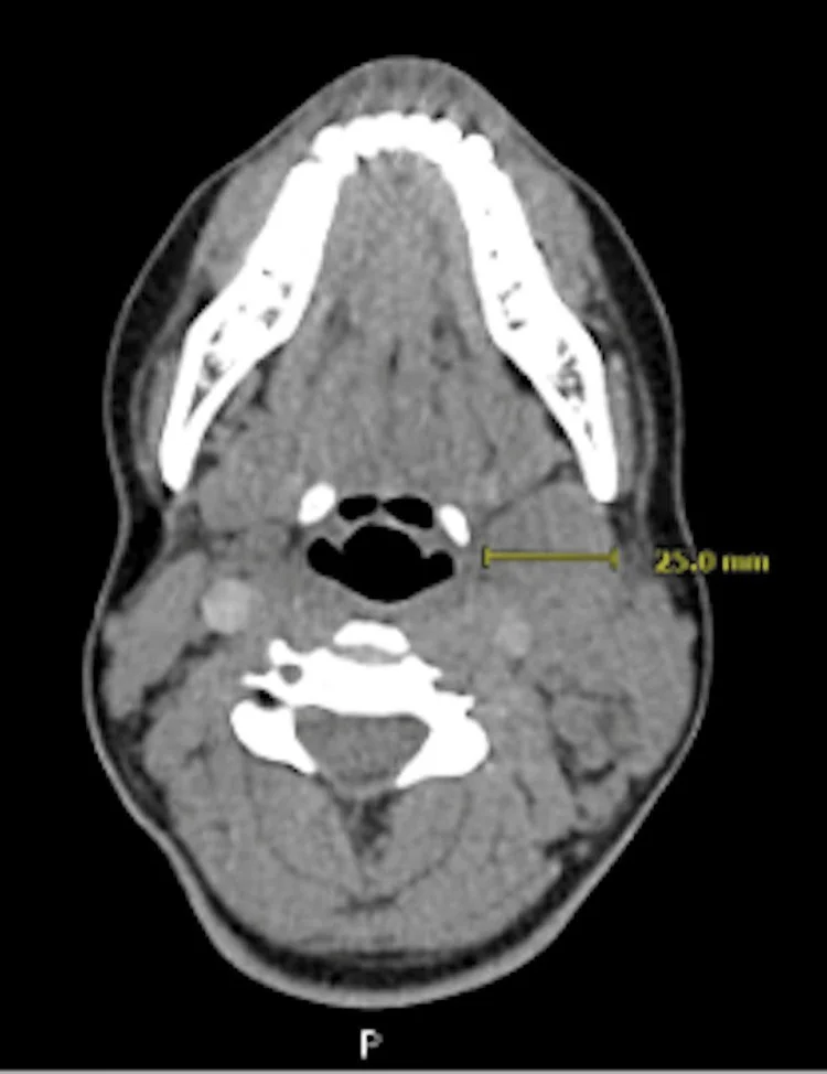
Servikal tüberküloz lenfadeniti, boyun lenf nodlarında granülomatöz enfeksiyon ve değişen derecelerde kazeöz nekroz oluşturan ekstrapulmoner tüberküloz tutulumudur. Kontrastlı boyun BT'si nodların dağılımını, merkezi nekrozu, çevre yumuşak dokularla ilişkisini ve birleşerek kitle oluşturup oluşturmadığını haritalar; görüntü tek başına etkeni kesinleştirmez. Kalın düzensiz halka, çok odacıklı düşük atenüasyon ve birleşik nod görünümü uygun klinik bağlamda tanıyı düşündürür. Kontrastlı BT ve MR, servikal lenf nodlarının yerini, tutulum paternini ve hastalığın uzamış bölgelerini göstererek yayılımın doğru değerlendirilmesine yardımcı olur.
Faz ve pencere
- Kontrastlı tanısal
- Kontrast sonrası nodlarda homojen boyanmadan santral düşük atenüasyonlu nekroz ve kalın, düzensiz çevresel boyanmaya kadar değişen görünüm izlenir; ilerlemiş hastalıkta çok odacıklı nekrotik nodlar birleşebilir ve çevre yağ planları silikleşebilir.
Önerilen pencereler: Yumuşak doku (G 400 / M 40), Mediasten (G 350 / M 50).
BT bulguları
- Bir veya birden fazla servikal zincirde, sıklıkla birbiriyle komşu büyümüş nodlar bulunur; dağılımı üstten alt boyuna kadar kesitler boyunca izle.
- Santral düşük atenüasyon kazeöz ya da liküefaktif nekrozu düşündürür; kontrastlanan periferik doku canlı granülasyon dokusunu temsil eder.
- Rim işareti — nekrotik merkezin çevresinde kalın veya düzensiz kontrastlanan nodal kenar görülür.
- Çok odacıklı merkezler ve arada seçilen ince septalar, tekdüze sıvı koleksiyonundan farklı nodal nekroz paterni oluşturur.
- Komşu nodlar konglomere olabilir; ileri olguda tek tek nod sınırları ve yağ planları silikleşir.
- Kronik veya tedavi sonrası nod içinde noktasal, kaba ya da kümelenmiş kalsifikasyon görülebilir.
- İleri evrede kapsül rüptürüne perilenfatik inflamasyon ve komşu nodların füzyonu eşlik edebilir.
Normalde
Normal servikal nodlar zincir boyunca ayrı seçilir, çevre yağ planları korunur ve kontrast sonrasında nod içi belirgin nekrotik merkez veya kalın halka bulunmaz. HIV pozitif hastalarda lenfadenopati sıklıkla simetrik ve çoklu iken, HIV negatif hastalarda fokal ve asimetrik prezentasyon daha sık görülür.
Normal BT ile kıyasla
Aynı bölgenin, kaynak veri setinde patoloji içermediği belirtilen bir BT incelemesini kesit kesit kaydırın; organ sınırlarını açarak anatomiyi hasta görüntüsüyle karşılaştırın. Küçük rastlantısal bulgular tümüyle dışlanmamıştır.
Kaynak: TotalSegmentator v2.01 veri seti (Wasserthal ve ark., Radiology: Artificial Intelligence 2023), CC BY 4.0, vaka s0622; organ sınırları veri setinin otomatik segmentasyonundan, birleştirilerek sadeleştirildi. Görüntüler 2,5 mm kesit aralığına yeniden örneklendi.
Ayırıcı tanı
- Metastatik lenf nodları primer tümörün lenfatik drenaj zincirlerinde ortaya çıkar; santral nekroz, düzensiz kortikal kalınlaşma ve ekstranodal uzanım ayırıcı tanıda dikkat gerektiren bulgulardır.
- Piyojenik apselerde merkezi non-enhanse alan ve çevrede hiperemik rim izlenirken, tüberkülozda düzensiz avasküler çekekler görülebilir.
- Lenfoma, servikal tüberküloz lenfadenitini taklit edebilen ayırıcı tanılardandır; BT ve MR bulguları tek başına kesin tanı koydurmaz.
- Atipik mikobakteriyel lenfadenit
- nekrotik ve rim boyanan nodlar oluşturur; görüntü örtüşebilir, mikrobiyolojik doğrulama gerekir.
- Sarkoidoz granülomatöz lenfadenitler arasında ayırıcı tanıya girer ve histopatolojik olarak non-kazeifiye granülomlarla karakterizedir.
Tuzaklar
- Kalın düzensiz rim tüberkülozu düşündürür fakat metastazı dışlamaz; primer tümör alanını ve diğer nodal istasyonları aynı incelemede tara.
- Birleşmiş nekrotik nodları tek derin boyun apsesi sanma; nodal zincir boyunca çok odacıklı yapıyı ve çevrede kalan nodları izle.
- Kalsifikasyon geçirilmiş veya tedavi görmüş tüberkülozla uyumlu olabilir; kalsifik nod tek başına aktif enfeksiyon kanıtı değildir.
- Tüberküloz lenfadenitte perinodal yumuşak doku değişikliği ve heterojenite izlenebilir; piyojenik apselerin merkezi non-enhanse alan ve çevresel hiperemi paterniyle bulgu örtüşmesi tanıyı zorlaştırabilir.
Kendini dene
Tüberkülozla uyumlu klinik bağlamda, birden fazla servikal nodda santral düşük atenüasyon, kalın düzensiz rim ve çok odacıklı görünüm var. En olası görüntüleme paterni nedir?
Cevabı göster
Tüberküloz lenfadeniti. Konglomere nodlar, santral nekroz ve kalın düzensiz rim tüberküloz lenfadenitini destekleyen bir patern oluşturur; ancak metastatik nodlar ve başka enfeksiyonlarla örtüşebilir. BT tek başına etkeni doğrulamaz; klinik ve mikrobiyolojik/histopatolojik bulgularla birlikte yorumlanır.
Servikal nodda nekroz var ve nod komşu yumuşak dokuda belirgin koleksiyonla devam ediyor. Hangi acil alternatif göz önünde tutulmalı?
Cevabı göster
Piyojenik nodal apse. Yaygın çevre inflamasyonu ve yumuşak dokuda sıvı koleksiyonuna devamlılık piyojenik apse olasılığını artırır. Kikuchi hastalığı ile reaktif hiperplazinin BT'de ayrımında konglomerasyon ve kontrastlanma paterni ayırt edici özellikler olarak bildirilmiştir.
İlgili konular
- Metastatik servikal lenf nodu
- Servikal lenfoma
- Lemierre sendromu ve septik internal juguler tromboflebit
Kaynaklar
Bu sayfadaki 36 cümle ve 2 quiz sorusu aşağıdaki kaynaklardan alıntılarla tek tek karşılaştırıldı; 9 cümle bu karşılaştırmada düzeltildi (2026-10-08).
- Diagnostic challenges in cervical tuberculous lymphadenitis: A reviewpmc.ncbi.nlm.nih.gov
- Fusing Radiomics and Ultrasonic Features: A Superior Nomogram for Diagnosing Cervical Tuberculous Lymphadenitispmc.ncbi.nlm.nih.gov
- Application of conventional and contrast-enhanced ultrasound in diagnosing cervical tuberculous lymphadenitispmc.ncbi.nlm.nih.gov
- Clinical Value of Contrast-Enhanced Ultrasound and Ultrasound Elastography in the Pathological Classification and Treatment Response Prediction of Cervical Tuberculous Lymphadenitispmc.ncbi.nlm.nih.gov
- A Rare Encounter of Cervical Tuberculous Lymphadenitis (Scrofula) in an Immunocompetent Adolescent Female: A Case Reportpmc.ncbi.nlm.nih.gov
- Coincidental diagnosis of tuberculous lymphadenitis: a case report.pubmed.ncbi.nlm.nih.gov
- Imaging of supra-thoracic tuberculosis.pubmed.ncbi.nlm.nih.gov
- [A Clinical Study on Cervical Tuberculous Lymphadenitis].pubmed.ncbi.nlm.nih.gov
- The diagnostic value of contrast-enhanced ultrasound for cervical tuberculous lymphadenitis.pubmed.ncbi.nlm.nih.gov
- Initial experience of a deep learning application for the differentiation of Kikuchi-Fujimoto's disease from tuberculous lymphadenitis on neck CECT.pubmed.ncbi.nlm.nih.gov
- Application of ultrasound multimodal imaging in the prediction of cervical tuberculous lymphadenitis rupture.pubmed.ncbi.nlm.nih.gov
- Calvarial Tuberculosis in Patient With Cervical Tuberculous Lymphadenitis: A Case Report.pubmed.ncbi.nlm.nih.gov
- [The surgical options of cervical tuberculous lymphadenitis].pubmed.ncbi.nlm.nih.gov
- Cervical Tuberculosis Lymphadenitis (Scrofula) in Wisconsin: Case Report, Pitfalls, and Challenges.pubmed.ncbi.nlm.nih.gov
- The efficacy of the interferon-γ release assay for diagnosing cervical tuberculous lymphadenitis: A prospective controlled study.pubmed.ncbi.nlm.nih.gov
- Exploratory study on the predictive value of ultrasound radiomics for cervical tuberculous lymphadenitis.pubmed.ncbi.nlm.nih.gov
- Disseminated tuberculous lymphadenitis presenting as cervical mass in patient with HIV infection, worsening after antiretroviral initiation: diagnosis and treatment challenges.pubmed.ncbi.nlm.nih.gov
- Observer- and radiomics model-based computed tomography classification of suppurative versus tuberculous lymphadenitis complicated with nodal necrosis of the neck in children.pubmed.ncbi.nlm.nih.gov
- Interleukin-15 correlates with cytotoxic immune networks in cervical tuberculous lymphadenitis.pubmed.ncbi.nlm.nih.gov
- Compartment-specific inflammation in tuberculous lymphadenitis: a case-control study.pubmed.ncbi.nlm.nih.gov
- Dual paradoxical reactions in tuberculous lymphadenitis during and after pregnancy: A case report.pubmed.ncbi.nlm.nih.gov
- Application of conventional and contrast-enhanced ultrasound in diagnosing cervical tuberculous lymphadenitis.pubmed.ncbi.nlm.nih.gov
- Primary cutaneous squamous cell carcinoma of the neck mimicking tuberculous lymphadenitis.pubmed.ncbi.nlm.nih.gov
- Diagnostic Imaging of Extrapulmonary Tuberculosis Across Organ Systems.pubmed.ncbi.nlm.nih.gov
- Extrapulmonary tuberculosıs: an old but resurgent problem.pubmed.ncbi.nlm.nih.gov
- The silent pandemic in South Africa: Extra-pulmonary tuberculosis from head to heel.pubmed.ncbi.nlm.nih.gov
- Imaging in tuberculosis.pubmed.ncbi.nlm.nih.gov
- Necrotizing (Abscessing) Lymphadenopathy and the Diagnostic Value of Contrast-Enhanced Ultrasound (CEUS): A Review with Clinical Vignettes.pubmed.ncbi.nlm.nih.gov
- Maxillofacial Squamous Cell Carcinoma With Cervical Metastasis and Tuberculous Lymphadenitis: Diagnostic Dilemmas and Clinical Insights.pubmed.ncbi.nlm.nih.gov
- Case report: Tumor-like mediastinal tuberculous lymphadenitis with paravertebral cold abscess under cumulative immunosuppression: a case highlighting altered host-pathogen interactions.pubmed.ncbi.nlm.nih.gov
- Rosai-Dorfman disease presenting as isolated cervical lymphadenopathy in a young adult: A case report.pubmed.ncbi.nlm.nih.gov
- Tularemia: A Unique Presentation for a Rare Disease.pubmed.ncbi.nlm.nih.gov
- Head and Neck Tuberculosis: A Rare Diagnosis and the Role of Surgical Biopsy and Histopathological Evaluation in Extrapulmonary Disease.pubmed.ncbi.nlm.nih.gov
- Perforated acute appendicitis on computed tomography revealing underlying tuberculosis.pubmed.ncbi.nlm.nih.gov
- Tuberculosis-Associated Hemophagocytic Syndrome Mimicking Malignant Lymphoma: A Case Report.pubmed.ncbi.nlm.nih.gov
- Tuberculous Peripancreatic Lymphadenitis Presenting as Obstructive Jaundice Due to Extrinsic Common Bile Duct Compression: A Case Report.pubmed.ncbi.nlm.nih.gov
- Mediastinal Lymphadenopathy Presenting With the "Medu Vada Sign".pubmed.ncbi.nlm.nih.gov
- Two Interesting Cases of Thyroid Gland Tuberculosis.pubmed.ncbi.nlm.nih.gov
- Ultrasound innovations in abdominal radiology: techniques and clinical applications in pediatric imaging.pubmed.ncbi.nlm.nih.gov
- Massive generalised lymphadenopathy: a rare presentation of tuberculosis with a favourable outcome.pubmed.ncbi.nlm.nih.gov
- Esophageal Tuberculosis: A Systematic Review.pubmed.ncbi.nlm.nih.gov
- Starry sky sign: A prevalent sonographic finding in mediastinal tuberculous lymph nodes.pubmed.ncbi.nlm.nih.gov
- Case Report: Concurrent thyroid tuberculosis and tuberculous lymphadenitis initially misdiagnosed as malignancy by <sup>18</sup>F-FDG PET/CT.pubmed.ncbi.nlm.nih.gov
- Recommendations of the International Consensus Meeting (ICM) 2025 on Spinal Infections: Part B Tubercular Spondylodiscitis.pubmed.ncbi.nlm.nih.gov
- Laboratory diagnosis of 44 cases of pediatric histiocytic necrotizing lymphadenitis.pubmed.ncbi.nlm.nih.gov
- Primary Pleural Tuberculosis Presenting With Noncaseating Granulomas: A Diagnostic Challenge.pubmed.ncbi.nlm.nih.gov
- Rare primary bone tuberculosis associated with ruxolitinib therapy: A case report and literature review.pubmed.ncbi.nlm.nih.gov
- Malignant lymph nodes of the head and neck: imaging pearls, pitfalls, and practical approach to reporting in the era of extranodal extension.pubmed.ncbi.nlm.nih.gov
- HPV-associated Oropharyngeal Squamous Cell Carcinoma: An Imaging Framework for AJCC Version 9 TNM Staging.pubmed.ncbi.nlm.nih.gov
- Kawasaki disease presenting with severe airway obstruction: a case report.pubmed.ncbi.nlm.nih.gov
- Lymph Node Assessment with Multiparametric Ultrasound: Normal Values, Morphologic Patterns, and Diagnostic Algorithms.pubmed.ncbi.nlm.nih.gov
- Brachytherapy use in cervical cancer: a narrative review.pubmed.ncbi.nlm.nih.gov
- A comprehensive review of endobronchial ultrasound-guided transbronchial needle aspiration (EBUS-TBNA): staging, techniques, and future directions.pubmed.ncbi.nlm.nih.gov
- Radiological Features of Human Papillomavirus (HPV)-Positive and HPV-Negative Oropharyngeal Squamous Cell Carcinoma (OPSCC)-Considerations for Multimodal Analysis.pubmed.ncbi.nlm.nih.gov
- Multiparametric MRI Assessment of Cervical Lymphadenopathy: Combined Diagnostic Performance of Morphological Features and Apparent Diffusion Coefficient.pubmed.ncbi.nlm.nih.gov
- Clinical characteristics and etiological profile of retropharyngeal space abnormalities in children: a nine-year retrospective analysis.pubmed.ncbi.nlm.nih.gov
- Antibiotic-Only Treatment of Pediatric Retropharyngeal and Parapharyngeal Abscess: A Case Report.pubmed.ncbi.nlm.nih.gov
- Lymphoma of the cervix invading the uterine body with distant lymphadenopathy: MRI and FDG-PET/CT findings.pubmed.ncbi.nlm.nih.gov
- Posterior mediastinal soft-tissue Rosai-Dorfman disease mimicking a neurogenic tumor.pubmed.ncbi.nlm.nih.gov
- Multifocal osseous and cutaneous Rosai-Dorfman-Destombes disease presenting as an aggressive intramedullary femoral lesion.pubmed.ncbi.nlm.nih.gov
- A Diagnostic Challenge in Regional Australia: Concurrent <i>Mycobacterium avium</i> Complex Lymphadenitis and Hairy Cell Leukemia.pubmed.ncbi.nlm.nih.gov
- Kikuchi-Fujimoto disease mimicking hemophagocytic lymphohistiocytosis in a young white male: a case report.pubmed.ncbi.nlm.nih.gov
- Unusual progression of Rosai-Dorfman disease in a pediatric patient.pubmed.ncbi.nlm.nih.gov
- A Hematologic Mimic: Sarcoidosis Presenting With Myelodysplastic Syndrome-Like Marrow Dysplasia, Massive Splenomegaly, and Pancytopenia.pubmed.ncbi.nlm.nih.gov
- Ocular TB as the only manifestation of TB: diagnostic uncertainty and treatment thresholds.pubmed.ncbi.nlm.nih.gov
- Pre-treatment and post-treatment nasopharyngeal carcinoma imaging: imaging updates, pearls and pitfalls.pubmed.ncbi.nlm.nih.gov
- US-Guided percutaneous core needle biopsy via the complete transhepatic approach: a reliable option for deep abdominal lesions.pubmed.ncbi.nlm.nih.gov
- Unusual manifestations of chest wall tuberculosis: diagnostic challenges in two cases from a low-incidence tuberculosis country.pubmed.ncbi.nlm.nih.gov
- Probable Primary Adrenal Insufficiency Associated With Granulomatous Adrenal Calcifications, Non-tuberculous Mycobacterial Ileitis, and Crohn's Disease: A Case Report.pubmed.ncbi.nlm.nih.gov
- Pulmonary tuberculosis and the surgeon.pubmed.ncbi.nlm.nih.gov
- Relationship among ultrasound features, pus Mycobacterium tuberculosis load, and efficacy of antituberculosis drugs in patients with necrotizing tuberculous lymphadenitis.pubmed.ncbi.nlm.nih.gov
- Refining the boundaries of the nodal CTV in the middle neck for nasopharyngeal carcinoma.pubmed.ncbi.nlm.nih.gov
- Optimization strategies for ultrasound zoning of cervical lymph nodes in esophageal cancer: a precision medicine approach.pubmed.ncbi.nlm.nih.gov
- Lymphatic metastasis of papillary thyroid carcinoma: mechanism and clinicopathological physiology.pubmed.ncbi.nlm.nih.gov
- Bacterial Granulomatous Lung Diseases: Radiological Findings and Differential Diagnosis.pubmed.ncbi.nlm.nih.gov
- Cervicofacial Actinomycosis Presenting as a Fistulized Neck Mass During Pregnancy: A Case Report.pubmed.ncbi.nlm.nih.gov
- Magnetic resonance imaging of musculoskeletal infections.pubmed.ncbi.nlm.nih.gov
- CT pattern analysis of necrotizing and nonnecrotizing lymph nodes in Kikuchi disease.pubmed.ncbi.nlm.nih.gov
- Tuberculosis of the Liver, Biliary Tract, and Pancreas.pubmed.ncbi.nlm.nih.gov
- Multimodal imaging of cervical lymphadenopathy: diagnostic value and clinical applications.pubmed.ncbi.nlm.nih.gov
- Cervical Paratracheal Bronchogenic Cyst Mimicking Nodal Metastatic Disease in a Colorectal Cancer Patient: Diagnostic Utility of Neck Ultrasound-Guided Fine Needle Aspiration.pubmed.ncbi.nlm.nih.gov
- Primary Mandibular Tuberculous Osteomyelitis Mimicking Ameloblastoma: A Case Report and Literature Review of Mandibular Tuberculous Osteomyelitis.pubmed.ncbi.nlm.nih.gov
- Diagnostic value of EBUS-TBNA for intrathoracic tuberculous lymphadenitis: a retrospective cohort analysis stratified by anti-tuberculosis treatment duration.pubmed.ncbi.nlm.nih.gov
- A diagnostic performance study of multiplex polymerase chain reaction-based targeted next-generation sequencing for the accurate identification of <i>Mycobacterium</i> tuberculosis in bronchoalveolar lavage fluid.pubmed.ncbi.nlm.nih.gov
- A rare invader on a cardiac stent: <i>Mycobacterium abscessus</i> mediastinitis managed with tedizolid after linezolid intolerance.pubmed.ncbi.nlm.nih.gov
- Nocardia brasiliensis infection causes skin abscesses around the eyes: A case-report and review of literature.pubmed.ncbi.nlm.nih.gov
- Acute suppurative thyroiditis in a 13-year-old boy: a rare pediatric case report.pubmed.ncbi.nlm.nih.gov
- Hidden in plain sight: Extrapulmonary manifestations of tuberculosis in Asia.pubmed.ncbi.nlm.nih.gov
- Navigating the labyrinth: Phenotypes of IgG4-related disease.pubmed.ncbi.nlm.nih.gov
- Diffuse lymphadenopathy as the presenting manifestation of systemic lupus erythematosus.pubmed.ncbi.nlm.nih.gov
- Cervical embryonal rhabdomyosarcoma beyond childhood: A case report and literature review.pubmed.ncbi.nlm.nih.gov
- CT and MRI imaging of mass in the carotid space: a pictorial review.pubmed.ncbi.nlm.nih.gov
- Gynecologic imaging errors and pitfalls: how to avoid misdiagnosis on pelvic MRI.pubmed.ncbi.nlm.nih.gov
- Fusing Radiomics and Ultrasonic Features: A Superior Nomogram for Diagnosing Cervical Tuberculous Lymphadenitis.pubmed.ncbi.nlm.nih.gov
- A Rare Clinical Presentation of Multifocal Tuberculous Osteomyelitis of the Right Parietal and Left Calcaneal Bones in an 11-Year-Old Male: A Case Report.pubmed.ncbi.nlm.nih.gov
- Efficacy and safety of an all-oral delamanid-containing regimen in the treatment of multidrug-resistant pulmonary tuberculosis complicated by extrapulmonary tuberculosis: Four case reports and review of the literature.pubmed.ncbi.nlm.nih.gov
- Ectopic calcitonin-secreting pulmonary neuroendocrine tumor mimicking medullary thyroid carcinoma.pubmed.ncbi.nlm.nih.gov
- Deep learning-based computed tomography detection of early lymph node metastasis in head and neck cancer.pubmed.ncbi.nlm.nih.gov
- ETV6-NTRK3 Fusion-Positive Late-Recurrent Parotid Carcinoma With Contralateral Axillary Metastasis and Early Response to Entrectinib: A Case Report.pubmed.ncbi.nlm.nih.gov
- Locally Advanced Cervical Cancer: Multiparametric MRI in Gynecologic Oncology and Precision Medicine.pubmed.ncbi.nlm.nih.gov
- Pictorial Review of Paediatric Limp.pubmed.ncbi.nlm.nih.gov
- Soft tissue masses of the epitrochlear region.pubmed.ncbi.nlm.nih.gov
- Diagnostic delay and histopathological subtyping challenges in idiopathic multicentric Castleman disease, not otherwise specified: a case report of a three-hospital odyssey.pubmed.ncbi.nlm.nih.gov
- Report of a case of renal hilum Castleman disease complicated with Paget's disease of the breast.pubmed.ncbi.nlm.nih.gov
- A rare case of unicentric Castleman disease in the posterior mediastinum: case report and integrated literature review.pubmed.ncbi.nlm.nih.gov
- Surgical approach for common carotid ligation at the level of the medial retropharyngeal lymph node in dogs: A comparative cadaveric study.pubmed.ncbi.nlm.nih.gov
- Imaging in Esophageal Cancer: A Comprehensive Review.pubmed.ncbi.nlm.nih.gov
- Comprehensive review of post-treatment imaging in head and neck cancers: from expected to unexpected and beyond.pubmed.ncbi.nlm.nih.gov
- Thyroid tuberculosis: Rare case report.pubmed.ncbi.nlm.nih.gov
- Indeterminate Lymph Nodes Assessment in Oral Squamous Cell Carcinoma Using CT, MRI, and PET-CT: A Retrospective Study.pubmed.ncbi.nlm.nih.gov
- Non-gestational choriocarcinoma presenting as cervical lymphadenopathy in a male patient: a case report.pubmed.ncbi.nlm.nih.gov
- PET/CT false-negative angioimmunoblastic T-cell lymphoma: a case report and literature review.pubmed.ncbi.nlm.nih.gov
- Tuberculosis Beyond the Lungs: A Pictorial Review of Key Diagnostic Imaging Insights.pubmed.ncbi.nlm.nih.gov
- Computed tomography imaging of acute neck inflammatory processes.pubmed.ncbi.nlm.nih.gov
- Spinal granulocytic sarcoma with both extradural and intradural involvement: a case report.pubmed.ncbi.nlm.nih.gov
Eğitim amaçlıdır; hasta başında tanı ve tedavi kararının yerine geçmez. İçerik yapay zekâ destekli bir süreçle güncel literatüre göre cümle cümle kaynaklarla karşılaştırıldı, ancak uzman radyolog incelemesinden geçmedi. Klinik kararlarda güncel kılavuzları, kurum protokollerini ve radyoloji raporunu esas alın.