Özet
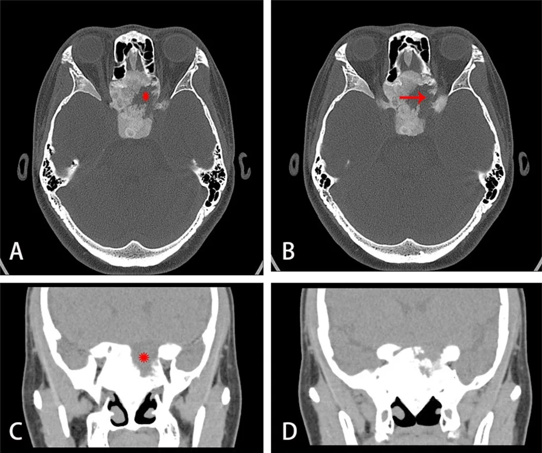
Kraniofasiyal fibröz displazi, normal kemik iliğinin fibröz doku ve düzensiz örgü kemikle yer değiştirdiği, çoğu kez yavaş seyirli benign bir kemik lezyonudur. Orbita duvarı veya sfenoid tutulduğunda kemik ekspansiyonu glob konumunu değiştirebilir ve optik kanalı daraltabilir; tanıda ve yayılım haritasında BT temel yöntemdir. BT’de tipik buzlu cam matriks ve komşu kemiğe geçişli sınırlar görülür, ancak matriks görünümü yaşla ve lezyonla değişken olabilir. Optik kanal daralmasını tek başına görme kaybı olarak yorumlamak veya eşlik eden yeni ekspansil lezyonu kaçırmak görsel işlev tehdidini yanlış sınıflandırabilir.
Faz ve pencere
- Kontrastsız tanısal
- İnce kesit kemik serisinde ekspansil kemik içi buzlu cam dansitesi, kortikal incelme ve sfenoid/optik kanal çevresinde daralma izlenir; damar içi kontrast tipik kemik matriksini göstermek için gerekmez.
Önerilen pencereler: Kemik (G 1800 / M 400), Yumuşak doku (G 400 / M 40).
BT bulguları
- Buzlu cam matriks: kemik iliği yerine geçen homojen veya hafif heterojen yüksek dansiteli mineralize doku.
- Ekspansil remodelling: sfenoid kanat, frontal kemik veya maksilla dış konturu düzgünce genişler.
- Kortikal kabuk incelir; çoğu tipik lezyonda dış kontur korunur ve keskin litik sınır beklenmez.
- Lezyon komşu kemikle silik geçiş yapabilir; ayrı, kapsüllü bir yumuşak doku kitlesi gibi görünmez.
- Optik kanal lümeni daralabilir veya çevresindeki kemik kalınlaşarak daralabilir; bulgu karşı taraftaki optik kanal ile kıyaslanmalıdır.
- Orbita duvarı kalınlaşması orbital hacmi azaltıp globda öne yer değiştirme veya distopi oluşturabilir.
- Yeni kistik/litik odak ya da hızlı kemik genişlemesi, tipik stabil matriksten farklı davranış gösterir ve ek değerlendirme gerektirir.
Ölçütler ve sınıflamalar
- Radyografik kraniofasiyal FD paterni
- BT'de homojen yoğun (buzlu cam) görünüm; hastanın yaş ilerledikçe gelişebilen karışık radyo-lusent ve radyo-dens alanlar; santral lusensi ve ince sklerotik kenarı olan kistik görünüm. Paternler değişken olabilir ve tek başına klinik evre değildir.
Normalde
Normal sfenoid kanat, frontal kemik ve maksilla trabeküler kemik görünümünü sürdürür; optik kanal kortike sınırları düzenli ve karşı tarafla yaklaşık simetriktir. Aynı aksiyel ve koronal düzeylerde kemik konturunun dışa bombeleşmesine, matriksin buzlu cam yoğunluğuna ve kanal çapındaki taraf farkına bak; yalnız kanalın dar görünmesi klinik optik nöropatiyi kanıtlamaz.
Normal BT ile kıyasla
Aynı bölgenin, kaynak veri setinde patoloji içermediği belirtilen bir BT incelemesini kesit kesit kaydırın; organ sınırlarını açarak anatomiyi hasta görüntüsüyle karşılaştırın. Küçük rastlantısal bulgular tümüyle dışlanmamıştır.
Kaynak: TotalSegmentator v2.01 veri seti (Wasserthal ve ark., Radiology: Artificial Intelligence 2023), CC BY 4.0, vaka s0849; organ sınırları veri setinin otomatik segmentasyonundan, birleştirilerek sadeleştirildi. Görüntüler 2,5 mm kesit aralığına yeniden örneklendi.
Ayırıcı tanı
- Ossifiye fibrom
- genellikle daha iyi sınırlı, ekspansif lezyon; radyolojik ve histolojik değerlendirmelerle fibröz displaziden ayırt edilmesi gereklidir.
- Paget hastalığı, fibröz displazinin olası taklitçilerinden biridir.
- Osteom
- kemik yüzeyinden köken alan, kompakt/kancellöz kemik proliferasyonuyla karakterize, iyi sınırlı ve yavaş büyüyen lezyon; fibröz displazinin geniş matriks yapısından farklıdır.
- Metastaz veya miyelom
- litik/destrüktif odaklar ve korteks aşımı; tipik geçişli FD matriksi beklenmez.
- Anevrizmal kemik kisti
- ekspansil, çoğunlukla lusent ve septalı lezyon; FD zemininde ikincil gelişebilir.
Tuzaklar
- Optik kanal daralması otomatik olarak görme kaybı anlamına gelmez; bu görüntü bulgusu klinik görme fonksiyonunun değerlendirilmesinin yerini tutmaz.
- Buzlu cam görünümünün her olguda aynı homojenlikte olmasını bekleme; yaşla veya lezyon bileşimiyle karışık lusent alanlar oluşabilir.
- Hızlı büyüyen kistik değişikliği sıradan FD matriksi kabul etme; ikincil kist veya başka agresif süreç olasılığını araştır.
Kendini dene
Yavaş gelişen tek taraflı glob distopisi olan kişide sfenoid kemik genişlemiş ve buzlu cam matriksli. En olası tanı nedir?
Cevabı göster
Fibröz displazi. Kemik boyunca geçişli buzlu cam matriks ve düzgün ekspansiyon fibröz displaziyi destekler. Ossifiye fibrom genellikle daha keskin sınırlıdır; osteom fokal yoğun kemik oluşumudur, metastaz ise çoğu kez destrüktif odak yapar.
Sfenoid fibröz displazide BT'de optik kanal dar, ancak hasta görme yakınması tarif etmiyor. Görüntü tek başına neyi kanıtlamaz?
Cevabı göster
Optik nöropatiyi. BT kanal lümeni ve kemik anatomisini gösterir; görme siniri işlev kaybı klinik ve oftalmolojik değerlendirmeyle belirlenir. Diğer seçenekler doğrudan kemik görüntüsünde gözlenebilir.
İlgili konular
Kaynaklar
Bu sayfadaki 38 cümle ve 2 quiz sorusu aşağıdaki kaynaklardan alıntılarla tek tek karşılaştırıldı; 7 cümle bu karşılaştırmada düzeltildi (2026-10-08).
- Clinical guidelines for the management of craniofacial fibrous dysplasiapmc.ncbi.nlm.nih.gov
- Best practice management guidelines for fibrous dysplasia/McCune-Albright syndrome: a consensus statement from the FD/MAS international consortiumpmc.ncbi.nlm.nih.gov
- Simultaneous decompression of the orbital lateral wall and optic canal for fibrous dysplasia in early adolescencepmc.ncbi.nlm.nih.gov
- Minimally invasive resection of a spheno-orbital tumor: Review of surgical techniques and a case reportpmc.ncbi.nlm.nih.gov
- Small ethmoid mucocele with intracranial extension associated with fibrous dysplasia: A rare case reportpmc.ncbi.nlm.nih.gov
- Craniofacial Fibrous Dysplasia Involving the Orbit: A Case Report and Literature Review.pubmed.ncbi.nlm.nih.gov
- Surgical Simulation and Custom-Made Implant for Cranial Fibrous Dysplasia.pubmed.ncbi.nlm.nih.gov
- Might Trauma Be a Triggering Factor for Craniofacial Fibrous Dysplasia?pubmed.ncbi.nlm.nih.gov
- The Natural History of Fibrous Dysplasia of the Orbit.pubmed.ncbi.nlm.nih.gov
- Complex skull defects reconstruction with САD/САМ titanium and polyetheretherketone (PEEK) implants.pubmed.ncbi.nlm.nih.gov
- Isolated orbital fibrous dysplasia associated with ipsilateral keratoconus.pubmed.ncbi.nlm.nih.gov
- Simultaneous decompression of the orbital lateral wall and optic canal for fibrous dysplasia in early adolescence.pubmed.ncbi.nlm.nih.gov
- Cherubism: report of three cases and literature review.pubmed.ncbi.nlm.nih.gov
- The indication and application of computer-assisted navigation in oral and maxillofacial surgery-Shanghai's experience based on 104 cases.pubmed.ncbi.nlm.nih.gov
- Orbital Involvement In Sinonasal Diseases.pubmed.ncbi.nlm.nih.gov
- [Application of computer-assisted navigation in oral and maxillofacial surgery: retrospective analysis of 104 consecutive cases].pubmed.ncbi.nlm.nih.gov
- Transconjunctival Orbital Decompression in Cherubism.pubmed.ncbi.nlm.nih.gov
- Innovative Surgical Treatment of Severe Cherubism.pubmed.ncbi.nlm.nih.gov
- Orbital tumours and tumour-like lesions: exploring the armamentarium of multiparametric imaging.pubmed.ncbi.nlm.nih.gov
- Small ethmoid mucocele with intracranial extension associated with fibrous dysplasia: A rare case report.pubmed.ncbi.nlm.nih.gov
- Intracranial aneurysmal bone cysts in association with fibrous dysplasia: Case report and scoping review of case reports.pubmed.ncbi.nlm.nih.gov
- Massive Mandibular Fibrous Dysplasia Mimicking Calcifying Epithelial Odontogenic Tumor Radiologically: A Case Report.pubmed.ncbi.nlm.nih.gov
- Pediatric triad of craniofacial fibrous dysplasia, Chiari malformation type I and syringomyelia: a case report.pubmed.ncbi.nlm.nih.gov
- Unusual Manifestations of Sinonasal Osteomas: A Narrative Review.pubmed.ncbi.nlm.nih.gov
- Polyostotic fibrous dysplasia: Unveiling the complexity of a rare bone disorder through a case report.pubmed.ncbi.nlm.nih.gov
- A Case Series and Literature Review of Craniofacial Fibrous Dysplasia.pubmed.ncbi.nlm.nih.gov
- Protein kinase A is a dependent factor and therapeutic target in mouse models of fibrous dysplasia.pubmed.ncbi.nlm.nih.gov
- Imaging findings of arrested pneumatisation and differentiation from other skull base lesions.pubmed.ncbi.nlm.nih.gov
- Orbital pseudotumor and skull base infiltration as the sole manifestation of granulomatosis with polyangiitis.pubmed.ncbi.nlm.nih.gov
- Cancer predisposition syndromes: an imaging review.pubmed.ncbi.nlm.nih.gov
- Salvaging Vision: A Study of Non-Traumatic Optic Neuropathies.pubmed.ncbi.nlm.nih.gov
- When plasmacytoma defies expectations: An unusual maxillary presentation with mixed imaging features in a young patient.pubmed.ncbi.nlm.nih.gov
- Ossifying fibroma of the maxilla: A case report with literature review.pubmed.ncbi.nlm.nih.gov
- Osteomyelitis of the Jaw Bones and Its Mimics: Resolving the Diagnostic Enigma.pubmed.ncbi.nlm.nih.gov
- The solitary skull mass: An atypical presentation of anaplastic large cell lymphoma.pubmed.ncbi.nlm.nih.gov
- Benign and malignant lesions of the mandible and maxilla: imaging findings and systematic approach.pubmed.ncbi.nlm.nih.gov
- Malignant Transformation of Musculoskeletal Lesions with Imaging-Pathology Correlation-Part 1: Bone Lesions.pubmed.ncbi.nlm.nih.gov
- Ossifying Fibroma of the Frontal Sinus: A Case Report and Literature Review.pubmed.ncbi.nlm.nih.gov
- Craniofacial fibrous dysplasia: Molecular insights, long-term institutional experience, and evolving surgical strategies.pubmed.ncbi.nlm.nih.gov
- Diagnostic and Treatment Strategies for Adult Non-Ossifying Fibromas: A Case Report and Literature Review.pubmed.ncbi.nlm.nih.gov
- Review of imaging modalities and radiological findings of calvarial lesions.pubmed.ncbi.nlm.nih.gov
- Surgical Management of a Case of Frontoparietal Fibrous Dysplasia.pubmed.ncbi.nlm.nih.gov
- Improving diagnostic clarity in fibro-osseous lesions using modified Gallego's, alizarin red, and von kossa stains: A pilot comparative study.pubmed.ncbi.nlm.nih.gov
- Benign Fibro-Osseous Lesions of the Head and Neck.pubmed.ncbi.nlm.nih.gov
- Histological Diversity, Diagnostic Challenges, and Surgical Treatment Strategies of Fibrous Dysplasia of Upper and Mid-Thirds of the Craniomaxillofacial Complex.pubmed.ncbi.nlm.nih.gov
- Multidisciplinary approach and clinical management of McCune-Albright syndrome.pubmed.ncbi.nlm.nih.gov
- Polyostotic Fibrous Dysplasia: A Case Report.pubmed.ncbi.nlm.nih.gov
- Foramen Magnum Stenosis in Achondroplasia: Imaging-Based Surgical Indications, Synchondrosis Fusion, and the Role of Ventriculomegaly.pubmed.ncbi.nlm.nih.gov
- Craniofacial fibrous dysplasia: Surgery and literature review.pubmed.ncbi.nlm.nih.gov
- Computer guided resection and reconstruction of intra-osseous zygomatic hemangioma: Case report and systematic review of literature.pubmed.ncbi.nlm.nih.gov
- Endoscopic management of uncommon benign maxillary sinus lesions: a case series.pubmed.ncbi.nlm.nih.gov
- Mucinous cystic neoplasms of the liver: current insights into epidemiology, diagnosis, and treatment.pubmed.ncbi.nlm.nih.gov
- Case Report: Novel <i>ADAMTSL2</i> compound heterozygous mutations in geleophysic dysplasia with bilateral glaucoma and keratoconus-like corneal ectasia.pubmed.ncbi.nlm.nih.gov
- Fibrous dysplasia of the head and neck in Southern Finland: a retrospective study on clinical characteristics, diagnostics, and treatment.pubmed.ncbi.nlm.nih.gov
- In Vivo Assessment of Retinal Phenotypes in Axenfeld-Rieger Syndrome.pubmed.ncbi.nlm.nih.gov
- Advances in origin, evolution, and pathogenesis of optic disc drusen: A narrative review.pubmed.ncbi.nlm.nih.gov
- Craniofacial disorders and dysplasias: Molecular, clinical, and management perspectives.pubmed.ncbi.nlm.nih.gov
- Expression of Beta-Catenin, Cadherins and P-Runx2 in Fibro-Osseous Lesions of the Jaw: Tissue Microarray Study.pubmed.ncbi.nlm.nih.gov
- Modality-specific quantitative radiologic characterization of diffuse sclerosing osteomyelitis of the mandible: a pilot study.pubmed.ncbi.nlm.nih.gov
- Performance of large language models on image-based oral pathology questions from the Japanese National Dental Examination.pubmed.ncbi.nlm.nih.gov
- Surgically Treated Benign Bone Tumors and Tumor-like Conditions in the Pediatric Population-A 10-Year Institutional Experience.pubmed.ncbi.nlm.nih.gov
- Lateral orbitotomy approach for removing hyperostosing en plaque sphenoid wing meningiomas. Description of surgical strategy and analysis of findings in a series of 88 patients with long-term follow up.pubmed.ncbi.nlm.nih.gov
- Long-term safety and effectiveness of the "OptEase" vena cava filter.pubmed.ncbi.nlm.nih.gov
- Orbital clock concept: A novel anatomical zoning system for endoscopic vs. external orbital approaches.pubmed.ncbi.nlm.nih.gov
- Modern technology complements classic technique: 3D-printed inter-locking cutting guides for frontal sinus osteoma resection and reconstruction with split-calvarial bone graft.pubmed.ncbi.nlm.nih.gov
- Xanthogranulomatous Osteomyelitis as a Bone Tumor Mimic: A Systematic Review of Pre-biopsy Diagnostic Clues and Management Decision Points.pubmed.ncbi.nlm.nih.gov
- Decoding calvarial lesions: Contemporary neurosurgical management integrating radiological and histopathological features.pubmed.ncbi.nlm.nih.gov
- A rare calvarial aneurysmal bone cyst in an adult: Case report and literature review.pubmed.ncbi.nlm.nih.gov
- Journey and Roadblocks in the Management of Aneurysmal Bone Cysts in Patients with Unusual Presentations.pubmed.ncbi.nlm.nih.gov
- The genetic basis of hydrocephalus: genes, pathways, mechanisms, and global impact.pubmed.ncbi.nlm.nih.gov
- Neoplasms and tumor-like lesions of the sellar region: imaging findings with correlation to pathology and 2021 WHO classification.pubmed.ncbi.nlm.nih.gov
- Management of Tibial Adamantinoma: Balancing Oncologic Cure and Reconstructive Longevity.pubmed.ncbi.nlm.nih.gov
- Fibrous dysplasia: Clinical features, deformities, management, and outcomes.pubmed.ncbi.nlm.nih.gov
- Aggressive Juvenile Trabecular Ossifying Fibroma in a 4-Year-Old: Case Report Bringing Diagnostic Challenges and Surgical Management.pubmed.ncbi.nlm.nih.gov
- Common cystic pancreatic neoplasms: A comprehensive magnetic resonance imaging-based review of typical and atypical imaging features.pubmed.ncbi.nlm.nih.gov
- Craniofacial Ossifying Fibromas in Children: Clinical Variability and Surgical Outcomes in a Case Series.pubmed.ncbi.nlm.nih.gov
- Conventional radiography for the assessment of focal bone lesions of the appendicular skeleton: fundamental concepts in the modern imaging era.pubmed.ncbi.nlm.nih.gov
- Magnetic resonance imaging features of craniofacial fibrous dysplasia.pubmed.ncbi.nlm.nih.gov
- Special Issue: EACR 2026 Congress: Innovative Cancer Science, 8-11 June 2026.pubmed.ncbi.nlm.nih.gov
- Intraoperative navigation in craniofacial surgery.pubmed.ncbi.nlm.nih.gov
- Surgical Measures Improving Functional Limitations of the Masticatory System, Aesthetic Deficits, and Skeletal Malformations in Neurofibromatosis Type 1-associated Hemifacial Diffuse Plexiform Neurofibroma Complicated by Rapidly Growing Midfacial Peripheral Nerve Sheath Tumor.pubmed.ncbi.nlm.nih.gov
- Current concepts of craniofacial fibrous dysplasia: pathophysiology and treatment.pubmed.ncbi.nlm.nih.gov
- Giant Fibrous Dysplasia of the Sixth Rib Managed in a Rural, Resource-Limited Setting: A Case Report.pubmed.ncbi.nlm.nih.gov
- Integration of Clinical, Radiographic, Histologic, and Molecular Findings to Diagnose Craniofacial Fibrous Dysplasia.pubmed.ncbi.nlm.nih.gov
- Imaging of the skull base and orbital tumors.pubmed.ncbi.nlm.nih.gov
- Cholesterol granuloma of the sphenoid wing with long-term growth dynamics: illustrative case.pubmed.ncbi.nlm.nih.gov
- A Review of Posteromedial Lesions of the Chest Wall: What Should a Chest Radiologist Know?pubmed.ncbi.nlm.nih.gov
- Osseous Manifestations of Primary Hyperparathyroidism: Imaging Findings.pubmed.ncbi.nlm.nih.gov
- Osteoblastic and hyperostotic craniofacial lesion detected by 99mTc-labeled methylene diphosphonate bone scintigraphy and single-photon emission computed tomography/computed tomography: a pictorial essay.pubmed.ncbi.nlm.nih.gov
- Radiological review of skull lesions.pubmed.ncbi.nlm.nih.gov
- Apparent diffusion coefficient can assist in differentiating between benign and malignant primary bone tumors in pediatric patients.pubmed.ncbi.nlm.nih.gov
- Benign incidental do-not-touch bone lesions.pubmed.ncbi.nlm.nih.gov
- Sclerotic Lesions of the Jaw: A Pictorial Review.pubmed.ncbi.nlm.nih.gov
- Imaging Features of Mandibular Ramus Osteoma on Computed Tomography: A Case Report With Literature Analysis.pubmed.ncbi.nlm.nih.gov
- Intraosseous tumours of the knee.pubmed.ncbi.nlm.nih.gov
- Radiologic and Pathologic Insights into Medication-Related Osteonecrosis of the Jaw in Myeloma Patients: A Report of 3 Cases.pubmed.ncbi.nlm.nih.gov
- Special Considerations in Pediatric Endoscopic Skull Base Surgery.pubmed.ncbi.nlm.nih.gov
- What's new in pediatric bone sarcomas and benign bone tumors.pubmed.ncbi.nlm.nih.gov
- Fibrous Dysplasia Presenting as an Exophytic Gingival Mass: A Rare Clinical Presentation.pubmed.ncbi.nlm.nih.gov
- Craniofacial Fibrous Dysplasia to Affect or Not the Optic Nerve in Long-Term Follow-Up of Three Cases.pubmed.ncbi.nlm.nih.gov
- Jansen metaphyseal chondrodysplasia: analysis of craniofacial manifestations.pubmed.ncbi.nlm.nih.gov
- Non-Arteritic Anterior Ischemic Optic Neuropathy (NA-AION): A Comprehensive Overview.pubmed.ncbi.nlm.nih.gov
- PET/CT and PET/MRI in ophthalmic oncology (Review).pubmed.ncbi.nlm.nih.gov
- Pagetoid polyostotic fibrous dysplasia.pubmed.ncbi.nlm.nih.gov
- Osteoid osteoma of the triquetrum: A rare cause of ulnar-sided wrist pain in an adolescent female.pubmed.ncbi.nlm.nih.gov
- Intraosseous lipoma of the sphenoid bone causing optic nerve compression: A case report.pubmed.ncbi.nlm.nih.gov
Eğitim amaçlıdır; hasta başında tanı ve tedavi kararının yerine geçmez. İçerik yapay zekâ destekli bir süreçle güncel literatüre göre cümle cümle kaynaklarla karşılaştırıldı, ancak uzman radyolog incelemesinden geçmedi. Klinik kararlarda güncel kılavuzları, kurum protokollerini ve radyoloji raporunu esas alın.