Özet
Orbital Langerhans hücreli histiyositoz (LHH), çoğunlukla çocuklarda görülen ve tek kemik odağından çok sistemli hastalığa uzanabilen histiyositik bir hastalıktır. Orbita tavanı veya superolateral duvarda litik kemik odağı ve komşu yumuşak doku kitlesi BT'de birlikte görülebilir; kemik penceresi kortikal yıkımın yerini ve yayılımını gösterir. Görüntüleme tanıyı düşündürür ve biyopsi/evreleme için anatomik harita sağlar, ancak histolojik doğrulamanın yerini tutmaz. Lezyonun orbital apeks, kafa içi ya da başka kemik alanlarına uzanımını gözden kaçırmak hastalığın yaygınlığını ve görme açısından önemli komşulukları eksik değerlendirmeye yol açabilir.
Faz ve pencere
- Kontrastsız tanısal
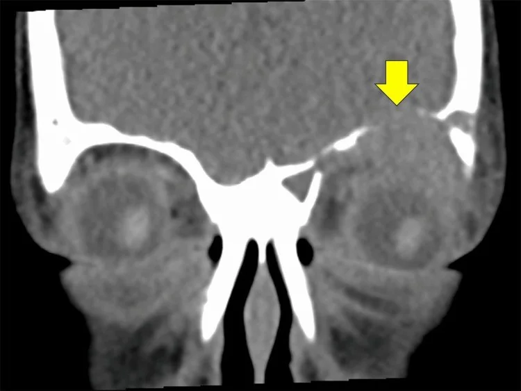
- Kontrastsız incelemede orbital tavan veya superolateral duvarda iyi sınırlı ya da güve yeniği görünümlü osteolitik defekt; eşlik edebilen ekstrakonal yumuşak doku bileşeni ve kortikal kenarların düzensiz/bevel biçimli yıkımı seçilir. HU eşiği tanımlı değildir; kemik lezyonu için kontrast fazı gerekmez.
- Kemik algoritması tanısal
- İç ve dış korteksin farklı derecede kaybı, bevel kenarlar, kalan küçük kemik parçası ve defektin orbital çatıdan komşu frontal ya da zigomatik kemiğe uzanımı kemik algoritmasında daha iyi haritalanır.
Önerilen pencereler: Kemik (G 1800 / M 400), Yumuşak doku (G 400 / M 40).
BT bulguları
- Orbital çatı veya superolateral duvarda fokal litik defekt; LHH orbital kemik tutulumu sıklıkla bu bölgede yerleşir.
- Keskin, oyulmuş ya da bevel kenar — iç ve dış korteksin eşit olmayan yıkımı aynı lezyonda farklı kenar seviyeleri oluşturabilir.
- Güve yeniği tarzı osteoliz — sınır boyunca küçük düzensiz erozyonlar daha geniş bir destrüksiyon alanına eşlik edebilir.
- Ekstrakonal yumuşak doku kitlesi — kemik defektiyle bitişik olup orbitaya, periorbital dokulara veya komşu temporal fossaya uzanabilir.
- Kalan kemik adacığı (button sequestrum) — osteolitik alan içinde izole yoğun kemik parçası görülebilir; yokluğu LHH'yi dışlamaz.
- Tek odaklı ya da çok odaklı kemik tutulumu — diğer kafatası kemikleri ve görüntü alanına giren kemik yapılarında ek litik odak aranır.
- Orbital hacim etkisi — kitle göz küresini öne itebilir; optik sinir, ekstraoküler kaslar ve orbital apeksle ilişkisi ayrıca izlenir.
Normalde
Normal orbital tavanda frontal kemiğin kortikal hatları kesintisizdir; superolateral duvarda frontal ve zigomatik kemik sınırları düzgündür ve ekstrakonal doku homojen görünür. Defektteki kemik yıkımının derecesini ve eşlik eden yumuşak doku kitlesinin orbitaya ya da temporal fossaya uzanımını değerlendirin.
Normal BT ile kıyasla
Aynı bölgenin, kaynak veri setinde patoloji içermediği belirtilen bir BT incelemesini kesit kesit kaydırın; organ sınırlarını açarak anatomiyi hasta görüntüsüyle karşılaştırın. Küçük rastlantısal bulgular tümüyle dışlanmamıştır.
Kaynak: TotalSegmentator v2.01 veri seti (Wasserthal ve ark., Radiology: Artificial Intelligence 2023), CC BY 4.0, vaka s0103; organ sınırları veri setinin otomatik segmentasyonundan, birleştirilerek sadeleştirildi. Görüntüler 2,5 mm kesit aralığına yeniden örneklendi.
Ayırıcı tanı
- Nöroblastom metastazı
- çocukta birden fazla kemik odağı, bilateral periorbital yumuşak doku veya eşlik eden kraniyal litik lezyonlar metastazı destekler; tek superolateral odak LHH ile örtüşebilir.
- Ewing sarkomu
- daha geniş geçiş zonlu agresif kemik yıkımı ve belirgin yumuşak doku bileşeni görülebilir; BT tek başına histolojik ayrım yaptıramaz.
- Enfekte veya rüptüre dermoid
- orbital sütür komşuluğunda yağ dansiteli içerik, yağ-kalsiyum bileşenleri ve inflamatuvar çevre değişikliği dermoid lehinedir.
- Orbital osteomiyelit
- kemik düzensizliğine eşlik eden sinüs enfeksiyonu, periost reaksiyonu veya yaygın inflamatuvar yağ dokusu değişikliği enfeksiyonu düşündürür.
- Rabdomiyosarkom
- orbital LHH'nin klinik olarak taklit edebildiği ayırıcı tanılar arasındadır; kesin tanı histopatolojik incelemeyle konur.
- Fibro-osseöz lezyon
- litik kortikal kayıptan ziyade kemikte ekspansiyon ve buzlu cam matriks mineralizasyonu tanıya yardımcıdır.
Tuzaklar
- Yalnızca yumuşak doku penceresine bakılırsa ince orbital çatı defekti gözden kaçabilir; şüpheli kitle düzeyini kemik algoritmasında yeniden değerlendirin.
- Superolateral yerleşim ve komşu kitle LHH için tipik olsa da özgül değildir; nöroblastom metastazı ve enfeksiyonla klinik/diğer görüntüleme bulguları üzerinden ayrım gerekir.
- Lezyon içindeki küçük kemik parçasını yabancı cisim sanma; multiplanar kesitlerde kortikal kökenini ve litik kavite içindeki konumunu doğrula.
- Orbital tek odağı izole hastalık kabul etme; BT'de görüntü alanına giren diğer kemikleri tara, kapsamlı hastalık sınıflaması için BT'nin tüm vücut değerlendirmesi olmadığını belirt.
Kendini dene
Çocukta orbital çatıda bevel kenarlı litik defekt ve bitişik ekstrakonal doku görülüyor. En olası süreç hangisidir?
Cevabı göster
Langerhans hücreli histiyositoz. Orbital çatıda odaklanmış osteoliz ile bitişik yumuşak doku kitlesinin birlikteliği LHH için tipik bir görüntüleme örüntüsüdür. Ewing sarkomu daha agresif, geniş geçiş zonlu olabilir; osteomiyelitte enfeksiyon odağı ve inflamatuvar değişiklikler, dermoidde ise yağlı içerik ipucu sağlar. Kesin tanı histopatolojiktir.
Çocukta bilateral periorbital ekimoz ve kafatasında birden çok litik kemik odağı saptanıyor. En olası tanı hangisidir?
Cevabı göster
Nöroblastom metastazı. Birden fazla kalvaryal litik odak gösteren çocukta metastatik nöroblastom öncelikli ayırıcı tanıdır; LHH tek odaklı veya bevel kenarlı lezyonlarla daha sık görülür ve çoklu odakta nöroblastom lehine değerlendirilir.
Kaynaklar
Bu sayfadaki 39 cümle ve 2 quiz sorusu aşağıdaki kaynaklardan alıntılarla tek tek karşılaştırıldı; 6 cümle bu karşılaştırmada düzeltildi (2026-10-08).
- Skeletal involvement in Langerhans cell histiocytosispmc.ncbi.nlm.nih.gov
- Clinical spectrum and management outcomes of Langerhans cell histiocytosis of the orbitpmc.ncbi.nlm.nih.gov
- Orbital Langerhans Cell Histiocytosis: A Systematic Review of 228 Casespmc.ncbi.nlm.nih.gov
- Orbital Langerhans Cell Histiocytosis in Young Pediatric Patients: Two Case Reports and Literature Reviewpmc.ncbi.nlm.nih.gov
- Orbital Langerhans Cell Histiocytosis: A Case Reportpmc.ncbi.nlm.nih.gov
- [Case of unifocal orbital langerhans cell histiocytosis in an adult].pubmed.ncbi.nlm.nih.gov
- Orbital Langerhans Cell Histiocytosis: A Systematic Review of 228 Cases.pubmed.ncbi.nlm.nih.gov
- MR imaging features of orbital Langerhans cell Histiocytosis.pubmed.ncbi.nlm.nih.gov
- Orbital Langerhans Cell Histiocytosis: A Case Report.pubmed.ncbi.nlm.nih.gov
- Langerhans cell histiocytosis mimicking hematoma of the lower eyelid: A case report.pubmed.ncbi.nlm.nih.gov
- Langerhans cell histiocytosis of the orbit: pediatric case series.pubmed.ncbi.nlm.nih.gov
- Atypical presentation of isolated orbital Langerhans cell histiocytosis.pubmed.ncbi.nlm.nih.gov
- Unifocal Langerhans cell histiocytosis of the superior oblique muscle.pubmed.ncbi.nlm.nih.gov
- [Unusual presentation of a Langerhans cell histiocytosis].pubmed.ncbi.nlm.nih.gov
- Ocular trauma as the first presentation of Langerhans cell histiocytosis.pubmed.ncbi.nlm.nih.gov
- A case report of orbital Langerhans cell histiocytosis presenting as a orbital cellulitis.pubmed.ncbi.nlm.nih.gov
- Successful treatment of orbital Langerhans cell histiocytosis with stereotactic radiosurgery: A case report and literature review.pubmed.ncbi.nlm.nih.gov
- Infiltrative and inflammatory systemic disorders involving the pituitary gland.pubmed.ncbi.nlm.nih.gov
- Zygomatic bone deformation caused by a periorbital epidermoid cyst in a child: a case report and literature review.pubmed.ncbi.nlm.nih.gov
- Pediatric orbital masses from a tertiary pediatric oncology referral center: clinical characteristics and survival outcomes from a 20-year single-center experience.pubmed.ncbi.nlm.nih.gov
- Targeted proteomics reveal histiocytosis-associated neurodegeneration signatures.pubmed.ncbi.nlm.nih.gov
- When plasmacytoma defies expectations: An unusual maxillary presentation with mixed imaging features in a young patient.pubmed.ncbi.nlm.nih.gov
- Collaborative Diagnostic Pathways for Unilateral Orbital Disease: A Clinicopathological Series.pubmed.ncbi.nlm.nih.gov
- An Unusual Presentation of Multisystem Langerhans Cell Histiocytosis in a Child: A Case Report.pubmed.ncbi.nlm.nih.gov
- Erdheim-Chester Disease with Breast and Axillary Involvement Diagnosed by Ultrasound-Guided Biopsy: A Case Report and Literature Review.pubmed.ncbi.nlm.nih.gov
- Unraveling Histiocytic Disorders: Two Cases From Resource-Limited Settings.pubmed.ncbi.nlm.nih.gov
- The Relevance and Potential Role of Orbital Fat in Inflammatory Orbital Diseases: Implications for Diagnosis and Treatment.pubmed.ncbi.nlm.nih.gov
- International expert consensus recommendations for the diagnosis and treatment of Langerhans cell histiocytosis in adults.pubmed.ncbi.nlm.nih.gov
- Incidence and radiological pattern of eosinophilic granuloma: a retrospective study in a Chinese tertiary hospital.pubmed.ncbi.nlm.nih.gov
- CT and MRI of pediatric skull lesions with fluid-fluid levels.pubmed.ncbi.nlm.nih.gov
- Review of imaging modalities and radiological findings of calvarial lesions.pubmed.ncbi.nlm.nih.gov
- Osteomyelitis of the Jaw Bones and Its Mimics: Resolving the Diagnostic Enigma.pubmed.ncbi.nlm.nih.gov
- Giant Cell Tumor of the Cranial Vault: A Rare Frontotemporal Case With Complete Neurological Recovery and Narrative Review.pubmed.ncbi.nlm.nih.gov
- Imaging features of lacrimal gland disease.pubmed.ncbi.nlm.nih.gov
- The solitary skull mass: An atypical presentation of anaplastic large cell lymphoma.pubmed.ncbi.nlm.nih.gov
- Unveiling the enigma: A rare case of eosinophilic granuloma.pubmed.ncbi.nlm.nih.gov
- Primary osseous kaposiform hemangioendothelioma confined to long bones: a retrospective imaging analysis of 12 pediatric cases.pubmed.ncbi.nlm.nih.gov
- Clinical spectrum and management outcomes of Langerhans cell histiocytosis of the orbit.pubmed.ncbi.nlm.nih.gov
- Extra Nodal Rosai-Dorfman Disease Originating in the Nasal and Paranasal Complex and Gnathic Bones: A Systematic Analysis of Seven Cases and Review of Literature.pubmed.ncbi.nlm.nih.gov
- Targeted Therapy in Erdheim-Chester Disease: A Case Report and Review of the Literature.pubmed.ncbi.nlm.nih.gov
- Clinicopathological insights into ophthalmic Rosai-Dorfman disease: A 15-year retrospective analysis.pubmed.ncbi.nlm.nih.gov
- Unusual sites of bone involvement in Langerhans cell histiocytosis: a systematic review of the literature.pubmed.ncbi.nlm.nih.gov
- Non-odontogenic tumors of the facial bones in children and adolescents: role of multiparametric imaging.pubmed.ncbi.nlm.nih.gov
- Skeletal involvement in Langerhans cell histiocytosis.pubmed.ncbi.nlm.nih.gov
- Literature review of pediatric Erdheim-Chester disease (ECD) and mixed histiocytosis (Langerhans cell histiocytosis/ECD).pubmed.ncbi.nlm.nih.gov
- Disease extent, not lesion location, determines relapse risk in single-system skeletal Langerhans cell histiocytosis.pubmed.ncbi.nlm.nih.gov
- Intravenous zoledronate for pediatric langerhans cell histiocytosis with bone involvement.pubmed.ncbi.nlm.nih.gov
- Orbital pseudotumor and skull base infiltration as the sole manifestation of granulomatosis with polyangiitis.pubmed.ncbi.nlm.nih.gov
- Rare mandibular metastasis of neuroblastoma in a child: a case report.pubmed.ncbi.nlm.nih.gov
- Neuroblastoma Metastasis to the Mandible in Children: A Case Report and Focused Narrative Review of Reported Cases.pubmed.ncbi.nlm.nih.gov
- Orbital Fungating Mass as a Rare Manifestation of Retinoblastoma: A Case Study in Palliative Care.pubmed.ncbi.nlm.nih.gov
- Multifocal osseous and cutaneous Rosai-Dorfman-Destombes disease presenting as an aggressive intramedullary femoral lesion.pubmed.ncbi.nlm.nih.gov
- Primary Ewing's Sarcoma of the Sinonasal Region: A Rare Clinical Encounter.pubmed.ncbi.nlm.nih.gov
- Synovial Sarcoma: Molecular Biology, Pathology, and Therapeutic Strategies.pubmed.ncbi.nlm.nih.gov
- Late finding of a dermoid cyst in a posterior fossa intra-axial lesion: A rare case report.pubmed.ncbi.nlm.nih.gov
- When Exophthalmos Reveals an Orbital Dermoid Cyst in a Child: A Case Report.pubmed.ncbi.nlm.nih.gov
- Imaging for Pediatric Head and Neck Emergencies: Critical Insights.pubmed.ncbi.nlm.nih.gov
- Bilateral Cavernous Sinus Thrombosis and Clival Osteomyelitis Complicating Pediatric Otitis Media.pubmed.ncbi.nlm.nih.gov
- Behind the Unilateral Rhinorrhea: Delayed Pediatric Intranasal Foreign Bodies Presenting as Chronic Sinonasal Disease and Severe Complications.pubmed.ncbi.nlm.nih.gov
- Application of imaging studies for orbital tumors: a practical review and representative cases.pubmed.ncbi.nlm.nih.gov
- Intracranial aneurysmal bone cysts in association with fibrous dysplasia: Case report and scoping review of case reports.pubmed.ncbi.nlm.nih.gov
- A rare case of odontogenic myxofibroma of the maxilla in a young patient: a challenging diagnostic and differential diagnosis.pubmed.ncbi.nlm.nih.gov
- Multimodal imaging in neuro-ophthalmology.pubmed.ncbi.nlm.nih.gov
- Coexistence of Autoantibodies against the Golgi Complex and Ro52 Antigen in a Patient with Nonspecific Interstitial Pneumonia.pubmed.ncbi.nlm.nih.gov
- A clinical update and radiologic review of pediatric orbital and ocular tumors.pubmed.ncbi.nlm.nih.gov
- Retinoblastoma and beyond: pediatric orbital mass lesions.pubmed.ncbi.nlm.nih.gov
- Juvenile Xanthogranuloma of the Head and Neck: Imaging Findings in 11 Cases.pubmed.ncbi.nlm.nih.gov
- International Society of Paediatric Surgical Oncology (IPSO) Surgical Practice Guidelines.pubmed.ncbi.nlm.nih.gov
- HMG-CoA reductase inhibitors as adjuvant treatment for hematologic malignancies: what is the current evidence?pubmed.ncbi.nlm.nih.gov
- Spontaneous bacteremia and spontaneous bacterial peritonitis share similar prognosis in patients with cirrhosis: a cohort study.pubmed.ncbi.nlm.nih.gov
- Proceedings of the 15th International Congress on Circumpolar Health.pubmed.ncbi.nlm.nih.gov
- CT Anatomy of the Skull in Lamb and Adult Romane Sheep.pubmed.ncbi.nlm.nih.gov
- Head of the Syrian Hamster (<i>Mesocricetus auratus</i>): Planar Anatomy with Correlative Micro-Computed Tomography and Magnetic Resonance Imaging.pubmed.ncbi.nlm.nih.gov
- Evaluation of treatment outcomes of zygomaticomaxillary complex (ZMC) fractures using Foley catheter ballooning technique.pubmed.ncbi.nlm.nih.gov
- Intracranial venous thrombosis imaging by anatomical location: an educational review with mimics and diagnostic pitfalls.pubmed.ncbi.nlm.nih.gov
- Prevalence of mineralization in the para-aural region in dogs with and without ear disease.pubmed.ncbi.nlm.nih.gov
- Perforator-Sparing Microsurgical Clipping of Tandem Dominant-Hemisphere Middle Cerebral Artery Aneurysms: Geometry-Guided Reconstruction of a Wide-Neck Bifurcation and Dorsal M1 Fusiform Lesion.pubmed.ncbi.nlm.nih.gov
- ESR Essentials: acute infections of the head and neck-practice recommendations by the European Society of Head and Neck Radiology.pubmed.ncbi.nlm.nih.gov
- Cavernous Sinus Thrombosis Associated With Contralateral Pansinusitis: A Case Report.pubmed.ncbi.nlm.nih.gov
- Invasive Aspergillosis with Intracranial Extension Initially Misdiagnosed as a Granulomatous Disease: A Case Report.pubmed.ncbi.nlm.nih.gov
- Imaging of Skull Base Tumors.pubmed.ncbi.nlm.nih.gov
- Craniofacial surgery for removing tumors in advanced stages that invade the anterior skull base.pubmed.ncbi.nlm.nih.gov
- Lateral transorbital neuroendoscopic approach using the superior eyelid incision: A straightforward, comprehensive, and illustrative step-by-step review.pubmed.ncbi.nlm.nih.gov
- Sinus pneumatisation and Keros type as predictors of anterior ethmoidal artery configuration: a 3D-reconstructed computed tomography analysis.pubmed.ncbi.nlm.nih.gov
- The utility of orbital imaging in the evaluation of orbital disease.pubmed.ncbi.nlm.nih.gov
- Autoimmune disease of head and neck, imaging, and clinical review.pubmed.ncbi.nlm.nih.gov
- Rare Diseases of the Orbit.pubmed.ncbi.nlm.nih.gov
- Pediatric Langerhans Cell Histiocytosis: Unifocal Localization in the Frontal Bone Presenting With Periorbital Swelling.pubmed.ncbi.nlm.nih.gov
- Radiological and Surgical Findings of Patients with Intradiploic Mass Lesion: Case Series and Systematic Review.pubmed.ncbi.nlm.nih.gov
- Treating metastatic extraocular retinoblastoma complicated with Langerhans cell histiocytosis.pubmed.ncbi.nlm.nih.gov
- The Diagnostic Dilemma of Pediatric Bilateral Proptosis in a Child With Juvenile Idiopathic Arthritis.pubmed.ncbi.nlm.nih.gov
- Pediatric orbital IgG4-related disease: A case series.pubmed.ncbi.nlm.nih.gov
- Orbital masses: a review of CT imaging characteristics.pubmed.ncbi.nlm.nih.gov
- Bilateral Proptosis as an Unusual Presentation of Adult-Onset Alveolar-Type Rhabdomyosarcoma of Ethmoid Sinus: A Case Report.pubmed.ncbi.nlm.nih.gov
- Orbital primary intraosseous hemangioma in a three-month old infant: A case report.pubmed.ncbi.nlm.nih.gov
- Special Considerations in Pediatric Endoscopic Skull Base Surgery.pubmed.ncbi.nlm.nih.gov
Eğitim amaçlıdır; hasta başında tanı ve tedavi kararının yerine geçmez. İçerik yapay zekâ destekli bir süreçle güncel literatüre göre cümle cümle kaynaklarla karşılaştırıldı, ancak uzman radyolog incelemesinden geçmedi. Klinik kararlarda güncel kılavuzları, kurum protokollerini ve radyoloji raporunu esas alın.