Özet
Aksesuar maksiller ostium, maksiller sinüs ile orta meatus arasında doğal ostiumdan ayrı bir açıklıktır ve çoğu kez tesadüfi bir anatomik varyant olarak saptanır. Doğal maksiller ostium etmoid infundibuluma açılarak primer drenaj yolunu oluşturur; aksesuar ostium ise genellikle unsinat çıkıntı ile alt konka arasında kalan fontanel bölgesinde bulunur; her iki yapıyı ayırt etmek sinüsün drenaj anatomisinin doğru değerlendirilmesi için gereklidir. İnce kesitli kontrastsız sinüs BT'si açıklığın yerini, kemik kenarlarını ve eşlik eden ostiomeatal hastalığı gösterir. İki ostiumun birbirinden bağımsız olması, sekresyonun sinüsten buruna çıkıp aksesuar açıklıktan yeniden sinüse döndüğü döngüsel akışa katkı sağlayabilir; ancak tek başına kronik sinüzit kanıtı değildir.
Faz ve pencere
- Kontrastsız tanısal
- Kontrastsız BT'de maksiller sinüs medial duvarındaki hava geçişini ve doğal ostiumdan bağımsızlığını en az iki ardışık koronal kesitte değerlendir; açıklığın bony defekt yapısı ve komşu kemik anatomisi tanımlanır.
- Kemik algoritması tanısal
- Unsinat çıkıntı, infundibulum ve medial sinüs duvarının ince kortikal sınırları ayrı seçilir; kemik algoritması aksesuar açıklığın cerrahi anatomiyle ilişkisini netleştirir.
Önerilen pencereler: Kemik (G 1800 / M 400), Yumuşak doku (G 400 / M 40).
BT bulguları
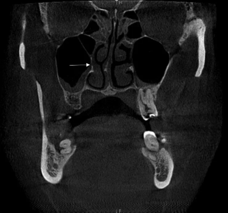
- Maksiller sinüs medial duvarında doğal ostiumdan ayrı ikinci bir açıklık izlenir.
- Açıklık çoğunlukla orta meatusa bağlı membranöz bölge olan fontanelde, unsinat çıkıntı ile alt konka arasında görülür.
- Doğal maksiller ostium sinüsün süperomedial duvarında yer alarak primer drenaj yolunu oluşturur; fontanel bölgesindeki aksesuar açıklıktan ayırt edilmelidir.
- Aksesuar ostium, unsinat çıkıntı ile alt konka arasındaki membranöz bölgenin kemik defekti olarak görünür; çevre kemikte patolojik destrüksiyon beklenmez.
- Aksesuar ostium varlığında maksiller sinüs mukozal kalınlaşması veya opasifikasyonu sıklıkla eşlik eder; ancak bu varyant tek başına kronik sinüzit tanısını doğrulamaz, mukoza bulguları ayrı değerlendirilmelidir.
- Aynı sinüs duvarında iki ostiumun açık olması, doğal ostiumdan çıkan sekresyonun aksesuar açıklığa geri dönebildiği anatomik düzeni oluşturabilir.
Normalde
Normal drenajda maksiller sinüsün doğal ostiumu arka fontanelin ön bölümüne hizalanır ve tek bir drenaj yolu oluşturur; ayrıca ayrı ikinci bir medial duvar açıklığı izlenmez. Koronal görüntüde doğal ostium-infundibulum sürekliliğini, aksiyel görüntüde olası aksesuar açıklığın bundan ayrı olup olmadığını kıyasla.
Normal BT ile kıyasla
Aynı bölgenin, kaynak veri setinde patoloji içermediği belirtilen bir BT incelemesini kesit kesit kaydırın; organ sınırlarını açarak anatomiyi hasta görüntüsüyle karşılaştırın. Küçük rastlantısal bulgular tümüyle dışlanmamıştır.
Kaynak: TotalSegmentator v2.01 veri seti (Wasserthal ve ark., Radiology: Artificial Intelligence 2023), CC BY 4.0, vaka s0849; organ sınırları veri setinin otomatik segmentasyonundan, birleştirilerek sadeleştirildi. Görüntüler 2,5 mm kesit aralığına yeniden örneklendi.
Ayırıcı tanı
- Doğal maksiller ostium
- sinüsün drenaj yolu olarak arka fontanelin ön bölümüne hizalanır; unsinat çıkıntı ile ilişkili birincil çıkıştır, aksesuar açıklık değildir.
- Cerrahi antrostomi
- geniş, düzgünleştirilmiş açıklık ve eşlik eden postoperatif kemik değişikliği bulunabilir.
- Oroantral fistül
- maksiller sinüs tabanı ile ağız boşluğu (diş alveolü) arasında bağlantı vardır; medial duvarda açılmaz.
Tuzaklar
- Doğal ostium her zaman tek kesitte görülmeyebilir; unsinat çıkıntı ve infundibulumu ardışık koronal kesitlerde takip et.
- Gerçek ostium, sinüs medial duvarında nazal kaviteye açılan ve BT'de ardışık kesitlerde izlenen bir kemik defektidir; sinüs lümenindeki sıvı düzeyi veya mukozal kalınlaşma ile karıştırılmamalıdır.
- Aksesuar ostium varlığından aktif ya da tekrarlayan sinüzit olduğu sonucunu çıkarma; mukoza ve ostiomeatal birim bulgularını ayrı tanımla.
Kendini dene
Maksiller sinüs medial duvarında, infundibulumdan ayrı ikinci hava açıklığı görülüyor. Bu yapı nedir?
Cevabı göster
Aksesuar ostium. Aksesuar ostium sinüs medial duvarında doğal drenaj yolundan ayrı açıklıktır. Haller hücresi etmoid kökenli hava hücresidir; kist ve polip lümeni yumuşak dokuyla doldurur.
Maksiller sinüs medial duvarından arka fontanelin ön bölümüne uzanan tek bir hava açıklığı izleniyor. En uygun yorum hangisidir?
Cevabı göster
Doğal maksiller ostium. Doğal maksiller ostium arka fontanelin ön bölümüne açılır ve sinüsün primer drenaj yolunu oluşturur. Tek bir açıklık görülmesi aksesuar ostium tanımını karşılamaz; aksesuar ostium doğal maksiller ostiumdan ayrı bir açıklıktır.
Kaynaklar
Bu sayfadaki 33 cümle ve 2 quiz sorusu aşağıdaki kaynaklardan alıntılarla tek tek karşılaştırıldı; 17 cümle bu karşılaştırmada düzeltildi (2026-10-08).
- Endoscopic visualization of CT-confirmed accessory maxillary ostium: a retrospective studypmc.ncbi.nlm.nih.gov
- Assessment of the Relationship Between Haller Cells, Accessory Maxillary Ostium, and Maxillary Sinus Pathologies: A Cross-Sectional CBCT Studypmc.ncbi.nlm.nih.gov
- Assessment of the impact of nasal septum deviation types on sinonasal variations and infundibulum morphometry in relation to gender and age using CT imagespmc.ncbi.nlm.nih.gov
- Anatomical Characteristics of the Accessory Maxillary Ostium in Three-Dimensional Analysispmc.ncbi.nlm.nih.gov
- Rare Variation of Accessory Maxillary Ostiumpmc.ncbi.nlm.nih.gov
- Maxillary sinus pain with radiolucent sinuses due to agenesis of the membranous ostium.pubmed.ncbi.nlm.nih.gov
- Anatomic variations associated with antrochoanal polyps.pubmed.ncbi.nlm.nih.gov
- Localization of the Maxillary Ostium in Relation to the Reduction of Depressed Nasomaxillary Fractures.pubmed.ncbi.nlm.nih.gov
- Investigation of the accessory maxillary ostium: a congenital variation or acquired defect?pubmed.ncbi.nlm.nih.gov
- Accessory Maxillary Ostia: Prevalence of an Anatomical Variant and Association with Chronic Sinusitis.pubmed.ncbi.nlm.nih.gov
- Anatomical Characteristics of the Accessory Maxillary Ostium in Three-Dimensional Analysis.pubmed.ncbi.nlm.nih.gov
- Cone-beam computed tomography analysis of accessory maxillary ostium and Haller cells: Prevalence and clinical significance.pubmed.ncbi.nlm.nih.gov
- CBCT analysis of haller cells: relationship with accessory maxillary ostium and maxillary sinus pathologies.pubmed.ncbi.nlm.nih.gov
- Radiological Study of Maxillary Sinus using CBCT: Relationship between Mucosal Thickening and Common Anatomic Variants in Chronic Rhinosinusitis.pubmed.ncbi.nlm.nih.gov
- The effect of the presence of the accessory maxillary ostium on the maxillary sinus.pubmed.ncbi.nlm.nih.gov
- A study on the association between accessory maxillary ostium and maxillary sinus mucosal thickening using cone beam computed tomography.pubmed.ncbi.nlm.nih.gov
- The potential clinical relevance of anatomical structures and variations of the maxillary sinus for planned sinus floor elevation procedures: A retrospective cone beam computed tomography study.pubmed.ncbi.nlm.nih.gov
- Relationship of the accessory maxillary ostium to mucosal thickening and maxillary sinus cysts: a 3D segmentation study.pubmed.ncbi.nlm.nih.gov
- Evaluation of the relationship between the presence of an accessory maxillary ostium and the presence and types of nasal septum deviation: A computed tomography study.pubmed.ncbi.nlm.nih.gov
- Endoscopic visualization of CT-confirmed accessory maxillary ostium: a retrospective study.pubmed.ncbi.nlm.nih.gov
- Anatomical Variations Favouring Antrochoanal Polyp Genesis.pubmed.ncbi.nlm.nih.gov
- The Infra-Bullar Groove: Assessing a Novel Surgical Landmark for Identifying the Natural Maxillary Ostium.pubmed.ncbi.nlm.nih.gov
- The agger nasi approach to the uncinate process: From top to bottom.pubmed.ncbi.nlm.nih.gov
- Anatomical variations and abnormalities of the maxillary region and clinical implications: A systematic review and metaanalysis.pubmed.ncbi.nlm.nih.gov
- Correlations between anatomical variations of the nasal cavity and ethmoidal sinuses on cone-beam computed tomography scans.pubmed.ncbi.nlm.nih.gov
- Morphological and Epidemiological Analysis of the Sphenoid Sinus Based on Computed Tomography and Magnetic Resonance Imaging: A Narrative Review.pubmed.ncbi.nlm.nih.gov
- Uncommon Nasal Mass Presentation: A Radiological Case Series.pubmed.ncbi.nlm.nih.gov
- Comprehensive microsurgical anatomy of the middle cranial fossa: Part I-Osseous and meningeal anatomy.pubmed.ncbi.nlm.nih.gov
- Accessory Maxillary Sinus Ostium Frequency and Correlation with Anatomical Variables and Sinus Mucosal Status: A CBCT Study.pubmed.ncbi.nlm.nih.gov
- Post-Traumatic Orbital Reconstruction Using Titanium Patient-Specific Implants: A Clinical and Radiological Cohort Study Focusing on Paranasal Sinuses Physiology.pubmed.ncbi.nlm.nih.gov
- Ostiomeatal complex preservation: submucosal inferior meatal antrostomy for maxillary sinus pathologies.pubmed.ncbi.nlm.nih.gov
- Association of sphenoid sinus pneumatization patterns, Onodi cells, and rostrum pneumatization with natural ostium location: A cross-sectional CT study.pubmed.ncbi.nlm.nih.gov
- Description of the Uncinate Process: A Computed Tomography Cross-Sectional Study.pubmed.ncbi.nlm.nih.gov
- Rhinosinusitis: Evidence and experience - 2024.pubmed.ncbi.nlm.nih.gov
- Management of Odontogenic Sinusitis: Results with Single-Step FESS and Dentoalveolar Surgery.pubmed.ncbi.nlm.nih.gov
- Automated Classification of Maxillary Sinus Ostium Patency Using a ConvNeXt-Tiny + DeiT Gated MLP-Based Hybrid Deep Learning Model: A Retrospective CBCT Study.pubmed.ncbi.nlm.nih.gov
- Comprehensive review on endonasal endoscopic sinus surgery.pubmed.ncbi.nlm.nih.gov
- Imaging diagnosis of paranasal sinus mucocele in a Yorkshire Terrier dog.pubmed.ncbi.nlm.nih.gov
- Descriptive CBCT Findings of Maxillary Sinus Mucosal Changes in Patients Undergoing Sinus Floor Elevation: A Retrospective Observational Study.pubmed.ncbi.nlm.nih.gov
- V Brazilian Consensus on Rhinitis - 2024.pubmed.ncbi.nlm.nih.gov
- Radioanatomy of the Silent Sinus Syndrome.pubmed.ncbi.nlm.nih.gov
- Operative Corridors in Endoscopic Skull Base Tumor Surgery.pubmed.ncbi.nlm.nih.gov
- Clinical anatomy of the paranasal sinuses and its terminology.pubmed.ncbi.nlm.nih.gov
- The maxillary sinus: physiology, development and imaging anatomy.pubmed.ncbi.nlm.nih.gov
- Surgical Instructions in Revision Endoscopic Sinus Surgery: Pearls and Pitfalls.pubmed.ncbi.nlm.nih.gov
- Systematic anatomical validation of the endoscopic mononostril transethmoid-paraseptal approach to the central skull base.pubmed.ncbi.nlm.nih.gov
- Persistent presumed allergic fungal rhinosinusitis during tezepelumab therapy with improvement after revision surgery: a case report.pubmed.ncbi.nlm.nih.gov
- Odontogenic Sinusitis: The Selective Role of a Modified Caldwell-Luc Approach in Two CT/CBCT-Documented Cases.pubmed.ncbi.nlm.nih.gov
- Foreign body-associated maxillary sinus aspergillosis caused by Endomethasone extrusion: diagnostic challenges, biological mechanisms, and surgical management.pubmed.ncbi.nlm.nih.gov
- Advanced Maxillary Squamous Cell Carcinoma With Extension to the Infratemporal Fossa Masquerading as Extensive Osteomyelitis: A Case Report.pubmed.ncbi.nlm.nih.gov
- Benign and malignant lesions of the mandible and maxilla: imaging findings and systematic approach.pubmed.ncbi.nlm.nih.gov
- Radiologic Evaluation of Paranasal Sinuses in Sickle Cell Anemia and Thalassemia: Case-Control Study.pubmed.ncbi.nlm.nih.gov
- From Displacement to Recovery: A Case Report on Surgical Root Retrieval in the Maxillary Sinus.pubmed.ncbi.nlm.nih.gov
- Acute Oroantral Communication Closure: Resorbable Collagen Membrane vs. Buccal Advancement Flap Outcomes: A Clinical Trial.pubmed.ncbi.nlm.nih.gov
- Clinical decision-making algorithm for the management of Oroantral fistula: A comprehensive guide.pubmed.ncbi.nlm.nih.gov
- Emerging and re-emerging infectious disease in otorhinolaryngology.pubmed.ncbi.nlm.nih.gov
- Diagnostic reliability and accuracy of the hydraulic contrast lift protocol in the radiographic detection of sinus lift and perforation: ex vivo randomized split-mouth study in an ovine model.pubmed.ncbi.nlm.nih.gov
- Analysis of risk variables for association with maxillary sinus mucosal thickenings: a cone-beam computed tomography-based retrospective study.pubmed.ncbi.nlm.nih.gov
- Diagnostic tools in Rhinology EAACI position paper.pubmed.ncbi.nlm.nih.gov
- Long-term safety and effectiveness of the "OptEase" vena cava filter.pubmed.ncbi.nlm.nih.gov
- Sinus Lift With Simultaneous Mucous Retention Cyst Drainage for Implant Rehabilitation: Clinical, Tomographic, and Histologic Assessments in a Six-Year Follow-Up Case Report.pubmed.ncbi.nlm.nih.gov
- Convolutional neural network for maxillary sinus segmentation based on the U-Net architecture at different planes in the Chinese population: a semantic segmentation study.pubmed.ncbi.nlm.nih.gov
- Association Between DMFT Index and Sinonasal Anatomical Variations in Children: A Retrospective Cone-Beam Computed Tomography Study.pubmed.ncbi.nlm.nih.gov
- Deep learning classification of nasal anatomical variants on computed tomography for endoscopic sinus surgery.pubmed.ncbi.nlm.nih.gov
- Antrochoanal Polyps: Nasal Anatomical and Pathological Variants as Risk Factors.pubmed.ncbi.nlm.nih.gov
- Effect of Le Fort I osteotomy on the sinonasal region: three-dimensional study.pubmed.ncbi.nlm.nih.gov
- Bifid inferior turbinate with inferomedial extension: a case report and updated review of reported cases.pubmed.ncbi.nlm.nih.gov
- Association of Deviated Nasal Septum as an Etiological Factor for Antrochoanal Polyp.pubmed.ncbi.nlm.nih.gov
- Infraorbital Ethmoid Cells (Haller Cells) and Their Role in Chronic Rhinosinusitis: An Evidence-Based Meta-Analysis.pubmed.ncbi.nlm.nih.gov
- Anatomical variations of the maxillary sinuses: A cross-sectional study.pubmed.ncbi.nlm.nih.gov
- Orbital Extension of Sinonasal Mucocele Presenting as Proptosis: A Report of Two Cases.pubmed.ncbi.nlm.nih.gov
- Maxillary Sinus Mucocele following Zygomaticomaxillary Complex Fracture Repair.pubmed.ncbi.nlm.nih.gov
- Intraorbital posttraumatic respiratory epithelial cyst: A case report and literature review.pubmed.ncbi.nlm.nih.gov
- Surgical approach to management of oroantral communications. Case report.pubmed.ncbi.nlm.nih.gov
- Endoscopic Management of a Right Maxillary Sinus Mucocele With Palatal and Medial Wall Erosion.pubmed.ncbi.nlm.nih.gov
- A functional Schneiderian membrane-preserving maxillectomy: leveraging mucosal bone-forming potential and clinical effectiveness verification.pubmed.ncbi.nlm.nih.gov
- Radiologic Evaluation of Odontogenic Sinusitis and Its Etiologic Factors: Lessons Learned from a Retrospective Study with a Proposed Imaging-Guided Management Pathway.pubmed.ncbi.nlm.nih.gov
- Maxillary Sinus Papillary Edema as a Predictor of Odontogenic Sinusitis.pubmed.ncbi.nlm.nih.gov
Eğitim amaçlıdır; hasta başında tanı ve tedavi kararının yerine geçmez. İçerik yapay zekâ destekli bir süreçle güncel literatüre göre cümle cümle kaynaklarla karşılaştırıldı, ancak uzman radyolog incelemesinden geçmedi. Klinik kararlarda güncel kılavuzları, kurum protokollerini ve radyoloji raporunu esas alın.