
Özet
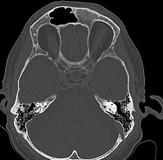
Sinüs hipoplazisi veya aplazisi gelişimsel bir varyasyon olabilir; tek taraflı olduğunda karşı tarafla belirgin boyut farkı oluşturabilir. Kontrastsız sinüs BT’si sinüs havalanmasını ve kemik anatomisini değerlendirmeye; maksiller sinüs aplazisinde uncinate proses ve infundibulum gibi eşlik eden anatomik farklılıkları göstermeye yardımcı olur. Frontal sinüs aplazisi gelişimsel bir varyasyon olabilir; maksiller sinüs hipoplazisi ise sinüs hacminde azalma ile tanımlanır. Gelişimsel maksiller sinüs varyantının tanınması, cerrahi planlamada anatomik sınırların ve diseksiyon alanının belirlenmesine yardımcı olabilir.
Faz ve pencere
- Kontrastsız tanısal
- Sinüs hacminin küçük veya havalanmanın yok olması hipoplazi ya da aplaziyi düşündürür; maksiller sinüs değerlendirmesinde uncinate proses ve infundibulum gibi eşlik eden anatomik farklılıklar da incelenebilir. Kontrast gerekmez.
Önerilen pencereler: Kemik (G 1800 / M 400).
BT bulguları
- Karşı tarafa göre daha küçük veya sığ maksiller sinüs boşluğu.
- Frontal sinüsün tek taraflı ya da iki taraflı havalanmaması ve frontal kemikte düzenli iç kontur.
- Sfenoid sinüs havalanması doğumda bulunmaz ve çocuklukta gelişmeye başlar; bu nedenle değerlendirmede yaş göz önünde bulundurulmalıdır.
- Sinüs duvarında keskin, korunmuş kortikal sınır; destrüktif kenar bulunmaması.
- Maksiller hipoplazide uncinate prosesin küçük, yer değiştirmiş veya seçilemez olması.
- Hipoplazi/aplazi olgularında genellikle sinüs içinde ekspansif kitle veya kemik yıkımı bulunmaz; mukosel ve tümörler ise kemik duvarları incelterek genişlemeye yol açabilir.
Ölçütler ve sınıflamalar
- Maksiller sinüs hipoplazisi, uncinate proses ve etmoid infundibulumun konumuyla birlikte değerlendirilir.
- Maksiller sinüs hipoplazisinde uncinate proses ve etmoid infundibulum laterale, orbita tabanının altına yer değiştirebilir; değerlendirmede bu yapıların konumu dikkate alınmalıdır.
Normalde
Paranazal sinüsler hava dolu boşluklardır; frontal sinüsler doğumda bulunmaz, çocuklukta gelişir ve erişkinlerde aplazi görülebilir. Sinüs boyutunu karşı tarafla kıyasla; maksiller sinüs hipoplazisinde uncinate proses ve etmoid infundibulumun orbita tabanına göre konumunu da değerlendir.
Normal BT ile kıyasla
Aynı bölgenin, kaynak veri setinde patoloji içermediği belirtilen bir BT incelemesini kesit kesit kaydırın; organ sınırlarını açarak anatomiyi hasta görüntüsüyle karşılaştırın. Küçük rastlantısal bulgular tümüyle dışlanmamıştır.
Kaynak: TotalSegmentator v2.01 veri seti (Wasserthal ve ark., Radiology: Artificial Intelligence 2023), CC BY 4.0, vaka s0849; organ sınırları veri setinin otomatik segmentasyonundan, birleştirilerek sadeleştirildi. Görüntüler 2,5 mm kesit aralığına yeniden örneklendi.
Ayırıcı tanı
- Kronik maksiller sinüs atelektazisi
- infundibulum tıkanıklığı ve içe çökmüş duvarlar, orbital tabanda aşağı yer değiştirme yapabilir.
- Sinüzitte mukozal kalınlaşma gibi inflamatuvar bulgular görülebilir; maksiller sinüs hipoplazisi ise sinüs hacminin azalmasıyla tanımlanır.
- Mukosel
- sinüs boşluğunu genişletir, duvarları inceltip dışa iter.
- Tümör
- düzensiz kemik yıkımı veya sinüs dışına taşan yumuşak doku oluşturabilir.
Tuzaklar
- Frontal sinüs asimetrisi çoğu kez normaldir; her iki frontal resesin havalanmasını ve kemik sınırını birlikte incele.
- Küçük maksiller sinüs silent sinus sendromunda da görülebilir; sinüs duvarlarının içe doğru yer değiştirmesi ve orbital tabanın çökmesi bu olasılığı düşündürür.
- Çocuklarda paranazal sinüsler çocukluk boyunca gelişmeye devam eder; erişkin ölçütlerini gelişmekte olan sinüslere doğrudan uygulama.
Kendini dene
Frontal sinüs aplazisi kaynakta nasıl tanımlanıyor?
Cevabı göster
Sinüs havalanmasının tamamen bulunmaması. Frontal sinüs aplazisi, sinüs havalanmasının tamamen bulunmaması olarak tanımlanır.
Çalışmada tek veya iki taraflı frontal sinüs varyasyonu olan gruplarda, kontrol grubuna kıyasla hangi ölçümlerde anlamlı düşüklük saptandı?
Cevabı göster
Sol ve sağ orbital genişlik, sol orbital yükseklik, biorbital genişlik, üst yüz genişliği ve bizigomatik genişlik. Tek ve iki taraflı frontal sinüs varyasyonu gruplarında belirtilen orbital ve üst yüz ölçümlerinin düzeltilmiş değerleri kontrol grubundan anlamlı olarak düşüktü; diğer seçeneklerdeki ölçümler için anlamlı grup farkı bildirilmedi.
Kaynaklar
Bu sayfadaki 32 cümle ve 2 quiz sorusu aşağıdaki kaynaklardan alıntılarla tek tek karşılaştırıldı; 17 cümle bu karşılaştırmada düzeltildi (2026-10-09).
- CraniofacialMorphometric Associations with Frontal Sinus Hypoplasia/Aplasia in Adults: Orbital and Upper Facial Differences on CTpmc.ncbi.nlm.nih.gov
- Pneumolacrimia: A Proposed Terminology and Classification System for Nasolacrimal Drainage System Aeration on Computed Tomographypmc.ncbi.nlm.nih.gov
- Fatal Presumed Rhino-Orbito-Sinus Mucormycosis in a Child With Fanconi Anemia: A Case Reportpmc.ncbi.nlm.nih.gov
- Intracranial venous thrombosis imaging by anatomical location: an educational review with mimics and diagnostic pitfallspmc.ncbi.nlm.nih.gov
- Delayed Diagnosis of Unilateral Choanal Atresia in an Adult: A Case Reportpmc.ncbi.nlm.nih.gov
- CHARGE Association.pubmed.ncbi.nlm.nih.gov
- Maxillary sinus aplasia.pubmed.ncbi.nlm.nih.gov
- Dyke-Davidoff-Masson syndrome.pubmed.ncbi.nlm.nih.gov
- Radiological and endoscopic findings in patients undergoing revision endoscopic sinus surgery.pubmed.ncbi.nlm.nih.gov
- Frontotemporal dementia: an unusual cause.pubmed.ncbi.nlm.nih.gov
- Rhinogenic and sinus headache - Literature review.pubmed.ncbi.nlm.nih.gov
- Inner ear malformations: a practical diagnostic approach.pubmed.ncbi.nlm.nih.gov
- Carotid canal and optic canal at sphenoid sinus.pubmed.ncbi.nlm.nih.gov
- [A modern approach to the diagnosis of nasal liquorrhea].pubmed.ncbi.nlm.nih.gov
- Normal cranial postmortem CT findings in children.pubmed.ncbi.nlm.nih.gov
- The clinico-radiological spectrum of Dyke-Davidoff-Masson syndrome in adults.pubmed.ncbi.nlm.nih.gov
- Lacrimal Drainage System Problems After Rhinoplasty.pubmed.ncbi.nlm.nih.gov
- A Systematic Review of Imaging Studies in Olfactory Dysfunction Secondary to COVID-19.pubmed.ncbi.nlm.nih.gov
- Midnasal stenosis in adults with normative values.pubmed.ncbi.nlm.nih.gov
- Maxillary sinus hypoplasia relevant to dental implant treatment: a narrative review.pubmed.ncbi.nlm.nih.gov
- The Use of CBCT in Evaluating the Health and Pathology of the Maxillary Sinus.pubmed.ncbi.nlm.nih.gov
- Anatomical Variations of the Nasal Cavities and Paranasal Sinuses: A Systematic Review.pubmed.ncbi.nlm.nih.gov
- Combined Radiological and Endoscopic Evaluation of Sino Nasal Anatomical Variations in Patients of Chronic Rhinosinusitis: A North Indian Study.pubmed.ncbi.nlm.nih.gov
- Anatomical variations and abnormalities of the maxillary region and clinical implications: A systematic review and metaanalysis.pubmed.ncbi.nlm.nih.gov
- Low-flow vascular malformations without arteriovenous shunting of the central nervous system: a pictorial review.pubmed.ncbi.nlm.nih.gov
- Anatomic Pulmonary Lobectomy via Video-Assisted Thoracoscopic Surgery in an Adult Patient With Congenital Lobar Emphysema: A Case Report.pubmed.ncbi.nlm.nih.gov
- Complex inferior vena cava malformation initially manifesting as a pulmonary space-occupying lesion: A case report.pubmed.ncbi.nlm.nih.gov
- Sphenoid sinus hyperpneumatization: anatomical variants, molecular blueprints, and AI-augmented roadmaps for skull base surgery.pubmed.ncbi.nlm.nih.gov
- Proceedings of the 15th International Congress on Circumpolar Health.pubmed.ncbi.nlm.nih.gov
- Frontal Sinus Fractures.pubmed.ncbi.nlm.nih.gov
- The maxillary sinus: physiology, development and imaging anatomy.pubmed.ncbi.nlm.nih.gov
- Neurofibromatosis Type 1 With Cherubism-like Phenotype, Multiple Osteolytic Bone Lesions of Lower Extremities, and Alagille-syndrome: Case Report With Literature Survey.pubmed.ncbi.nlm.nih.gov
- Imaging for Pediatric Head and Neck Emergencies: Critical Insights.pubmed.ncbi.nlm.nih.gov
- Primary Mastoid Cholesteatoma: A Case Report and Review of the Literature.pubmed.ncbi.nlm.nih.gov
- Therapy options in the management of endolymphatic sac tumors: A case series and literature review.pubmed.ncbi.nlm.nih.gov
- Anatomical Changes in a Case with Asymmetrical Bilateral Maxillary Sinus Hypoplasia.pubmed.ncbi.nlm.nih.gov
- Cone-beam computed tomographic imaging of silent sinus syndrome: A case series and a literature review.pubmed.ncbi.nlm.nih.gov
- Retinoblastoma and beyond: pediatric orbital mass lesions.pubmed.ncbi.nlm.nih.gov
- A unique case of multiple calvarial hemangiomas with one large symplastic hemangioma.pubmed.ncbi.nlm.nih.gov
- Zero-TE MRI: principles and applications in the head and neck.pubmed.ncbi.nlm.nih.gov
- Assessment of Paranasal Sinus Growth with 3D Volumetric Measurements and the Effect of Anatomic Variations on Sinus Volume in a Pediatric Population.pubmed.ncbi.nlm.nih.gov
- Impact of RAMPA Therapy on Nasal Cavity Expansion and Paranasal Drainage: Fluid Mechanics Analysis, CAE Simulation, and a Case Study.pubmed.ncbi.nlm.nih.gov
- Clinician-Guided Deep Learning Segmentation of Skull Base Pneumatization on Computed Tomography Using 3D Slicer and MONAI Label.pubmed.ncbi.nlm.nih.gov
- Investigating paranasal sinuses developmental disorders and septum deviation angle effects on olfactory fossa depth using cone-beam computed tomography.pubmed.ncbi.nlm.nih.gov
- Anatomical and Pathological Assessment of the Maxillary Sinus Using CBCT Imaging: A Retrospective Descriptive Study.pubmed.ncbi.nlm.nih.gov
- Silent Sinus Syndrome: Interesting Computed Tomography and Magnetic Resonance Imaging Findings.pubmed.ncbi.nlm.nih.gov
- Uncovering Cystic Fibrosis Carrier: Insights From a Heterozygous CFTR-F508del Rabbit Model.pubmed.ncbi.nlm.nih.gov
- Radiologic Evaluation of Odontogenic Sinusitis and Its Etiologic Factors: Lessons Learned from a Retrospective Study with a Proposed Imaging-Guided Management Pathway.pubmed.ncbi.nlm.nih.gov
- Multimodal imaging in neuro-ophthalmology.pubmed.ncbi.nlm.nih.gov
- Small ethmoid mucocele with intracranial extension associated with fibrous dysplasia: A rare case report.pubmed.ncbi.nlm.nih.gov
- Incidentally discovered asymptomatic extrasinusal mucocele of the sphenoid greater wing: A case report and review of differential diagnoses.pubmed.ncbi.nlm.nih.gov
- Endoscopic Management of a Right Maxillary Sinus Mucocele With Palatal and Medial Wall Erosion.pubmed.ncbi.nlm.nih.gov
- Cavernous venous malformation of the frontal sinus: CT and MRI imaging findings.pubmed.ncbi.nlm.nih.gov
- Advanced Maxillary Squamous Cell Carcinoma With Extension to the Infratemporal Fossa Masquerading as Extensive Osteomyelitis: A Case Report.pubmed.ncbi.nlm.nih.gov
- Rhino-orbital-cerebral mucormycosis with skull base involvement mimicking an intracranial tumor in an immunocompetent patient.pubmed.ncbi.nlm.nih.gov
- Morphological and Epidemiological Analysis of the Sphenoid Sinus Based on Computed Tomography and Magnetic Resonance Imaging: A Narrative Review.pubmed.ncbi.nlm.nih.gov
- The Developing Human Sphenoid Bone: Linking Embryological Development to Adult Morphology.pubmed.ncbi.nlm.nih.gov
- International Harmonization of Nomenclature and Diagnostic Criteria (INHAND): Nonproliferative and Proliferative Lesions of the Rabbit.pubmed.ncbi.nlm.nih.gov
- Incidental findings from panoramic radiographs: A systematic review with meta-analysis.pubmed.ncbi.nlm.nih.gov
- Multidisciplinary Management of Pediatric Craniopharyngioma: From Diagnosis to Long-Term Outcomes.pubmed.ncbi.nlm.nih.gov
- Mechanism patterns and age variations in pediatric cranio-maxillofacial trauma: a 5-year analysis of the national trauma data bank.pubmed.ncbi.nlm.nih.gov
- Cone Beam Computed Tomography (CBCT) Image Based Volumetric and Dimension Measurements of Maxillary Sinus in Correlation to the Age and Gender of a Lebanese Population Sample.pubmed.ncbi.nlm.nih.gov
- Novel TCOF1 Frameshift Variant and Phenotypic Heterogeneity in a Chinese Family With Treacher Collins Syndrome.pubmed.ncbi.nlm.nih.gov
- Maxillary Sinus Assessment: A Computed Tomography Analysis and Classification.pubmed.ncbi.nlm.nih.gov
- Nasal cavity perforation by implant fixtures: case series with emphasis on panoramic imaging of nasal cavity extending posteriorly.pubmed.ncbi.nlm.nih.gov
- Association of Chiropractic Colleges Educational Conference and Research Agenda Conference 2025: Transformative Trends in Healthcare.pubmed.ncbi.nlm.nih.gov
- Non-Thrombotic Filling Defects in Cerebral Veins and Sinuses: When Normal Structures Mimic a Disease.pubmed.ncbi.nlm.nih.gov
- Long-term safety and effectiveness of the "OptEase" vena cava filter.pubmed.ncbi.nlm.nih.gov
- Unravelling the Complex Relationship Between Frontal Sinus Pneumatization and Paranasal Sinus Anatomical Variants: Insights from CT Imaging.pubmed.ncbi.nlm.nih.gov
- CraniofacialMorphometric Associations with Frontal Sinus Hypoplasia/Aplasia in Adults: Orbital and Upper Facial Differences on CT.pubmed.ncbi.nlm.nih.gov
- Morphometric Characterization of Frontal Sinus and Outflow Tract Anatomy: Implications for Surgical Planning.pubmed.ncbi.nlm.nih.gov
- Pneumolacrimia: A Proposed Terminology and Classification System for Nasolacrimal Drainage System Aeration on Computed Tomography.pubmed.ncbi.nlm.nih.gov
- Imaging More than Skin-Deep: Radiologic and Dermatologic Presentations of Systemic Disorders.pubmed.ncbi.nlm.nih.gov
- Imaging review of the anterior skull base.pubmed.ncbi.nlm.nih.gov
- Unilateral Maxillary Sinus Aplasia With Hypoplasia of the Inferior Concha and Absence of the Infundibulum and Uncinate Process: A Case Report.pubmed.ncbi.nlm.nih.gov
- Perioperative Airway Management in Treacher Collins Syndrome With Obstructive Sleep Apnea: A Case Report.pubmed.ncbi.nlm.nih.gov
- CBCT Volumetric Changes in Combined Nasal Cavity and Paranasal Sinuses Following RAMPA-ROA Therapy: A Retrospective Cohort Study with Reference to Longitudinal Growth Data.pubmed.ncbi.nlm.nih.gov
- Patterns of injury and violence in Yaoundé Cameroon: an analysis of hospital data.pubmed.ncbi.nlm.nih.gov
- Total laparoscopic radical hysterectomy for cervical cancer in prolapsed uterus.pubmed.ncbi.nlm.nih.gov
- Pathogenesis and Management in Cerebrovenous Outflow Disorders.pubmed.ncbi.nlm.nih.gov
- Automated detection and classification of maxillary sinus variations using slice-based and full-volume CBCT deep learning models.pubmed.ncbi.nlm.nih.gov
- Isolated Maxillary Extracranial Metastasis of a Suprasellar Germinoma.pubmed.ncbi.nlm.nih.gov
- Imaging hematomas in unforeseen sites - unveiling 2 cases of pediatric scurvy.pubmed.ncbi.nlm.nih.gov
- Assessment of the impact of nasal septum deviation types on sinonasal variations and infundibulum morphometry in relation to gender and age using CT images.pubmed.ncbi.nlm.nih.gov
- Rhinosinusitis: Evidence and experience - 2024.pubmed.ncbi.nlm.nih.gov
- Monolateral sinonasal complications of dental disease or treatment: when does endoscopic endonasal surgery require an intraoral approach?pubmed.ncbi.nlm.nih.gov
- Danger points, complications and medico-legal aspects in endoscopic sinus surgery.pubmed.ncbi.nlm.nih.gov
- Ataxia-Telangiectasia and Associated Bronchiectasis: Case Report and Literature Review.pubmed.ncbi.nlm.nih.gov
- A review of the oral health of individuals with disabilities in Puerto Rico and among U.S. Hispanics.pubmed.ncbi.nlm.nih.gov
- Anatomical and Functional Alterations in Nasolacrimal Duct Obstruction: A Comprehensive Review.pubmed.ncbi.nlm.nih.gov
- Rare Diseases of the Orbit.pubmed.ncbi.nlm.nih.gov
- Incidental Detection of a Posterior Inferior Cerebellar Artery (PICA)-Terminating Vertebral Artery Variant During CT Evaluation of Venous Sinus Thrombosis: A Case Report.pubmed.ncbi.nlm.nih.gov
- Arrested pneumatization of the sphenoid sinus mimicking intraosseous lesions of the skull base.pubmed.ncbi.nlm.nih.gov
- Relationship Between Maxillary Transverse Deficiency and Respiratory Problems: A Systematic Review of the Effectiveness of Devices over the Past Decade.pubmed.ncbi.nlm.nih.gov
- Imaging in rhabdomyosarcoma: a patient journey.pubmed.ncbi.nlm.nih.gov
- V Brazilian Consensus on Rhinitis - 2024.pubmed.ncbi.nlm.nih.gov
- Diseases of the nose and paranasal sinuses in child.pubmed.ncbi.nlm.nih.gov
- Abnormally wide eustachian tubes involving the sphenoid bone: A collection.pubmed.ncbi.nlm.nih.gov
- Review of imaging modalities and radiological findings of calvarial lesions.pubmed.ncbi.nlm.nih.gov
- Dentofacial manifestations of a Paediatric patient with Goltz-Gorlin Syndrome.pubmed.ncbi.nlm.nih.gov
- Comprehensive review on endonasal endoscopic sinus surgery.pubmed.ncbi.nlm.nih.gov
- Radioanatomy of the Silent Sinus Syndrome.pubmed.ncbi.nlm.nih.gov
- Pediatric Maxillary Strut Anatomy: A CT-Based Analysis of Morphometry, Shape, and Pneumatization.pubmed.ncbi.nlm.nih.gov
- Radioanatomical assessment of the subtympanic sinus and its relationship to facial nerve position and temporal bone pneumatization in adults: CT study.pubmed.ncbi.nlm.nih.gov
- Surgical Measures Improving Functional Limitations of the Masticatory System, Aesthetic Deficits, and Skeletal Malformations in Neurofibromatosis Type 1-associated Hemifacial Diffuse Plexiform Neurofibroma Complicated by Rapidly Growing Midfacial Peripheral Nerve Sheath Tumor.pubmed.ncbi.nlm.nih.gov
Eğitim amaçlıdır; hasta başında tanı ve tedavi kararının yerine geçmez. İçerik yapay zekâ destekli bir süreçle güncel literatüre göre cümle cümle kaynaklarla karşılaştırıldı, ancak uzman radyolog incelemesinden geçmedi. Klinik kararlarda güncel kılavuzları, kurum protokollerini ve radyoloji raporunu esas alın.