Özet
Maksiller sinüs mukozasındaki retansiyon kisti/psödokisti, seröz sıvının submukozada birikmesiyle veya mukus bezi kanalının tıkanmasıyla oluşabilen ve çoğu kez rastlantısal saptanan bir oluşumdur. BT'de sinüs tabanından yükselen düzgün kubbe görünümü verir; lümeni dolduran inflamatuvar sekresyondan ve ostiuma uzanan polipten ayrılmasına yardım eder. Tipik, sınırlı görünümde BT tanımlayıcıdır; tek başına bu bulgunun semptomların nedenini kanıtlamadığı akılda tutulmalıdır. Kubbeyi agresif kitle ya da sinüs tıkanıklığı sanmak gereksiz ileri yoruma, eşlik eden gerçek diş veya mukozal hastalığı atlamak ise eksik değerlendirmeye yol açabilir.
Faz ve pencere
- Kontrastsız tanısal
- İnce kesit sinüs BT'sinde tabandan lümene uzanan, düzgün sınırlı, homojen yumuşak doku yoğunluklu kubbe seçilir; hava içeren sinüsün geri kalanı ve kemik kontur değerlendirilir. Tanıda belirli bir yoğunluk eşiğine başvurulmaz; Hounsfield birimi ölçümleri lezyon karakterizasyonu için yeterli bilgi vermez.
Önerilen pencereler: Yumuşak doku (G 400 / M 40), Kemik (G 1800 / M 400).
BT bulguları
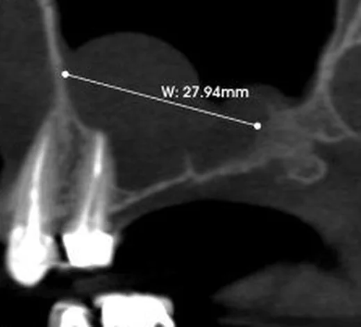
- Maksiller sinüs tabanından veya yan duvarından yükselen, geniş tabanlı kubbe biçimli oluşum.
- Düzgün ve keskin sınırlı kontur; iç yapı çoğunlukla homojen görünür.
- Sinüs duvarına bağlı olarak belirgin kubbe oluşturur; lezyonun kenarı ile sinüs duvarı arasındaki ilişkisi tanıya yön verir.
- Maksiller sinüs hacmi korunur; belirgin ekspansiyon ya da kortikal yıkım tipik değildir.
- Maksiller ostium üzerinden orta meatusa veya koanaya uzanım göstermez; bu özellik onu antrokoanal poliplerden ayırt eder.
- Aynı sinüste ayrı mukozal kalınlaşma veya diş kaynaklı odak eşlik edebilir ve ayrı tarif edilmelidir.
Normalde
Normal maksiller sinüs lümeni havalıdır; izolasyonlu mukozal kalınlaşmalar genellikle semptomla ilişkili değildir ve maksiller sinüste 4 mm'den büyükse raporlanması önerilir. Retansiyon kistlerinin karakteristik duvara bağlı konumu ve keskin sınırları, diğer yaygın maksiller patolojilerle karşılaştırılarak doğrulanır.
Normal BT ile kıyasla
Aynı bölgenin, kaynak veri setinde patoloji içermediği belirtilen bir BT incelemesini kesit kesit kaydırın; organ sınırlarını açarak anatomiyi hasta görüntüsüyle karşılaştırın. Küçük rastlantısal bulgular tümüyle dışlanmamıştır.
Kaynak: TotalSegmentator v2.01 veri seti (Wasserthal ve ark., Radiology: Artificial Intelligence 2023), CC BY 4.0, vaka s0849; organ sınırları veri setinin otomatik segmentasyonundan, birleştirilerek sadeleştirildi. Görüntüler 2,5 mm kesit aralığına yeniden örneklendi.
Ayırıcı tanı
- Maksiller sinüste izole mukozal kalınlaşmalar genellikle klinik belirtilerle ilişkili değildir ve 4 mm'yi aşması durumunda rapor edilmesi önerilir.
- Antrokoanal polip
- maksiller ostiumdan orta meatusa ve posterior koanaya uzanan devamlı yumuşak doku vardır.
- Odontojenik kist
- diş kökü veya gömülü diş çevresinde merkezlenebilir, alveoler kemiği genişletebilir ve sinüse komşu defekt yapabilir.
- Maksiller sinüs mukoseli
- sinüs hacmini genişleten yaygın opasifikasyon ve basınç remodelingi beklenir.
- Kronik enfalmatuvar süreçler, ostiomeatal kompleks veya fundibüler obstrüksiyonlara bağlı olarak sinüslerde mukozal kalınlaşma veya opasifikasyon oluşturur.
Tuzaklar
- Kubbe biçimi tek başına histolojik olarak gerçek kisti kanıtlamaz; BT'de benzer görünebilen mukozal psödokistler de aynı biçimde tarif edilir.
- Diş kökü çevresindeki periapikal lezyon sinüs tabanına komşuysa kubbe görünümünü otomatik olarak retansiyon kisti sayma; alveoler kesitleri ve kök apeksini izle.
- Büyük görünmesi tek başına agresiflik anlamına gelmez; ekspansiyon, kemik kesintisi veya ostiuma uzanım olup olmadığını ayrı değerlendir.
Kendini dene
Sinüs duvarına bağlı, belirgin kenarları olan ve konveks şekilli bir oluşum izleniyor; hasta asemptomatik. En olası radyolojik bulgu nedir? En olası radyolojik bulgu nedir?
Cevabı göster
Mukozal retansiyon kisti/psödokisti. Duvara bağlı düzgün kubbe ve korunmuş sinüs hacmi retansiyon kisti görünümündedir. Mukozal kalınlaşma ve tutulmuş sıvılar sinüsü opasifiye eder; antrokoanal polip maksiller sinüsten burun boşluğuna ve koanaya doğru uzanır; odontojenik sinüzit ise izolasyonlu maksiller tutulum ve periapikal inflamatuvar lezyonlarla karakterizedir.
Maksiller sinüsten burun boşluğuna ve ileriye doğru koanaya uzanan izole bir yumuşak doku lezyonu izleniyor. Hangi tanı öne çıkar? Hangi tanı öne çıkar?
Cevabı göster
Antrokoanal polip. Maksiller sinüsten burun boşluğuna ve koanaya uzanan büyüme deseni, izole polipoid yapıyla birlikte antrokoanal polip tanısını destekler. Retansiyon kisti sinüs duvarına bağlı konveks bir yapıdadır; fungal top unilateral maksiller sinüste hiperdensite ile seyrederken, mukosel sinüs hacmini genişletir ve kemik yeniden modellemeye yol açar.
İlgili konular
Kaynaklar
Bu sayfadaki 37 cümle ve 2 quiz sorusu aşağıdaki kaynaklardan alıntılarla tek tek karşılaştırıldı; 11 cümle bu karşılaştırmada düzeltildi (2026-10-08).
- Flying through congested airspaces: imaging of chronic rhinosinusitispmc.ncbi.nlm.nih.gov
- The effect of anatomic variations and maxillary sinus volume in antrochoanal polyp formation.pubmed.ncbi.nlm.nih.gov
- Perforating internal root resorption repaired with mineral trioxide aggregate caused complete resolution of odontogenic sinus mucositis: a case report.pubmed.ncbi.nlm.nih.gov
- Correlation of quantitative MR imaging findings with symptoms in patients with incidentally detected inflammatory sinonasal disease.pubmed.ncbi.nlm.nih.gov
- Painful Trigeminal Neuropathy Attributed to a Space-occupying Lesion Presenting as a Toothache: A Report of 4 Cases.pubmed.ncbi.nlm.nih.gov
- Symptomatic Maxillary Sinus Retention Cyst Following a Prior Sinus Perforation: A Case Report.pubmed.ncbi.nlm.nih.gov
- The anatomical variations of paranasal sinuses may be related to the formation of antrochoanal polyp by computed tomography imaging study.pubmed.ncbi.nlm.nih.gov
- Unilateral sinonasal symptoms.pubmed.ncbi.nlm.nih.gov
- Investigation of the accessory maxillary ostium: a congenital variation or acquired defect?pubmed.ncbi.nlm.nih.gov
- Anatomic variations associated with antrochoanal polyps.pubmed.ncbi.nlm.nih.gov
- [Rehabilitation by hollow obturator prosthesis immediately after total maxillectomy for malignant tumor].pubmed.ncbi.nlm.nih.gov
- The contribution of computed tomography to nasal septoplasty.pubmed.ncbi.nlm.nih.gov
- [Retention cysts: a new approach to treatment].pubmed.ncbi.nlm.nih.gov
- Paranasal sinus disease could be a reason for unsuccessful immunotherapy of inhalant allergy in children.pubmed.ncbi.nlm.nih.gov
- Fabrication of a 3D Printing Definitive Obturator Prosthesis: a Clinical Report.pubmed.ncbi.nlm.nih.gov
- International Harmonization of Nomenclature and Diagnostic Criteria (INHAND): Nonproliferative and Proliferative Lesions of the Rabbit.pubmed.ncbi.nlm.nih.gov
- Sinus Lift With Simultaneous Mucous Retention Cyst Drainage for Implant Rehabilitation: Clinical, Tomographic, and Histologic Assessments in a Six-Year Follow-Up Case Report.pubmed.ncbi.nlm.nih.gov
- Automated detection and classification of maxillary sinus variations using slice-based and full-volume CBCT deep learning models.pubmed.ncbi.nlm.nih.gov
- A Retrospective Study of the Prevalence of Maxillary Sinus Cysts Incidentally Detected on MRI Among Non-Symptomatic Caucasian Population.pubmed.ncbi.nlm.nih.gov
- Anatomical and Pathological Assessment of the Maxillary Sinus Using CBCT Imaging: A Retrospective Descriptive Study.pubmed.ncbi.nlm.nih.gov
- A de novo start-loss variant in the NIPBL gene causing mild type 1 Cornelia de Lange syndrome in an Iranian family: A Case Report.pubmed.ncbi.nlm.nih.gov
- Endoscopic management of uncommon benign maxillary sinus lesions: a case series.pubmed.ncbi.nlm.nih.gov
- Chronic maxillary sinusitis masquerading as a maxillary sinus neoplasm: A case report.pubmed.ncbi.nlm.nih.gov
- Ultrasound of the Cheek: Examination Technique, Anatomy, and Abnormal Findings.pubmed.ncbi.nlm.nih.gov
- Facial pain as a presenting symptom of a left maxillary sinus cyst: Case report and literature review.pubmed.ncbi.nlm.nih.gov
- Magnetic resonance imaging of the orbit, Part 2: Characterization of orbital pathologies.pubmed.ncbi.nlm.nih.gov
- Value of MRI signal intensity in evaluation of allergic fungal rhinosinusitis compared with CT Hounsfield units: Retrospective study.pubmed.ncbi.nlm.nih.gov
- Impact of small antral pseudocyst interventions on bone remodeling during sinus floor elevation: a retrospective analysis.pubmed.ncbi.nlm.nih.gov
- Simplified Vertical Ridge Augmentation in Severely Resorbed Alveolar Ridges Using a Novel Wide-Head Tenting Pole Screw: Clinical and Histomorphometric Analysis-A Case Series.pubmed.ncbi.nlm.nih.gov
- Radiologic overview of sinonasal lesions.pubmed.ncbi.nlm.nih.gov
- Review of imaging modalities and radiological findings of calvarial lesions.pubmed.ncbi.nlm.nih.gov
- Multidisciplinary approach and clinical management of McCune-Albright syndrome.pubmed.ncbi.nlm.nih.gov
- Spontaneous Unilateral Intrasphenoidal Meningocele.pubmed.ncbi.nlm.nih.gov
- Rare Diseases of the Nose, the Paranasal Sinuses, and the Anterior Skull Base.pubmed.ncbi.nlm.nih.gov
- Clinical presentation of leptospirosis: a retrospective study of 34 patients admitted to a single institution in metropolitan France.pubmed.ncbi.nlm.nih.gov
- Different Approaches to the Treatment of Radicular and Related Cysts Associated with Nasal Mucosa in the Maxilla: A Case Series.pubmed.ncbi.nlm.nih.gov
- Expert consensus on the management of third molar health.pubmed.ncbi.nlm.nih.gov
- Combined Soft and Hard Tissue Facial Lesions In Early Childhood Pioneering the Diagnosis of the Tumor Predisposition Syndrome Neurofibromatosis Type 1.pubmed.ncbi.nlm.nih.gov
- Accessory Maxillary Sinus Ostium Frequency and Correlation with Anatomical Variables and Sinus Mucosal Status: A CBCT Study.pubmed.ncbi.nlm.nih.gov
- Collaboration between otolaryngologists and oral surgeons in maxillary sinus elevation planning.pubmed.ncbi.nlm.nih.gov
- Rhinosinusitis: Evidence and experience - 2024.pubmed.ncbi.nlm.nih.gov
- Radiologic Evaluation of Odontogenic Sinusitis and Its Etiologic Factors: Lessons Learned from a Retrospective Study with a Proposed Imaging-Guided Management Pathway.pubmed.ncbi.nlm.nih.gov
- The maxillary sinus: physiology, development and imaging anatomy.pubmed.ncbi.nlm.nih.gov
- Effect of Intracapsular Pressure on Pulp Sensitivity in Teeth Affected by Jaw Cysts: A Clinical Study Combined With Finite Element Analysis.pubmed.ncbi.nlm.nih.gov
- Deep learning detection and classification of fungal and non-fungal calcifications on paranasal sinus CT imaging.pubmed.ncbi.nlm.nih.gov
- Maxillary sinus pathologies in dental implant candidates: CBCT-based prevalence and odontogenic risk factors.pubmed.ncbi.nlm.nih.gov
- Giant Radicular Cyst of the Pediatric Anterior Maxilla: A Rare Case.pubmed.ncbi.nlm.nih.gov
- Alveolar Recess of the Maxillary Sinus Mimicking a Radicular Cyst: A Diagnostic Challenge and Systematic Literature Search.pubmed.ncbi.nlm.nih.gov
- Surgical Management of a Large Dentigerous Cyst Associated With an Impacted Supernumerary Tooth: A Rare Case Report.pubmed.ncbi.nlm.nih.gov
- Persistent presumed allergic fungal rhinosinusitis during tezepelumab therapy with improvement after revision surgery: a case report.pubmed.ncbi.nlm.nih.gov
- Small ethmoid mucocele with intracranial extension associated with fibrous dysplasia: A rare case report.pubmed.ncbi.nlm.nih.gov
- Orbital Extension of Sinonasal Mucocele Presenting as Proptosis: A Report of Two Cases.pubmed.ncbi.nlm.nih.gov
- Accuracy of Signs and Symptoms for the Diagnosis of Acute Rhinosinusitis and Acute Bacterial Rhinosinusitis.pubmed.ncbi.nlm.nih.gov
- Masses of developmental and genetic origin affecting the paediatric craniofacial skeleton.pubmed.ncbi.nlm.nih.gov
- International Society of Paediatric Surgical Oncology (IPSO) Surgical Practice Guidelines.pubmed.ncbi.nlm.nih.gov
- Unusual Manifestations of Sinonasal Osteomas: A Narrative Review.pubmed.ncbi.nlm.nih.gov
- "Dry Tap" Fine-Needle Aspiration Biopsy as a Diagnostic Clue in Cyst-like Juvenile Jaw Lesions Mimicking Dentigerous Cysts on Panoramic Radiography and Cone-Beam Computed Tomography.pubmed.ncbi.nlm.nih.gov
- Imaging Evaluation for Jaw Deformities: Diagnostic Workup and Pre-Treatment Imaging Checklist for Orthognathic Surgery.pubmed.ncbi.nlm.nih.gov
- Clinical utility of dental cone-beam computed tomography: current perspectives.pubmed.ncbi.nlm.nih.gov
- Coexistence of Autoantibodies against the Golgi Complex and Ro52 Antigen in a Patient with Nonspecific Interstitial Pneumonia.pubmed.ncbi.nlm.nih.gov
- Ostiomeatal complex inflammation with a rare ethmoid sinolith utilizing cone-beam computed tomography: A clinical and radiological approach to diagnosis.pubmed.ncbi.nlm.nih.gov
- The Frontal Sinus and Frontal Recess: Anatomical, Radiological and Surgical Concepts.pubmed.ncbi.nlm.nih.gov
- Maxillary Sinus Mucocele following Zygomaticomaxillary Complex Fracture Repair.pubmed.ncbi.nlm.nih.gov
- Antrochoanal Polyps: Nasal Anatomical and Pathological Variants as Risk Factors.pubmed.ncbi.nlm.nih.gov
- Evaluations of maxillary sinus pathologies using CBCT imaging.pubmed.ncbi.nlm.nih.gov
- Spontaneous bone formation after sinus graft infection control: a case report and review of the literature.pubmed.ncbi.nlm.nih.gov
- Detection of maxillary sinusitis of endodontic origin in cone-beam CT images using deep learning algorithms.pubmed.ncbi.nlm.nih.gov
- Anatomical Proximity of Posterior Maxillary Tooth Roots as an Independent Predictor of Maxillary Sinus Mucosal Thickening: A CBCT-Based Study.pubmed.ncbi.nlm.nih.gov
- Surgical management of massive calcium silicate-based sealer extrusion into the maxillary sinus with palatal necrosis in an adolescent.pubmed.ncbi.nlm.nih.gov
- Retrospective analysis of pathological changes in the maxillary sinus with CBCT.pubmed.ncbi.nlm.nih.gov
- Automatic maxillary sinus segmentation and pathology classification on cone-beam computed tomographic images using deep learning.pubmed.ncbi.nlm.nih.gov
- Oral Rehabilitation of Severely Atrophic Alveolar Ridge Using Sinus Augmentation and Ridge Split Technique: A Case Report.pubmed.ncbi.nlm.nih.gov
- CBCT-guided Caldwell-Luc management of a dentigerous cyst associated with an ectopic maxillary third molar in the maxillary sinus: a case report.pubmed.ncbi.nlm.nih.gov
- When plasmacytoma defies expectations: An unusual maxillary presentation with mixed imaging features in a young patient.pubmed.ncbi.nlm.nih.gov
Eğitim amaçlıdır; hasta başında tanı ve tedavi kararının yerine geçmez. İçerik yapay zekâ destekli bir süreçle güncel literatüre göre cümle cümle kaynaklarla karşılaştırıldı, ancak uzman radyolog incelemesinden geçmedi. Klinik kararlarda güncel kılavuzları, kurum protokollerini ve radyoloji raporunu esas alın.