Özet
Sinonazal osteosarkom, maksilla veya paranazal sinüs kemiklerinden gelişen ve lümene ya da komşu yumuşak dokulara büyüyebilen nadir malign kemik tümörüdür. Kontrastsız ince kesit BT tümör matriks mineralizasyonunu, başlangıç kemiğini ve kortikal yıkımı gösterir; kontrastlı BT yumuşak doku uzanımını göstermeye yardımcı olur. Osteoid matriks görülmesi olasılığı artırır ancak histolojik tanı yerine geçmez. Orbita, kafa tabanı veya infratemporal uzanımın gözden kaçması cerrahi sınır ve lokal yayılım değerlendirmesini eksik bırakır.
Faz ve pencere
- Kontrastsız tanısal
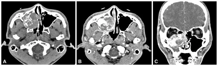
- Kemik algoritmasında kemik kaynaklı kitle içinde düzensiz bulutumsu osteoid mineralizasyon, kortikal destrüksiyon ve ekstraosseöz yumuşak doku komponenti izlenebilir; kontrast zorunlu değildir.
Önerilen pencereler: Kemik (G 1800 / M 400), Yumuşak doku (G 400 / M 40).
BT bulguları
- Maksilla, etmoid veya başka sinüs duvarında merkezlenen agresif kemik kaynaklı kitle.
- Kitle içinde bulutumsu, amorf veya yoğun osteoid matriks mineralizasyonu.
- Başlangıç kemiğinde kortikal kesinti ve sinüs/nazal kaviteye uzanan yumuşak doku komponenti.
- Sinüs duvarının dışına orbita, damak veya infratemporal fossaya ilerleme.
- Eşlik eden lamina papyracea ya da kafa tabanı destrüksiyonu.
- Lezyon çevresinde reaktif kemik üretimi veya düzensiz periostal yeni kemik oluşumu.
- Matriks mineralizasyonu olmayan bölümlerde kıkırdak veya yumuşak doku yoğunluğunda tümör.
Normalde
Normal sinüs duvarı ince kortike kemik hattıdır ve lümen havası homojendir; kemik içinde matriks opasitesi veya kortikal kesinti bulunmaz. Karşı yüz kemikleriyle kıyasla lezyonun hangi kemikten çıktığını ve mineralize kısmın sinüs içindeki yumuşak dokuya devam edip etmediğini belirle.
Normal BT ile kıyasla
Aynı bölgenin, kaynak veri setinde patoloji içermediği belirtilen bir BT incelemesini kesit kesit kaydırın; organ sınırlarını açarak anatomiyi hasta görüntüsüyle karşılaştırın. Küçük rastlantısal bulgular tümüyle dışlanmamıştır.
Kaynak: TotalSegmentator v2.01 veri seti (Wasserthal ve ark., Radiology: Artificial Intelligence 2023), CC BY 4.0, vaka s0849; organ sınırları veri setinin otomatik segmentasyonundan, birleştirilerek sadeleştirildi. Görüntüler 2,5 mm kesit aralığına yeniden örneklendi.
Ayırıcı tanı
- Osteom
- yoğun, düzenli kemik dansiteli ve iyi sınırlı çıkıntı yapar; agresif kortikal yıkım beklenmez.
- Kondrosarkom
- kıkırdak matriksinde halka-yay biçimli kalsifikasyonlar görülebilir.
- Fibro-osseöz lezyon
- çevre kemikle harmanlanan matürasyon paterni ve daha düzenli sınırlar olabilir.
- Ossifiye fibrom
- iyi sınırlı ekspansil lezyon ve çevresel kortike kenar yapabilir.
- Kalsifiye fungal içerik
- sinüs lümeninde mineral yoğunluğu vardır ancak kemik kökeni ve ekstraosseöz kitle bağlantısı yoktur.
Tuzaklar
- Sinüs içi kalsifikasyon otomatik osteoid matriks değildir; mineralizasyonun komşu kemik yıkımıyla ve yumuşak doku kitlesiyle ilişkisini izle.
- Kondrosarkom da mineralize matriks yapabilir; kalsifikasyon biçimi tek başına histolojik alt tipi kesinleştirmez.
- Diş materyali veya antrolit maksiller sinüs içinde yoğun görünür; başlangıç kemiğiyle süreklilik olup olmadığını koronal ve sagittal planda doğrula.
Kendini dene
Etmoid kemikten kaynaklanan kitle içinde amorf yoğun matriks var; korteks yıkılmış ve sinüse yumuşak doku uzanmış. En olası tanı?
Cevabı göster
Osteosarkom. Kemik merkezli agresif lezyonda osteoid benzeri mineralizasyon ve ekstraosseöz komponent osteosarkom olasılığını destekler; kesin tanı patolojiyle konur.
Sinüs duvarına bağlı, düzenli yoğun kemik çıkıntısı var; kortikal yıkım veya yumuşak doku kitlesi yok. En olası lezyon?
Cevabı göster
Osteom. Düzenli ve yoğun kemik dansitesindeki duvar kökenli çıkıntı osteomdur; osteosarkom daha agresif yıkım ve yumuşak doku uzanımı gösterir.
Kaynaklar
Bu sayfadaki 33 cümle ve 2 quiz sorusu aşağıdaki kaynaklardan alıntılarla tek tek karşılaştırıldı (2026-10-08).
- Sinonasal osteosarcoma of the ethmoid bone with decade-later recurrence presenting as early orbital apex syndrome: an 18-year case reportpmc.ncbi.nlm.nih.gov
- A Huge Intraosseous Hemangioma in the Maxillary Sinus Misdiagnosed as Osteosarcoma: A Case Reportpmc.ncbi.nlm.nih.gov
- Intranasal ectopic tooth: An unusual diagnostic finding with review of literaturepmc.ncbi.nlm.nih.gov
- Unusual Manifestations of Sinonasal Osteomas: A Narrative Reviewpmc.ncbi.nlm.nih.gov
- Clinical Outcomes of Once-Weekly Hypofractionated Intensity-Modulated Radiation Therapy with Concurrent α-Sulfoquinovosyl-Acylpropanediol for Modified Adams Stage 4 Canine Intranasal Tumors: A Retrospective Case Seriespmc.ncbi.nlm.nih.gov
- Sinonasal osteosarcoma of the ethmoid bone with decade-later recurrence presenting as early orbital apex syndrome: an 18-year case report.pubmed.ncbi.nlm.nih.gov
- Chondrosarcomas of the head and neck.pubmed.ncbi.nlm.nih.gov
- Primary sinonasal Ewing sarcoma: A case report.pubmed.ncbi.nlm.nih.gov
- Spatiotemporal stability of Cu-ATSM and FLT positron emission tomography distributions during radiation therapy.pubmed.ncbi.nlm.nih.gov
- Molecular imaging biomarkers of resistance to radiation therapy for spontaneous nasal tumors in canines.pubmed.ncbi.nlm.nih.gov
- Radiation induced malignancy in retinoblastoma: new pathology in a case report.pubmed.ncbi.nlm.nih.gov
- Skull Base Primary Ewing Sarcoma: A Radiological Experience of a Rare Disease in an Atypical Location.pubmed.ncbi.nlm.nih.gov
- Computed tomographic appearance of equine sinonasal neoplasia.pubmed.ncbi.nlm.nih.gov
- Sinonasal Ewing Sarcoma: A Case Report and Literature Review.pubmed.ncbi.nlm.nih.gov
- Characterization of Sinonasal Lesions on CT, MRI, and PET/CT.pubmed.ncbi.nlm.nih.gov
- [Applications of vascularized pericranial flaps in endoscopic skull base surgeries].pubmed.ncbi.nlm.nih.gov
- CT Findings of Feline Nasal Hamartomas Mimicking Malignant Growth.pubmed.ncbi.nlm.nih.gov
- Unique Imaging Findings of a Calvarial Ossifying Fibroma Successfully Managed With Surgery and Definitive Intent Radiotherapy.pubmed.ncbi.nlm.nih.gov
- Chondromyxoid Fibroma: An Updated Review.pubmed.ncbi.nlm.nih.gov
- Unveiling the biochemical signature of tumor-induced osteomalacia: implications for distinguishing it from primary osteoporosis.pubmed.ncbi.nlm.nih.gov
- Sculpting Solutions: A Case Report of Resection and Reconstruction in an Aggressive Mandibular Juvenile Ossifying Fibroma and Review of Literature.pubmed.ncbi.nlm.nih.gov
- Imaging in Rare and Atypical Sinonasal Masses: An Interesting Case Series.pubmed.ncbi.nlm.nih.gov
- The combined compartmental maxillary resection: technique codification and preliminary multicenter series analysis.pubmed.ncbi.nlm.nih.gov
- Midface Degloving Approach for Total Maxillectomy Without Orbital Exenteration: A Case Series.pubmed.ncbi.nlm.nih.gov
- Imaging of Skull Base Tumors.pubmed.ncbi.nlm.nih.gov
- Imaging appearance of bone tumors of the maxillofacial region.pubmed.ncbi.nlm.nih.gov
- Endoscopic Endonasal Resection of Fibro-Osseous Lesions Involving the Nasal Turbinates: A Report of Two Cases and a Literature Review.pubmed.ncbi.nlm.nih.gov
- Chinese expert consensus on imaging examination and diagnosis of nasal cavity and paranasal sinus tumors.pubmed.ncbi.nlm.nih.gov
- Sinonasal Phosphaturic Mesenchymal Tumor: A Case Report.pubmed.ncbi.nlm.nih.gov
- Orbital Exenteration in Maxillary Osteosarcoma: A Case Report.pubmed.ncbi.nlm.nih.gov
- Patterns of lymph node metastasis in 441 patients with sinonasal squamous cell carcinoma.pubmed.ncbi.nlm.nih.gov
- Primary Ewing's Sarcoma of the Sinonasal Region: A Rare Clinical Encounter.pubmed.ncbi.nlm.nih.gov
- Small Round Blue Cell Tumours of the Sinonasal Area: Our 5 year Experience in a Tertiary Care Centre in India.pubmed.ncbi.nlm.nih.gov
- Unusual Manifestations of Sinonasal Osteomas: A Narrative Review.pubmed.ncbi.nlm.nih.gov
- Osteomyelitis of the Jaw Bones and Its Mimics: Resolving the Diagnostic Enigma.pubmed.ncbi.nlm.nih.gov
- Clinical and histological characterization of 19 chondro-osseous respiratory epithelial adenomatoid hamartomas and 2 respiratory epithelial adenomatoid hamartomas in dogs.pubmed.ncbi.nlm.nih.gov
- Malignant Bone-Forming Neoplasm With NIPBL::BEND2 Fusion.pubmed.ncbi.nlm.nih.gov
- A practical overview of CT and MRI features of developmental, inflammatory, and neoplastic lesions of the sphenoid body and clivus.pubmed.ncbi.nlm.nih.gov
- The Role of Computed Tomography in Imaging Non-neurologic Disorders of the Head in Equine Patients.pubmed.ncbi.nlm.nih.gov
- Cross Sectional Imaging of the Ear and Temporal Bone.pubmed.ncbi.nlm.nih.gov
- Human infection with Fusobacterium necrophorum (Necrobacillosis), with a focus on Lemierre's syndrome.pubmed.ncbi.nlm.nih.gov
- Cavernous venous malformation of the frontal sinus: CT and MRI imaging findings.pubmed.ncbi.nlm.nih.gov
- A case of giant fronto-ethmoidal osteoma (45 × 42 mm) with intracranial and orbital extension: a case report.pubmed.ncbi.nlm.nih.gov
- Head and neck manifestations of extramedullary plasmacytomas and their differential diagnoses: a pictorial review.pubmed.ncbi.nlm.nih.gov
- Imaging of the skull base and orbital tumors.pubmed.ncbi.nlm.nih.gov
- An unusual encounter in ophthalmology: Fronto-Ethemoido-Orbital Osteoma with the art of precise referral and management of PEEK implant infection- A rare case report.pubmed.ncbi.nlm.nih.gov
- Navigating Fibrous Dysplasia of the Middle Turbinate: A Decade-Long Journey and Comprehensive Management Strategies.pubmed.ncbi.nlm.nih.gov
- Bone-Preserving Management of Maxillary Cemento-Ossifying Fibroma With Orbital Floor Thinning: A Case Report.pubmed.ncbi.nlm.nih.gov
- A Huge Intraosseous Hemangioma in the Maxillary Sinus Misdiagnosed as Osteosarcoma: A Case Report.pubmed.ncbi.nlm.nih.gov
- Bilateral rhinoliths causing long-standing nasal obstruction: a case report and review of the literature.pubmed.ncbi.nlm.nih.gov
- Uncommon Nasal Mass Presentation: A Radiological Case Series.pubmed.ncbi.nlm.nih.gov
- Ostiomeatal complex inflammation with a rare ethmoid sinolith utilizing cone-beam computed tomography: A clinical and radiological approach to diagnosis.pubmed.ncbi.nlm.nih.gov
- Synovial Sarcoma: Molecular Biology, Pathology, and Therapeutic Strategies.pubmed.ncbi.nlm.nih.gov
- Current topics and management of head and neck sarcomas.pubmed.ncbi.nlm.nih.gov
- Benign and malignant lesions of the mandible and maxilla: imaging findings and systematic approach.pubmed.ncbi.nlm.nih.gov
- Review of imaging modalities and radiological findings of calvarial lesions.pubmed.ncbi.nlm.nih.gov
- Cemento-ossifying Fibroma Of Paranasal Sinus Presenting Acutely As Orbital Cellulitis.pubmed.ncbi.nlm.nih.gov
- Inflammation-driven periostin in ECRS has contrasting effects on tissue structural integrity and osteitis.pubmed.ncbi.nlm.nih.gov
- Giant Cell Tumor of the Central Skull Base.pubmed.ncbi.nlm.nih.gov
- Imaging review of the anterior skull base.pubmed.ncbi.nlm.nih.gov
- Endoscopic Management of Malignant Tumors of Paranasal Sinus and Nasal Cavity: An Institutional Experience.pubmed.ncbi.nlm.nih.gov
- Infra-Temporal and Pterygo-Palatine Fossae Tumors: A Frontier in Endoscopic Endonasal Surgery-Description of the Surgical Anatomy of the Approach and Report of Illustrative Cases.pubmed.ncbi.nlm.nih.gov
- Spontaneous oronasal ejection of neoplastic and non-neoplastic nodules by 21 dogs, 2000-2024.pubmed.ncbi.nlm.nih.gov
- Sinonasal schwannoma masquerading as an IgG4-related nasal mass.pubmed.ncbi.nlm.nih.gov
- Nasal sinus leiomyosarcoma in a patient with history of non-hereditary unilateral treated retinoblastoma.pubmed.ncbi.nlm.nih.gov
Eğitim amaçlıdır; hasta başında tanı ve tedavi kararının yerine geçmez. İçerik yapay zekâ destekli bir süreçle güncel literatüre göre cümle cümle kaynaklarla karşılaştırıldı, ancak uzman radyolog incelemesinden geçmedi. Klinik kararlarda güncel kılavuzları, kurum protokollerini ve radyoloji raporunu esas alın.