Özet
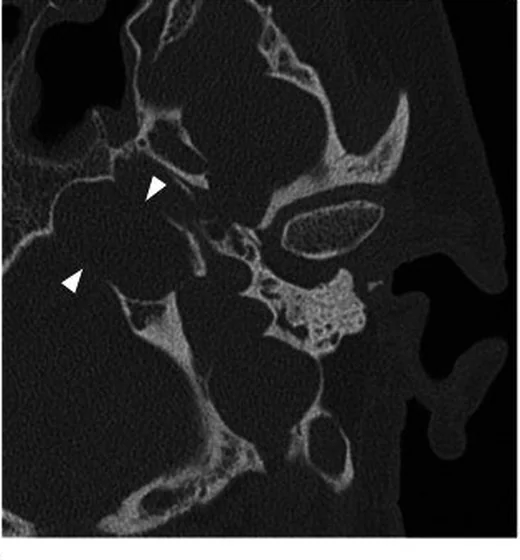
Petroz kemik kolesteatomu, keratin içeren ve petroz kemiğin derin bölümlerine ilerleyerek komşu kemik yapıları aşındırabilen nadir bir lezyondur. İnce kesit kontrastsız temporal kemik BT, kemik yıkımının sınırını ve otik kapsül ile kafa tabanı kanallarına uzanımını gösterir; BT'deki yumuşak doku görünümü tek başına kolesteatom tanısını kanıtlamaz. MRI'da difüzyon kısıtlaması kolesteatomu destekler ve BT'nin sınırlı doku karakterizasyonunu tamamlar. Koklea, fasiyal sinir kanalı veya kafa içi yapı komşuluğundaki erozyonun atlanması, hastalığın yayılımının eksik tanımlanmasına yol açar.
Faz ve pencere
- Kontrastsız tanısal
- Petroz kemik içinde sınırlı yumuşak doku yoğunluklu odak, ekspansiyon ve çevre trabeküllerde erozyon görülebilir. Kontrastlı BT kolesteatom içeriğini özgül olarak göstermez; tanısal belirsizlikte MRI difüzyon sekansları kullanılır.
- Kemik algoritması tanısal
- İnce kesit ve çok düzlemli kemik rekonstrüksiyonları erozyonun petroz apeks, otik kapsül, karotis kanalı ve internal akustik kanal ile temasını ve olası kemik duvarı kaybını ortaya koyar.
Önerilen pencereler: Kemik (G 1800 / M 400), Yumuşak doku (G 400 / M 40).
BT bulguları
- Petroz kemikte eroziv odak — kemik iliği veya havalı hücrelerin yerini alan yumuşak doku yoğunluklu lezyon trabekülleri aşındırır.
- Sınırlı ekspansiyon — lezyon petroz kemiğin hacmini artırabilir; şekil ve sınır çok düzlemli incelenir.
- Kortikal kemik kaybı — lezyonun dış kenarında incelme, kesinti veya yeniden şekillenme bulunabilir.
- Otik kapsül komşuluğu — koklea, vestibül ve semisirküler kanalların kemik duvarlarında incelme ya da açıklık aranır.
- Karotis kanal duvarı ilişkisi — petroz internal karotid arter kanalının çevre korteksinin korunup korunmadığı kaydedilir.
- Sinir çıkış yollarına uzanım — iç akustik kanal ve juguler foramen sınırlarındaki lezyon uzanımı kaydedilir.
- Yumuşak doku karakterizasyonu sınırı — BT erozyon haritasını verir, ancak granülasyon veya tümöral dokudan ayırımı her zaman yapamaz.
Normalde
Petroz kemiğin havalanma derecesi bireysel farklılıklar gösterir; lezyon yokken petroz apeksin kemik bütünlüğü ve komşu iç kulak/kranial yapı sınırları kesintisiz olarak izlenir. Yüksek çözünürlüklü BT, lezyonun petroz apeksteki boyutunu, çevre kemik bütünlüğünü ve koklea, vestibül, karotis kanalı veya iç akustik kanal gibi komşu yapılarla ilişkisini belirlemek için tanısal standarttır.
Normal BT ile kıyasla
Aynı bölgenin, kaynak veri setinde patoloji içermediği belirtilen bir BT incelemesini kesit kesit kaydırın; organ sınırlarını açarak anatomiyi hasta görüntüsüyle karşılaştırın. Küçük rastlantısal bulgular tümüyle dışlanmamıştır.
Kaynak: TotalSegmentator v2.01 veri seti (Wasserthal ve ark., Radiology: Artificial Intelligence 2023), CC BY 4.0, vaka s0103; organ sınırları veri setinin otomatik segmentasyonundan, birleştirilerek sadeleştirildi. Görüntüler 2,5 mm kesit aralığına yeniden örneklendi.
Ayırıcı tanı
- Kolesterol granülomu
- ekspansil ve trabekülleri silen görünüm benzer olabilir; MRI'da T1/T2 sinyali genellikle yüksek, difüzyon kısıtlaması beklenmez.
- Petroz apeks efüzyonu, petroz kemik lezyonlarının ayırıcı tanısında sık karşılaşılan nedenlerdendir.
- Petroz apisit
- enfeksiyon bağlamında apikal kemik sınırında düzensiz değişiklik ve komşu otomastoid inflamasyon görülebilir.
- Jugulotimpanik paragangliom
- juguler foramen merkezli kemik erozyonu ve yoğun kontrastlanan yumuşak doku kitlesi ile ayrılır.
- Petroz kemik metastazı
- kemik iliği infiltrasyonu ve destrüksiyon yapabilir; primer malignite ve diğer kemik odakları tanısal bağlam sağlar.
Tuzaklar
- BT'deki her petroz kemik yumuşak doku odağını kolesteatom olarak adlandırmayın; BT kemik sınırlarını iyi gösterirken doku karakterizasyonunda sınırlıdır.
- Ekspansiyon görünümünü tek başına kolesterol granülomundan ayırıcı kabul etmeyin; erozyon haritasını MRI sinyali ve difüzyon bulgusuyla birlikte yorumlayın.
- Lezyonun en geniş aksiyel kesitiyle yetinmeyin; koronal ve sagittal kemik görüntülerde koklea, kanal duvarları ve kafa içi tabanla temasını takip edin.
Kendini dene
Petroz apeks odağı kemik erozyonu yapıyor ve MRI'da difüzyon kısıtlıyor. En olası tanı hangisidir?
Cevabı göster
Kolesteatom. Keratin içeren kolesteatomda difüzyon kısıtlaması beklenir ve petroz kemikte erozyon eşlik edebilir. Kolesterol granülomunda tipik T1/T2 sinyali yüksektir; efüzyonda kemik trabekülleri korunur; apisit inflamatuvar klinik ve kemik değişiklikleriyle değerlendirilir.
Petroz kemikte yumuşak doku odağı görülüyor; BT'de erozyon var ancak içerik ayırt edilemiyor. En yararlı ek MR bulgusu nedir?
Cevabı göster
Difüzyon kısıtlaması. Difüzyon kısıtlaması kolesteatomun keratin içeriğini destekler ve BT'nin sınırlı özgüllüğünü tamamlar. Akım boşluğu vasküler lezyonları düşündürür; yağ baskılanması ve kemik iliği ödemi tek başına kolesteatom tanısı koydurmaz.
Kaynaklar
Bu sayfadaki 37 cümle ve 2 quiz sorusu aşağıdaki kaynaklardan alıntılarla tek tek karşılaştırıldı; 6 cümle bu karşılaştırmada düzeltildi (2026-10-08).
- Beyond the otoscope: an imaging review of congenital cholesteatomapmc.ncbi.nlm.nih.gov
- A Case of Acquired Pars Flaccida Cholesteatoma Extending to the Supralabyrinthine Petrous Apex With Preservation of the Otic Capsulepmc.ncbi.nlm.nih.gov
- Extensive Temporal Bone Necrosis in Petrous Bone Cholesteatoma Complicated by Actinomycosis: A Case Reportpmc.ncbi.nlm.nih.gov
- Combined microscope–endoscopy resection of petrous bone cholesteatoma with temporary facial nerve transposition versus nontranspositionpmc.ncbi.nlm.nih.gov
- Quantitative Analysis of Cochlear-Carotid Recesses Based on HRCT in Endoscope-Assisted Management of Massive Petrous Bone Cholesteatomapmc.ncbi.nlm.nih.gov
- Beyond the otoscope: an imaging review of congenital cholesteatoma.pubmed.ncbi.nlm.nih.gov
- Management of Cholesteatoma: Extension Beyond Middle Ear/Mastoid.pubmed.ncbi.nlm.nih.gov
- Case analysis of temporal bone lesions with facial paralysis as main manifestation and literature review.pubmed.ncbi.nlm.nih.gov
- Tumors of the temporal bone.pubmed.ncbi.nlm.nih.gov
- An analysis of petrous bone cholesteatomas treated with translabyrinthine transotic petrosectomy.pubmed.ncbi.nlm.nih.gov
- Petrous Bone Cholesteatoma: Facial and Hearing Preservation.pubmed.ncbi.nlm.nih.gov
- Endoscope-Assisted Surgery for Petrous Bone Cholesteatoma with Hearing Preservation.pubmed.ncbi.nlm.nih.gov
- Management of petrous bone cholesteatoma: open versus obliterative techniques.pubmed.ncbi.nlm.nih.gov
- Exteriorization of Petrous Bone Cholesteatoma by Endonasal Endoscopic Approach: A Case Report.pubmed.ncbi.nlm.nih.gov
- Petrous bone cholesteatoma removal with hearing preservation.pubmed.ncbi.nlm.nih.gov
- [Analysis of revision surgery for petrous bone cholesteatoma].pubmed.ncbi.nlm.nih.gov
- Petrous bone cholesteatoma: our experience of 51 patients with emphasis on cochlea preservation and use of endoscope.pubmed.ncbi.nlm.nih.gov
- Petrous Bone Cholesteatoma- A Comprehensive Management Algorithm and Outcomes.pubmed.ncbi.nlm.nih.gov
- Congenital Mastoid Cholesteatoma With Giant Intracranial Extension: A Case Report.pubmed.ncbi.nlm.nih.gov
- Endoscopic Marsupialization With Periodic Debridement for Petrous Apex Cholesteatoma.pubmed.ncbi.nlm.nih.gov
- Cerebellar abscess complicating chronic suppurative otitis media: An unusual route of intracranial spread.pubmed.ncbi.nlm.nih.gov
- The preoperative temporal bone CT: a mnemonic for reducing surgical risk.pubmed.ncbi.nlm.nih.gov
- Is CT Still the Gold Standard in Semicircular Canal Dehiscence? Diagnostic Value of MRI in Poschl and Stenver Planes.pubmed.ncbi.nlm.nih.gov
- "Leave me alone": anatomical structures and variations seen on computed tomography of the temporal bone.pubmed.ncbi.nlm.nih.gov
- Diagnosis and Management of Middle Ear Neuroendocrine Tumour (MeNET).pubmed.ncbi.nlm.nih.gov
- Primary Intracranial Cholesteatoma Presenting as a Cerebellar Mass Without Otologic Symptoms.pubmed.ncbi.nlm.nih.gov
- Role of the Ear in Meningitis: A Narrative Review of Neuroimaging.pubmed.ncbi.nlm.nih.gov
- Extensive Temporal Bone Necrosis in Petrous Bone Cholesteatoma Complicated by Actinomycosis: A Case Report.pubmed.ncbi.nlm.nih.gov
- A Case of Acquired Pars Flaccida Cholesteatoma Extending to the Supralabyrinthine Petrous Apex With Preservation of the Otic Capsule.pubmed.ncbi.nlm.nih.gov
- Endoscopic assisted minimally invasive resection of petrous bone epidermoid cyst via a presigmoid burr hole approach: illustrative case.pubmed.ncbi.nlm.nih.gov
- Imaging of Skull Base Tumors.pubmed.ncbi.nlm.nih.gov
- Radiological review of skull lesions.pubmed.ncbi.nlm.nih.gov
- A compartment-based approach for the imaging evaluation of tinnitus.pubmed.ncbi.nlm.nih.gov
- Temporal Bone Fractures on High-Resolution CT: Bridging Radiologic Detail with Otologic Anatomy and Surgical Implications.pubmed.ncbi.nlm.nih.gov
- Multidisciplinary Exploration of Computed-Tomographic and Ancient-DNA Data of an Iron Age Skull From Latvia With Multiple Lytic Bone Lesions: Differential Diagnosis Between Metastatic Carcinoma, Multiple Myeloma and Skeletal Tuberculosis.pubmed.ncbi.nlm.nih.gov
- Patient cases with malignant otitis externa at the university clinic of Ludwig Maximilians University Munich from 2009 until 2020.pubmed.ncbi.nlm.nih.gov
- Translabyrinthine Resection of Posterior Petrous Face Meningiomas: Surgical, Functional, and Oncological Outcomes and Associations with Anatomical Extension and Tumor Burden.pubmed.ncbi.nlm.nih.gov
- Radioanatomical assessment of the subtympanic sinus and its relationship to facial nerve position and temporal bone pneumatization in adults: CT study.pubmed.ncbi.nlm.nih.gov
- Structural Phenotype of High-Frequency Recurrent Facial Palsy.pubmed.ncbi.nlm.nih.gov
- Tympanic Membrane Involvement of Multiple Myeloma: Report of a Case.pubmed.ncbi.nlm.nih.gov
- Surgical Techniques and Materials Used in the Treatment of Complicated Otomastoiditis: A Systematic Review.pubmed.ncbi.nlm.nih.gov
- Third Window Syndrome: An Up-to-Date Systematic Review of Causes, Diagnosis, and Treatment.pubmed.ncbi.nlm.nih.gov
- A Case Study and Concise Literature Review: Adult Patient's Initial Manifestation of Complicated Acute Otitis Media Presenting as Jugular Foramen Syndrome.pubmed.ncbi.nlm.nih.gov
- Magnetic Resonance Imaging in Otosyphilis: A Rare Manifestation of Neurosyphilis.pubmed.ncbi.nlm.nih.gov
- Non-invasive imaging modalities for diagnosing pulsatile tinnitus: a comprehensive review and recommended imaging algorithm.pubmed.ncbi.nlm.nih.gov
- Cross Sectional Imaging of the Ear and Temporal Bone.pubmed.ncbi.nlm.nih.gov
- Skull Base Metastases: A Pictorial Review.pubmed.ncbi.nlm.nih.gov
- Imaging for Pediatric Head and Neck Emergencies: Critical Insights.pubmed.ncbi.nlm.nih.gov
- Comprehensive Review of Inner Ear Anatomy on Photon-Counting CT.pubmed.ncbi.nlm.nih.gov
- Pre- and post-operative imaging of cochlear implants: a pictorial review.pubmed.ncbi.nlm.nih.gov
- Surgery of the ear and the lateral skull base: pitfalls and complications.pubmed.ncbi.nlm.nih.gov
- Cholesterol granuloma of the sphenoid wing with long-term growth dynamics: illustrative case.pubmed.ncbi.nlm.nih.gov
- Frontal Cholesterol Granuloma in a Pediatric Patient: A Case Report.pubmed.ncbi.nlm.nih.gov
- Nocardia Otomastoiditis in a Chemotherapy-Induced Neutropenic Patient With Diffuse Large B-cell Lymphoma: Successful Medical Management.pubmed.ncbi.nlm.nih.gov
- Emerging and re-emerging infectious disease in otorhinolaryngology.pubmed.ncbi.nlm.nih.gov
- An Atypical Pediatric Headache: Outpatient Presentation of Petrous Apicitis With Cerebral Venous Sinus Thrombosis.pubmed.ncbi.nlm.nih.gov
- Raeder's Syndrome: An Uncommon Cause of Partial Horner Syndrome Secondary to Petromastoid Inflammation.pubmed.ncbi.nlm.nih.gov
- Pulsatile Tinnitus: A Comprehensive Clinical Approach to Diagnosis and Management.pubmed.ncbi.nlm.nih.gov
- Solitary Plasmacytoma of the Jugular Foramen: A Case Report and Review of the Differential Diagnosis.pubmed.ncbi.nlm.nih.gov
- Case report: endolymphatic sac tumor masquerading as Meniere's disease.pubmed.ncbi.nlm.nih.gov
- Giant Cell Tumor of the Cranial Vault: A Rare Frontotemporal Case With Complete Neurological Recovery and Narrative Review.pubmed.ncbi.nlm.nih.gov
- Utility and Challenges of Imaging in Peripheral Vestibular Disorder Diagnosis: A Narrative Review.pubmed.ncbi.nlm.nih.gov
- Key pre-operative CT parameters for cochlear implantation: a focused radiologic review with surgical relevance.pubmed.ncbi.nlm.nih.gov
- A Diagnostic Enigma: A Rare Case of Cholesterol Granuloma of Mastoid Masquerading as Acute Mastoiditis in a Young Female.pubmed.ncbi.nlm.nih.gov
- Anterolateral, lateral, and posterior corridors to complex skull base lesions in sphenocavernous and petroclival regions: microsurgical anatomy with three-dimensional reconstructions and illustrative cases.pubmed.ncbi.nlm.nih.gov
- Intraosseous vascular malformation of the skull: a case report and literature review.pubmed.ncbi.nlm.nih.gov
- Anatomical Step-by-Step Dissection of Complex Skull Base Approaches for Trainees: Surgical Anatomy of the Translabyrinthine and Transcochlear Approaches.pubmed.ncbi.nlm.nih.gov
- Comprehensive Review of External and Middle Ear Anatomy on Photon-Counting CT.pubmed.ncbi.nlm.nih.gov
- Lateral transorbital neuroendoscopic approach using the superior eyelid incision: A straightforward, comprehensive, and illustrative step-by-step review.pubmed.ncbi.nlm.nih.gov
- Operative Corridors in Endoscopic Skull Base Tumor Surgery.pubmed.ncbi.nlm.nih.gov
- Pediatric Cholesteatoma Presenting as Persistent Otorrhea: A Case of Delayed Diagnosis Across Multiple ENT Specialists.pubmed.ncbi.nlm.nih.gov
- A Case of Petrous Apex Cholesteatoma Removed Using an Exoscope and an Endoscope Without Posterior Rerouting of the Facial Nerve.pubmed.ncbi.nlm.nih.gov
- Pre-treatment and post-treatment nasopharyngeal carcinoma imaging: imaging updates, pearls and pitfalls.pubmed.ncbi.nlm.nih.gov
- The cavernous sinus: An anatomic study with clinical implication.pubmed.ncbi.nlm.nih.gov
- Gradenigo's syndrome presenting as IX and X cranial nerve palsy without clinically apparent ear infection: A case report and review of literature.pubmed.ncbi.nlm.nih.gov
- Image-based deep learning in otology: a state-of-the-art review.pubmed.ncbi.nlm.nih.gov
- High-resolution Computed Tomography of Temporal Bone as a Diagnostic Technique for Early Diagnosis of Cholesteatoma and Prevention of Its Severe Complications.pubmed.ncbi.nlm.nih.gov
- Deep learning for automated diagnosis and differentiation of otitis media on temporal bone CT.pubmed.ncbi.nlm.nih.gov
- Multimodal imaging in conductive hearing loss: Optimising CT, MRI and CTA for accurate diagnosis and management.pubmed.ncbi.nlm.nih.gov
- Successful resection of an extensive type C1 glomus jugulare tumor involving the jugular foramen and petrous bone: A case report and literature review.pubmed.ncbi.nlm.nih.gov
- Repeated transitions between intracranial hypotension and intracranial hypertension in Gorham-Stout disease: illustrative case.pubmed.ncbi.nlm.nih.gov
Eğitim amaçlıdır; hasta başında tanı ve tedavi kararının yerine geçmez. İçerik yapay zekâ destekli bir süreçle güncel literatüre göre cümle cümle kaynaklarla karşılaştırıldı, ancak uzman radyolog incelemesinden geçmedi. Klinik kararlarda güncel kılavuzları, kurum protokollerini ve radyoloji raporunu esas alın.