Özet
Kronik otit veya iletim tipi işitme kaybında kemikçiklerin incelmesi, erozyonu ya da çevre dokulara yapışarak hareket edemez hale gelmesi araştırılır. Yüksek çözünürlüklü kontrastsız BT, kemikçik konturunu, eklem ilişkisini ve çevredeki kalsifik ya da kemiksi köprüleri gösterir; yalnız yumuşak doku örtüsü fiksasyonu kanıtlamaz. Timpanoskleroz ve postinflamatuvar fibrozis farklı BT yoğunluklarında görünse de gerçek hareket kısıtlılığı görüntülemede kesinleştirilemeyebilir. İnkus erozyonunun veya stapes çevresi fiksasyonunun atlanması işitme kaybının yapısal açıklamasını ve cerrahi anatomi bilgisini eksik bırakır.
Faz ve pencere
- Kontrastsız tanısal
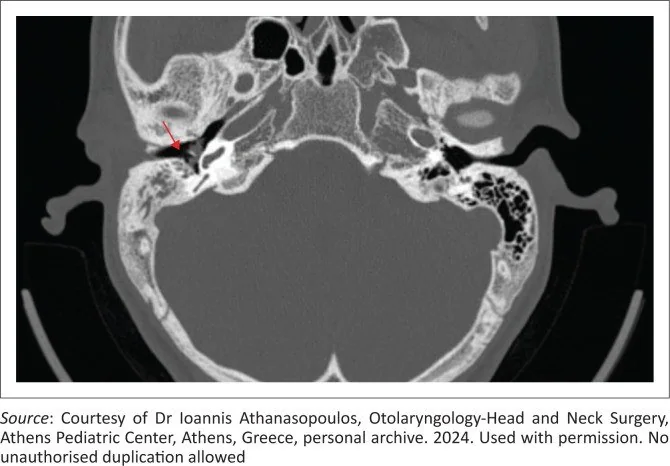
- Orta kulakta yüksek dansiteli noktasal veya ağsı kalsifikasyonlar, kemikçik yüzeylerinde kalsifikasyonlar, stapes krur ve tabanında kalınlaşma, askı bağları ve kas tendonlarında artmış dansite ile yeni kemik oluşumu aranır. Yüksek çözünürlüklü temporal kemik BT, timpanosklerozun yerleşimini değerlendirmede kullanılabilir.
- Kemik algoritması tanısal
- Aksiyel kesitler inkus uzun kolu ve lentiküler çıkıntıyı, koronal kesitler epitympanum ile malleus başı ve stapes/oval pencere çevresini gösterir; eklem aralığında kemiksi köprü veya çevre duvara uzanan kalsifikasyon seçilir.
Önerilen pencereler: Kemik (G 1800 / M 400).
BT bulguları
- Timpanosklerotik plak — timpanik kavitede veya membranda noktasal, çizgisel ya da ağsı yüksek dansiteli kalsifikasyon.
- Kemikçik kılıflanması — malleus/inkus kompleksini çevreleyen kalsifiye dansite ve tegmen ya da kavite duvarına uzanan sabitleyici köprü.
- Oval pencere plağı — stapes tabanı veya oval pencere nişinde yoğunluk artışı ve nişin daralması.
- Peristapedial yumuşak doku — oval pencere nişinde stapes çevresini saran, kemik yoğunluğu göstermeyen fibrotik doku.
- Yeni kemik oluşumu — özellikle attikusta kemikçik çevresinde kalın web veya kemiksi çevrelenme.
- İnkus uzun kolu erozyonu — lentiküler çıkıntıya doğru incelme, kısalma veya devamlılığın kaybı.
- Zincir deformasyonu — malleus-inkus ekleminde kabalaşma veya kemikçiklerin eklem yüzlerinde düzensiz kontur.
Normalde
Koronal rekonstrüksiyon, özellikle attikustaki fiksasyonun değerlendirilmesini sağlar. Kemikçik zinciri olası timpanoskleroz tutulumu açısından aksiyel kesitlerde ardışık kesitler boyunca değerlendirilir.
Ayırıcı tanı
- Otoskleroz
- oval pencere ve stapes tabanı çevresinde odaklanır; timpanik kavitede yaygın plak veya kemikçik çevrelenmesi tipik değildir.
- Kolesteatom
- orta kulakta kitle benzeri yumuşak doku ile scutum, inkus veya başka kemikte erozyon yapar; kalsifiye ağsı plak beklenmez.
- Akut kemikçik luksasyonu
- kemikçikler ve eklem arası yeni yer değiştirme gösterir; çevreye yapışık plak/köprü paterni yoktur.
- Kronik efüzyon/granülasyon
- kaviteyi yumuşak doku dansitesinde doldurabilir ancak ince kemikçik çevresinde mineralize köprü oluşturmaz.
- Doğuştan kemikçik anomalisi
- kronik enfeksiyon bulgusu olmadan saptanan, gelişimsel eklem devamsızlığı veya yapı bozukluğu içeren nadir bir durumdur.
Tuzaklar
- Yumuşak doku örtüsünü kesin fiksasyon olarak raporlamayın; fibrozis ve efüzyon BT'de benzer görünebilir, kemiksi/kalsifiye köprü daha doğrudan ipucudur.
- İnkusun uzun kolu normalde incedir; gerçek erozyon için lentiküler çıkıntıya kadar devamlılığını ardışık kesitlerde ve karşı tarafla kıyaslayın.
- Stapes çevresindeki çok küçük kalsifikasyonlar hacim ortalaması nedeniyle kaybolabilir; oval pencere nişini farklı düzlem rekonstrüksiyonlarında gözden geçirin.
- Miringoskleroz yalnız timpanik membranda kalabilir; orta kulak kemikçiklerine uzanmadıkça zincir fiksasyonu anlamına gelmez.
Kendini dene
Kronik otitli hastada malleus ve inkus çevresinde ağsı kalsifikasyon, eklem aralığında kemiksi köprü izleniyor. Bu görünüm neyi destekler?
Cevabı göster
Ossiküler timpanoskleroz. Kalsifik plak ve eklem köprüleşmesi timpanosklerotik fiksasyonu destekler. Luksasyonda eklem ayrılması ve yer değiştirme olur; kolesteatom yumuşak doku ile erozyon yapar; otoskleroz oval pencere/stapes tabanına odaklanır.
İletim tipi işitme kaybında inkus uzun kolunun devamlılığı yok, Prussak aralığında eroziv yumuşak doku görülüyor. En uygun tanı hangisidir?
Cevabı göster
Edinilmiş kolesteatom. Prussak aralığı yumuşak dokusu ve komşu kemikçik erozyonu kolesteatom lehinedir. Fiksasyonda kemikçiğin sürekliliği çoğu kez korunur ve çevre dokuda plak/köprü aranır; stapes tabanı lezyonu sınırlı kalır; gelişimsel anomali kronik eroziv kitle oluşturmaz.
İlgili konular
Kaynaklar
Bu sayfadaki 38 cümle ve 2 quiz sorusu aşağıdaki kaynaklardan alıntılarla tek tek karşılaştırıldı; 5 cümle bu karşılaştırmada düzeltildi (2026-10-08).
- Reliability of high‐resolution CT scan in diagnosis of ossicular tympanosclerosispmc.ncbi.nlm.nih.gov
- Psoriatic arthritis: imaging techniques.pubmed.ncbi.nlm.nih.gov
- Imaging of gout: New tools and biomarkers?pubmed.ncbi.nlm.nih.gov
- Imaging in early rheumatoid arthritis.pubmed.ncbi.nlm.nih.gov
- Emerging MRI methods in rheumatoid arthritis.pubmed.ncbi.nlm.nih.gov
- Esophageal Erosion of Adjustable Gastric Band, Endoscopic Solution of an Unreported Complication.pubmed.ncbi.nlm.nih.gov
- Neuroimaging in Central Nervous System Infections.pubmed.ncbi.nlm.nih.gov
- Gouty arthropathy: Review of clinico-pathologic and imaging features.pubmed.ncbi.nlm.nih.gov
- Cross-polarization optical coherence tomographic assessment of in situ simulated erosive tooth wear.pubmed.ncbi.nlm.nih.gov
- Cervical spine involvement early in the course of rheumatoid arthritis.pubmed.ncbi.nlm.nih.gov
- Isolated malleus fixation: A pediatric case series.pubmed.ncbi.nlm.nih.gov
- CT findings in temporal bone osteoradionecrosis.pubmed.ncbi.nlm.nih.gov
- HR-pQCT in vivo imaging of periarticular bone changes in chronic inflammatory diseases: Data from acquisition to impact on treatment indications.pubmed.ncbi.nlm.nih.gov
- Imaging in gout: insights into the pathological features of disease.pubmed.ncbi.nlm.nih.gov
- Computed tomography in rheumatology - From DECT to high-resolution peripheral quantitative CT.pubmed.ncbi.nlm.nih.gov
- Isolated Congenital Ossicular Chain Anomalies.pubmed.ncbi.nlm.nih.gov
- The preoperative temporal bone CT: a mnemonic for reducing surgical risk.pubmed.ncbi.nlm.nih.gov
- Prognostic Scoring Systems in Ossiculoplasty: A Comprehensive Review and Introduction of a Novel Predictive Index.pubmed.ncbi.nlm.nih.gov
- Pediatric Jugular Bulb Anomalies: Management Insights From Two Cases.pubmed.ncbi.nlm.nih.gov
- Diagnosis and Management of Middle Ear Neuroendocrine Tumour (MeNET).pubmed.ncbi.nlm.nih.gov
- Intelligent diagnosis of ossicular chain malformations on CT: development and clinical efficacy of a cascaded AI framework.pubmed.ncbi.nlm.nih.gov
- Improved visualization of malpositioned middle ear prostheses using photon-counting detector CT: a cadaver study.pubmed.ncbi.nlm.nih.gov
- Hearing results following endoscopic type I tympanoplasty in medium and large perforations.pubmed.ncbi.nlm.nih.gov
- Role of the Ear in Meningitis: A Narrative Review of Neuroimaging.pubmed.ncbi.nlm.nih.gov
- Diagnostic Value of Cone-Beam Computed Tomography in Conductive or Mixed Hearing Loss with Intact Tympanic Membrane.pubmed.ncbi.nlm.nih.gov
- Bilateral Congenital Stapes Footplate Fixation With Absence of the Stapedial Tendon.pubmed.ncbi.nlm.nih.gov
- Photon-counting detector computed tomography for temporal bone: does higher resolution matter?pubmed.ncbi.nlm.nih.gov
- Can inflammatory indices predict myringosclerosis?pubmed.ncbi.nlm.nih.gov
- Slow-Flow Venous Vascular Malformation in the Carotid Sheath.pubmed.ncbi.nlm.nih.gov
- Clinical situations for which 3D printing is considered an appropriate representation or extension of data contained in a medical imaging examination: neurosurgical and otolaryngologic conditions.pubmed.ncbi.nlm.nih.gov
- Otosclerosis under microCT: New insights into the disease and its anatomy.pubmed.ncbi.nlm.nih.gov
- Association of Radiologic Findings of Otosclerosis Using Photon-Counting Computed Tomography With Hearing.pubmed.ncbi.nlm.nih.gov
- External and middle ear diseases: radiological diagnosis based on clinical signs and symptoms.pubmed.ncbi.nlm.nih.gov
- Middle ear malformations combined with cerebrospinal fluid otorrhea: A case report.pubmed.ncbi.nlm.nih.gov
- Multimodal imaging in conductive hearing loss: Optimising CT, MRI and CTA for accurate diagnosis and management.pubmed.ncbi.nlm.nih.gov
- The Retrotympanum Revisited By a Volumetric Approach Using Synchrotron-Based X-Ray Phase-Contrast Imaging.pubmed.ncbi.nlm.nih.gov
- Exploration of Automated Measurement for Ossicular Chains Based on 3-Dimensional Geometric Information.pubmed.ncbi.nlm.nih.gov
- Photon-counting CT for preoperative assessment of stapes footplate fixation in otosclerosis.pubmed.ncbi.nlm.nih.gov
- Endoscopic stapes surgery for otosclerosis: clinical, radiological, and audiological outcomes in a retrospective series of 40 patients.pubmed.ncbi.nlm.nih.gov
- Prognostic Factors in Cochlear Implant in Otosclerosis: Radiologic and Clinical Variables: Cochlear Implant Predictors in Otosclerosis.pubmed.ncbi.nlm.nih.gov
- External auditory canal cholesteatoma presenting with facial nerve palsy: rare clinical entity.pubmed.ncbi.nlm.nih.gov
- Pediatric Cholesteatoma Presenting as Persistent Otorrhea: A Case of Delayed Diagnosis Across Multiple ENT Specialists.pubmed.ncbi.nlm.nih.gov
- A Recurrent Tympanicum Paraganglioma With a SDHD Pathogenic Variant: First Reported Case.pubmed.ncbi.nlm.nih.gov
- Traumatic stapediovestibular luxation: two contrasting cases highlighting the importance of early treatment decision: A case report and literature review.pubmed.ncbi.nlm.nih.gov
- Traumatic perilymphatic fistula with stapes luxation into the vestibule and pneumolabyrinth: a case report.pubmed.ncbi.nlm.nih.gov
- Petrous bone fractures and ossicular chain lesions detected by PMCT in burnt bodies.pubmed.ncbi.nlm.nih.gov
- Chronic Suppurative Otitispubmed.ncbi.nlm.nih.gov
- [Clinical analysis of 10 cases of congenital middle ear cholesteatoma with concurrent congenital ossicular malformation].pubmed.ncbi.nlm.nih.gov
- Reconstructive flap surgery in head and neck cancer patients: an interdisciplinary view of the challenges encountered by radiation oncologists in postoperative radiotherapy.pubmed.ncbi.nlm.nih.gov
- Brazilian Society of Otology task force - Otosclerosis: evaluation and treatment.pubmed.ncbi.nlm.nih.gov
- Rare Diseases of the Middle Ear and Lateral Skull Base.pubmed.ncbi.nlm.nih.gov
- Imaging Features of Juvenile Xanthogranuloma of the Pediatric Head and Neck.pubmed.ncbi.nlm.nih.gov
- Different clinical presentation and management of temporal bone fibrous dysplasia in children.pubmed.ncbi.nlm.nih.gov
- Active middle ear implantation: imaging in the pre-operative planning and post-operative assessment of the Vibrant Soundbridge TM .pubmed.ncbi.nlm.nih.gov
- Bilateral Oval and Round Window Atresia on CT Temporal Bone: A Rare Anomaly Clinically Mimicking Otosclerosis in an Adult.pubmed.ncbi.nlm.nih.gov
- Post traumatic deafness: a pictorial review of CT and MRI findings.pubmed.ncbi.nlm.nih.gov
- Case Report: Simultaneous repair of cerebrospinal fluid otorrhea and cochlear implantation in a patient with single-sided deafness and inner ear anomaly.pubmed.ncbi.nlm.nih.gov
- Tuberculous Otomastoiditis: A Diagnostic Dilemma.pubmed.ncbi.nlm.nih.gov
- Temporal Bone Cholesteatoma: Typical Findings and Evaluation of Diagnostic Utility on High Resolution Computed Tomography.pubmed.ncbi.nlm.nih.gov
- Mesna in Otologic Surgery: Efficacy and Safety-A Scoping Review.pubmed.ncbi.nlm.nih.gov
- The bone conduction threshold pattern may help to estimate the pathology underlying conductive hearing loss.pubmed.ncbi.nlm.nih.gov
- Malleostapedotomy in Patients With Stapes Fixation: A Systematic Review.pubmed.ncbi.nlm.nih.gov
- Fusion of Middle Ear Optical Coherence Tomography and Computed Tomography.pubmed.ncbi.nlm.nih.gov
- Sigmoid and Transverse Sinus Thrombosis as a Complication of Chronic Suppurative Otitis Media in a Child: A Case Report and Review of Literature.pubmed.ncbi.nlm.nih.gov
- A case of congenital cholesteatoma in adult patient with intermittent episodes of facial palsy.pubmed.ncbi.nlm.nih.gov
- Persistent Stapedial Artery, Facial Nerve Overhang, and Calcified Stapedius Tendon Leading to Aborted Stapedotomy: An Unusual Middle Ear Anatomy.pubmed.ncbi.nlm.nih.gov
- Tartrazine clearing of the tympanic membrane for improved visualization of the middle ear.pubmed.ncbi.nlm.nih.gov
- Impact of prosthesis position on hearing outcomes in otosclerosis patients based on ultra-high-resolution CT.pubmed.ncbi.nlm.nih.gov
- Endoscopic Findings on Facial Nerve Anatomy During Exclusive Endoscopic Stapedotomy: Clinical Considerations and Impact on Surgical Results.pubmed.ncbi.nlm.nih.gov
- Diagnosing Middle Ear Malformation by Pure-Tone Audiometry Using a Three-Dimensional Finite Element Model: A Case-Control Study.pubmed.ncbi.nlm.nih.gov
- Efficacy of endoscopic removal of anterior malleolar ligament calcification combined with tympanic membrane repair for the treatment of conductive hearing loss.pubmed.ncbi.nlm.nih.gov
- Middle ear lymphoepithelioma-like carcinoma with cerebellopontine angle invasion misdiagnosed as meningioma: A case report and literature review.pubmed.ncbi.nlm.nih.gov
- Case Report: A case of craniopharyngioma in the cerebellopontine angle.pubmed.ncbi.nlm.nih.gov
- Primary intracranial adenocarcinoma associated with an epidermoid cyst: a case report and literature review.pubmed.ncbi.nlm.nih.gov
- Image-based deep learning in otology: a state-of-the-art review.pubmed.ncbi.nlm.nih.gov
- Audiometric Trends Among Children With Congenital Aural Atresia or External Auditory Canal Stenosis.pubmed.ncbi.nlm.nih.gov
- Success rates and predictors of outcomes of type I tympanoplasty performed by residents in a teaching tertiary hospital.pubmed.ncbi.nlm.nih.gov
- Implementation of Wideband Acoustic Immittance in Clinical Practice: Relationships among Audiologic and Otologic Findings.pubmed.ncbi.nlm.nih.gov
- Aural Manifestations of Antineutrophil Cytoplasmic Antibody (ANCA)-Associated Vasculitis-Diagnosis, Symptoms, Treatment.pubmed.ncbi.nlm.nih.gov
- Accessing the stapedius muscle via novel surgical retrofacial approach during cochlear implantation surgery: Intraoperative results on feasibility and safety.pubmed.ncbi.nlm.nih.gov
- Rare Cause of Congenital Unilateral Facial Palsy: High-Resolution Computed Tomography (HRCT) Findings of Isolated Facial Canal Hypoplasia and Aplasia.pubmed.ncbi.nlm.nih.gov
- Anatomic Malformations of the Middle and Inner Ear in 22q11.2 Deletion Syndrome: Case Series and Literature Review.pubmed.ncbi.nlm.nih.gov
- Chronically Discharging Ears: Evalution with High Resolution Computed Tomography.pubmed.ncbi.nlm.nih.gov
- Diseases of the middle ear in childhood.pubmed.ncbi.nlm.nih.gov
- High Resolution Computed Tomography Atlas of the Porcine Temporal Bone and Skull Base: Anatomical Correlates for Traumatic Brain Injury Research.pubmed.ncbi.nlm.nih.gov
Eğitim amaçlıdır; hasta başında tanı ve tedavi kararının yerine geçmez. İçerik yapay zekâ destekli bir süreçle güncel literatüre göre cümle cümle kaynaklarla karşılaştırıldı, ancak uzman radyolog incelemesinden geçmedi. Klinik kararlarda güncel kılavuzları, kurum protokollerini ve radyoloji raporunu esas alın.