Özet
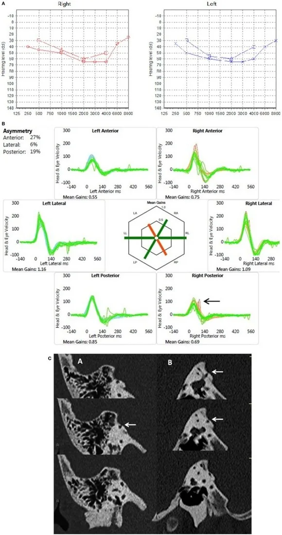
İşitsel veya vestibüler yakınmaları olan ve temporal kemik BT'sinde yüksek yerleşimli juguler bulbus bulunan kişide posterior semisirküler kanalın kemik sınırı incelenir. Kontrastsız yüksek çözünürlüklü BT, kanal ile juguler bulbus veya arka kafa çukuru (posterior fossa) durası arasındaki kemik örtüyü gösterir. Bu anatomik açıklık görüntüde tek başına semptomların kaynağını kanıtlamaz; yüksek juguler bulbus ve divertikül gibi varyantlar dehisansı taklit edebilir.
Faz ve pencere
- Kontrastsız tanısal
- Posterior semisirküler kanalın kemik hattı ile yüksek juguler bulbus arasındaki ince kemik şeridi, ince aksiyel ve koronal yüksek çözünürlüklü BT kesitlerinde değerlendirilir.
- Kemik algoritması tanısal
- Aksiyel ve koronal ince kesitlerde posterior kanalın özellikle ampuller bölümü ile juguler bulbus veya arka kafa çukuru (posterior fossa) dura plağı arasındaki kemik duvar izlenir; olası açıklığın farklı düzlemlerde sürekliliği sınanır.
Önerilen pencereler: Kemik (G 1800 / M 400).
BT bulguları
- Posterior kanal-juguler bulbus arasında kemik kesintisi — kanal lümeni ile juguler bulbusa ait venöz lümen arasında ayırıcı korteks görülmez.
- Yüksek yerleşimli juguler bulbus — bulbus tepesi posterior kanalın alt/ampuller segmentine yaklaşır ve komşuluk alanını daraltır.
- Defekt dışsal (arka kafa çukuru yönü) veya içsel (mastoid yönü) lokasyonda olabilir; çok düzlemli incelemelerle yeri belirlenir.
- Juguler bulbusa komşu kanal incelmesi — kemik şerit görünür kalır ancak belirgin biçimde incelmiş olabilir.
- Venöz komşuluk boyunca defektin devamı — kemik açıklığının juguler bulbus konturuyla aynı yerde ve seri boyunca izlendiği doğrulanır.
Normalde
Normalde posterior semisirküler kanalın kortikal halkası kesintisizdir; yüksek juguler bulbus bulunsa bile kanal ile venöz lümen arasında kemik ayırıcı tabaka seçilir. Posterior kanalın arka kafa çukuru (posterior fossa) durası ile ilişkili kemik plağı aynı çok düzlemli incelemede takip edilir.
Ayırıcı tanı
- Juguler bulbus dehisansı
- kemik kaybı orta kulak tabanı/hipotimpanumda venöz lümen ile timpanik kavite arasındadır; posterior kanal lümeni açık kalır.
- Üst semisirküler kanal dehisansı
- açıklık posterior kanal-juguler bulbus sınırında değil, üst kanal çatısında orta kafa çukuruna doğrudur.
- İnce posterior kanal duvarı
- korteks belirgin incelmiş olsa da ardışık kesit ve oblik düzlemlerde kemik hattı süreklilik gösterir.
- Juguler bulb divertikülü
- venöz yapı yukarı veya laterale uzanır; tek başına posterior kanal korteksinin kaybı anlamına gelmez.
Tuzaklar
- Yüksek juguler bulbusun kanala yakınlığını doğrudan dehisans diye adlandırma; aradaki kortikal şeridi birkaç düzlemde doğrula.
- Sadece koronal kesitte üst üste binmeyi kemik açıklığı sanma; aksiyel görüntüde kanal ve venöz lümen sınırlarını ayrı ayrı izle.
- Posterior kanal komşuluğundaki her juguler bulb varyantını semptom nedeni sayma; görüntü anatomik ilişkiyi gösterir, klinik korelasyon gerekir.
Kendini dene
Yüksek juguler bulbus yanında posterior kanal ile venöz lümen arasında kemik seçilmiyor. Bulguyu en iyi açıklayan tanı hangisidir?
Cevabı göster
Posterior kanal dehisansı. Kanal korteksinin juguler bulbusla komşu bölümde kaybı posterior kanal dehisansını düşündürür. Juguler bulbus divertikülü bulbusun dışa uzanan bir çıkıntısıdır; üst ve posterior semisirküler kanal dehisansları farklı kanalları tutar.
Juguler bulbus timpanik kavite tabanına yükseliyor ve aradaki kemik örtü seçilmiyor; posterior kanalın kortikal halkası korunmuş. Hangi varyant daha olası?
Cevabı göster
Juguler bulbus dehisansı. Timpanik kavite tabanındaki kemik örtünün venöz bulbusla ilişkili kaybı juguler bulbus dehisansına uyar. Bu bulgu juguler bulbus dehisansını tarif eder; posterior kanal dehisansı ise posterior semisirküler kanalın kemik örtüsünün yokluğudur.
Kaynaklar
Bu sayfadaki 33 cümle ve 2 quiz sorusu aşağıdaki kaynaklardan alıntılarla tek tek karşılaştırıldı; 13 cümle bu karşılaştırmada düzeltildi (2026-10-08).
- Anatomical Variations of the Jugular Bulb: A Critical and Comprehensive Reviewpmc.ncbi.nlm.nih.gov
- Prevalence and classification of posterior semicircular canal dehiscence in HRCTs of the petrous part of the temporal bonepmc.ncbi.nlm.nih.gov
- Posterior Semicircular Canal Dehiscence with Vestibulo-Ocular Reflex Reduction for the Affected Canal at the Video-Head Impulse Test: Considerations to Pathomechanismspmc.ncbi.nlm.nih.gov
- Is CT Still the Gold Standard in Semicircular Canal Dehiscence? Diagnostic Value of MRI in Poschl and Stenver Planespmc.ncbi.nlm.nih.gov
- Diagnosis and management of bilateral and multiple semicircular canal dehiscence: a case reportpmc.ncbi.nlm.nih.gov
- Multiple dehiscences of bony labyrinthine capsule. A rare case report and review of the literature.pubmed.ncbi.nlm.nih.gov
- [A case of adult temporal bone langerhans cell histiocytosis presenting as posterior canal dehiscence syndrome and literature review].pubmed.ncbi.nlm.nih.gov
- Posterior semicircular canal dehiscence in asymptomatic ears.pubmed.ncbi.nlm.nih.gov
- Rare otologic presentation of cat eye syndrome.pubmed.ncbi.nlm.nih.gov
- Jugular bulb abnormalities in patients with Meniere's disease using high-resolution computed tomography.pubmed.ncbi.nlm.nih.gov
- Children with posterior semicircular canal dehiscence: A case series.pubmed.ncbi.nlm.nih.gov
- Pediatric Semicircular Canal Dehiscence: Radiographic and Histologic Prevalence, With Clinical Correlation.pubmed.ncbi.nlm.nih.gov
- Audiovestibular Quantification in Rare Third Window Disorders in Children.pubmed.ncbi.nlm.nih.gov
- Posterior semicircular canal dehiscence: first reported case series.pubmed.ncbi.nlm.nih.gov
- Posterior semicircular canal dehiscence following endolymphatic sac surgery.pubmed.ncbi.nlm.nih.gov
- Posterior semicircular canal dehiscence: a diagnostic and surgical conundrum.pubmed.ncbi.nlm.nih.gov
- Posterior semicircular canal dehiscence: CT prevalence and clinical symptoms.pubmed.ncbi.nlm.nih.gov
- Congenital dehiscence in the posterior semicircular canal.pubmed.ncbi.nlm.nih.gov
- Repair of posterior semicircular canal dehiscence from a high jugular bulb.pubmed.ncbi.nlm.nih.gov
- The preoperative temporal bone CT: a mnemonic for reducing surgical risk.pubmed.ncbi.nlm.nih.gov
- Key pre-operative CT parameters for cochlear implantation: a focused radiologic review with surgical relevance.pubmed.ncbi.nlm.nih.gov
- The Possible Relationship Between Sigmoid Dehiscence, Degree of Mastoid Pneumatization, and Sigmoid Sinus Position in Patients with Pulsatile Tinnitus.pubmed.ncbi.nlm.nih.gov
- Anatomical Reasons for an Impaired Internal Jugular Flow.pubmed.ncbi.nlm.nih.gov
- "Leave me alone": anatomical structures and variations seen on computed tomography of the temporal bone.pubmed.ncbi.nlm.nih.gov
- Spontaneous SSCD Auto-Plugging: Clinical, Electrophysiological and Radiological Evidence.pubmed.ncbi.nlm.nih.gov
- Posterior Semicircular Canal Dehiscence with Vestibulo-Ocular Reflex Reduction for the Affected Canal at the Video-Head Impulse Test: Considerations to Pathomechanisms.pubmed.ncbi.nlm.nih.gov
- Beyond Tympanomastoidectomy: A Review of Less Common Postoperative Temporal Bone CT Findings.pubmed.ncbi.nlm.nih.gov
- Radioanatomical assessment of the subtympanic sinus and its relationship to facial nerve position and temporal bone pneumatization in adults: CT study.pubmed.ncbi.nlm.nih.gov
- NIR Indocyanine-White Light Overlay Visualization for Neuro-Oto-Vascular Preservation During Anterior Transpetrosal Approaches: A Technical Note.pubmed.ncbi.nlm.nih.gov
- Ear Surgery Training: A Comprehensive Review.pubmed.ncbi.nlm.nih.gov
- Vestibular Aqueduct Hypoplasia Identifies Semicircular Canal Dehiscence Patients Who Progress to Meniere's Disease.pubmed.ncbi.nlm.nih.gov
- Audio-Vestibular Evaluation of Pediatric Pseudo-Conductive Hearing Loss: Third Window Syndromes.pubmed.ncbi.nlm.nih.gov
- Single-Stage Surgical Treatment of Concurrent Meniere's Disease and Superior Semicircular Canal Dehiscence: A Case Report.pubmed.ncbi.nlm.nih.gov
- Extradural anterior transpetrosal approach: how I do it.pubmed.ncbi.nlm.nih.gov
- Pre-operative High-Resolution CT and MRI Evaluation in Pediatric Cochlear Implant Candidates: Correlation With Surgical Findings and Outcomes.pubmed.ncbi.nlm.nih.gov
- Non-invasive imaging modalities for diagnosing pulsatile tinnitus: a comprehensive review and recommended imaging algorithm.pubmed.ncbi.nlm.nih.gov
- Characteristics of the Facial Canal-Lateral Canal Dehiscence: The Under-recognized Third Window.pubmed.ncbi.nlm.nih.gov
- Endoscopic assisted minimally invasive resection of petrous bone epidermoid cyst via a presigmoid burr hole approach: illustrative case.pubmed.ncbi.nlm.nih.gov
- Functional and imaging anomalies of the vestibular system in motion sickness: a clinical observation study.pubmed.ncbi.nlm.nih.gov
- A multicorridor strategy including the infralabyrinthine route for extensive skull base chondrosarcoma: illustrative case.pubmed.ncbi.nlm.nih.gov
- Tympanic Bone: Small Neglected Bone with Extreme Anatomic Importance in Lateral Skull-Base Surgery.pubmed.ncbi.nlm.nih.gov
- Pulsatile Tinnitus: A Comprehensive Clinical Approach to Diagnosis and Management.pubmed.ncbi.nlm.nih.gov
- Anatomical Variations of the Jugular Bulb: A Critical and Comprehensive Review.pubmed.ncbi.nlm.nih.gov
- Chronic otitis media and middle ear variants: Is there relation?pubmed.ncbi.nlm.nih.gov
- Unique Anatomical Imaging Findings in Patients With Patulous Eustachian Tube.pubmed.ncbi.nlm.nih.gov
- Superior Semicircular Canal Dehiscence Repair With Hydroxyapetite Cement via a Transmastoid Approach.pubmed.ncbi.nlm.nih.gov
- A Practical Diagnostic Approach to Pediatric Episodic Vestibular Syndrome.pubmed.ncbi.nlm.nih.gov
- Surgical Techniques and Materials Used in the Treatment of Complicated Otomastoiditis: A Systematic Review.pubmed.ncbi.nlm.nih.gov
- Role of the Ear in Meningitis: A Narrative Review of Neuroimaging.pubmed.ncbi.nlm.nih.gov
- Acute Otomastoiditis in Children: An Observational Study on the Role of Mastoid Morphology in the Development of Intracranial Complications.pubmed.ncbi.nlm.nih.gov
- Beyond the otoscope: an imaging review of congenital cholesteatoma.pubmed.ncbi.nlm.nih.gov
- Low Frequency Air-Bone Gap in Meniere's Disease: Relationship With Magnetic Resonance Imaging Features of Endolymphatic Hydrops.pubmed.ncbi.nlm.nih.gov
- Third Window Syndrome: Surgical Management of Cochlea-Facial Nerve Dehiscence.pubmed.ncbi.nlm.nih.gov
- High-frequency pseudo-hypofunction of the superior semicircular canal dehiscence syndrome: a vHIT - oVEMP dissociation explained by hydrodynamic energy loss.pubmed.ncbi.nlm.nih.gov
- Otic Capsule Dehiscences Simulating Other Inner Ear Diseases: Characterization, Clinical Profile, and Follow-Up-Is Ménière's Disease the Sole Cause of Vertigo and Fluctuating Hearing Loss?pubmed.ncbi.nlm.nih.gov
- Flat-panel conebeam CT in the clinic: history and current state.pubmed.ncbi.nlm.nih.gov
- Endovascular treatment of pulsatile tinnitus associated with venous sinus diverticulum: a scoping review.pubmed.ncbi.nlm.nih.gov
- Cranial nerves involvement in craniosynostosis: a systematic review.pubmed.ncbi.nlm.nih.gov
- Mastoid cortex osteoma associated with middle-ear and mastoid cholesteatoma: a case report.pubmed.ncbi.nlm.nih.gov
- Third Window Syndrome: An Up-to-Date Systematic Review of Causes, Diagnosis, and Treatment.pubmed.ncbi.nlm.nih.gov
- Anterolateral, lateral, and posterior corridors to complex skull base lesions in sphenocavernous and petroclival regions: microsurgical anatomy with three-dimensional reconstructions and illustrative cases.pubmed.ncbi.nlm.nih.gov
- Anatomical Characteristics of the Facial Nerve in Patients With Cochlear Hypoplasia.pubmed.ncbi.nlm.nih.gov
- Inter- and Intrarater Reliability of the Manjila and Semaan Classification System when Assessing Jugular Bulb Position.pubmed.ncbi.nlm.nih.gov
- Double dehiscence (Superior semicircular canal and tegmen tympani) in the epicampaniform period (Arboli type).pubmed.ncbi.nlm.nih.gov
- Sheep Head Cadaveric Model for the Transmeatal Extensions of the Retrosigmoid Approach.pubmed.ncbi.nlm.nih.gov
- Comparison of Outcomes in Superior Canal Dehiscence Surgery Using Either Transmastoid or Middle Fossa Surgical Approaches: A Systematic Review and Meta-Analysis.pubmed.ncbi.nlm.nih.gov
- Morphological and Epidemiological Analysis of the Sphenoid Sinus Based on Computed Tomography and Magnetic Resonance Imaging: A Narrative Review.pubmed.ncbi.nlm.nih.gov
- Managing Complex Chronic Otitis Media: Insights from Subtotal Petrosectomy with Blind Sac Closure.pubmed.ncbi.nlm.nih.gov
- Imaging of Skull Base Tumors.pubmed.ncbi.nlm.nih.gov
- Identifying central positional vertigo to prevent diagnostic pitfalls: A case series and review of literature.pubmed.ncbi.nlm.nih.gov
- Is CT Still the Gold Standard in Semicircular Canal Dehiscence? Diagnostic Value of MRI in Poschl and Stenver Planes.pubmed.ncbi.nlm.nih.gov
- A Case of Acquired Pars Flaccida Cholesteatoma Extending to the Supralabyrinthine Petrous Apex With Preservation of the Otic Capsule.pubmed.ncbi.nlm.nih.gov
Eğitim amaçlıdır; hasta başında tanı ve tedavi kararının yerine geçmez. İçerik yapay zekâ destekli bir süreçle güncel literatüre göre cümle cümle kaynaklarla karşılaştırıldı, ancak uzman radyolog incelemesinden geçmedi. Klinik kararlarda güncel kılavuzları, kurum protokollerini ve radyoloji raporunu esas alın.