Özet
Anterior yerleşimli sigmoid sinüs, sinüs oluğunun tipik anatomik konumuna göre mastoid bölgesine doğru uzandığı varyanttır; temporal kemik havalanması ile sinüs konumu arasında ilişki bulunabilir. Kontrastsız yüksek çözünürlüklü temporal kemik BT'sinde kemik pencerede sigmoid oluğun kemik konturu değerlendirilir; bu inceleme cerrahi planlamaya katkı sağlayabilir. Varyant özellikle mastoidektomi ve arka kulak cerrahisi planlanırken önem kazanır. Sinüsün önde konumlanması cerrahi ekspojuru ve damar yaralanması riskini etkileyebilir; sigmoid plate'deki kemik örtü kaybı ise dehisans olarak ayrı değerlendirilir.
Faz ve pencere
- Kontrastsız tanısal
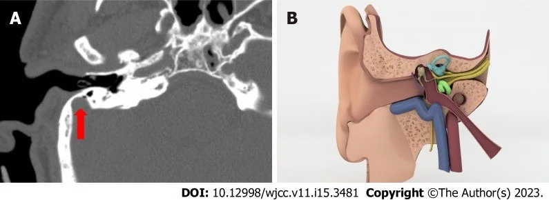
- Aksiyel ince kesit kontrastsız temporal kemik BT'sinin kemik algoritmalı rekonstrüksiyonunda sigmoid oluğun en belirgin anterior konveksitesi değerlendirilir. Temporal kemik pnömatizasyonu sigmoid sinüsle ilişkisi içinde değerlendirilir; kemik pencerede sigmoid oluğun kemik konturu venöz lümenden ayrı incelenir.
Önerilen pencereler: Kemik (G 1800 / M 400).
BT bulguları
- Anterior sigmoid oluk — sinüsün kemik oluğu mastoidin ön bölümüne yaklaşır ve postauriküler cerrahi çalışma alanını daraltır.
- Mastoid hücrelerle komşuluk — hava hücreleri sigmoid konturun önüne kadar uzanamayabilir; azalmış pnömatizasyon eşlik edebilir.
- Kortikal sigmoid plate — kemik pencerede sigmoid oluğun kortikal sınırları izlenir; kemik sınırdaki defekt sigmoid plate dehisansı olarak değerlendirilir.
- Dış kulak yolu arka duvarı — sinüsün ön konturuyla kanalın arka duvarı arasındaki mesafe aksiyel kesitlerde cerrahi erişim açısından gözden geçirilir.
- Taraflar arası konum farkı — sağ ve sol sigmoid olukların öne uzanma derecesi simetrik olmayabilir; karşı mastoid yalnız yardımcı kıyastır.
- Yetersiz havalanmış veya sklerotik temporal kemik, yeterli presigmoid ekspojuru güçleştirip zaman alıcı hale getirebilir; sigmoid oluğun kemik konturu ayrıca değerlendirilmelidir.
- Aksiyel temporal kemik BT'de sigmoid sinüs oluğunun en anterior konveksitesini belirleyin; sigmoid sinüs ile dış kulak yolu arasındaki mesafeyi cerrahi planlama için değerlendirin.
Normalde
Tipik aksiyel temporal kemik BT'sinde sigmoid oluk mastoidin daha arka bölümünde seyreder ve mastoid hava hücreleri ile dış kulak yolu arka duvarı arasında cerrahi boşluk bırakır. Aynı seviyede sigmoid plate'in dış kulak yolu arka duvarına yaklaşmasını ve mastoid hücrelerin ne kadar öne uzandığını karşı tarafla kıyaslayın; kortike sınırın sürekliliğini ayrıca kontrol edin.
Normal BT ile kıyasla
Aynı bölgenin, kaynak veri setinde patoloji içermediği belirtilen bir BT incelemesini kesit kesit kaydırın; organ sınırlarını açarak anatomiyi hasta görüntüsüyle karşılaştırın. Küçük rastlantısal bulgular tümüyle dışlanmamıştır.
Kaynak: TotalSegmentator v2.01 veri seti (Wasserthal ve ark., Radiology: Artificial Intelligence 2023), CC BY 4.0, vaka s0103; organ sınırları veri setinin otomatik segmentasyonundan, birleştirilerek sadeleştirildi. Görüntüler 2,5 mm kesit aralığına yeniden örneklendi.
Ayırıcı tanı
- Sigmoid plate dehisansı
- sinüs önde yerleşebilir ancak aradaki kortike kemik örtüde de fokal kayıp vardır.
- Sigmoid sinüs divertikülü
- sinüs duvarından dışa doğru sınırlı keseleşme venöz konturda fokal çıkıntı oluşturur.
- Hipopnömatize mastoid, ayırıcı tanıdan çok cerrahi ekspojuru etkileyebilecek bir anatomik özelliktir; yetersiz havalanmış veya sklerotik temporal kemikte yeterli ekspojur güçleşip zaman alabilir.
- Sigmoid sinüs trombozu
- lümen dolum kusuru veya sinüs çapında değişiklik kontrastlı venöz görüntüleme ile değerlendirilir; kemik penceredeki öne konumdan farklıdır.
Tuzaklar
- Komşu mastoid hava hücrelerinin kortikal duvarları, sinüsün öne bombeleşen konturu ile karıştırılmamalıdır; tüm kesitlerde sinüs oluğu ve sigmoid plate sürekliliği izlenmelidir.
- Azalmış mastoid pnömatizasyonu sinüsü göreceli olarak önde gösterebilir; varyantı tek bir kesitte değil, maksimum anterior kontur boyunca tanımla.
- Sinüsün öne yerleşimi ile sigmoid plate dehisansını aynı bulgu gibi yazma; birincisi konum, ikincisi kemik örtü bütünlüğüdür.
Kendini dene
Temporal kemik BT’sinde sigmoid sinüsün çevre kemik yapılara göre konumunu değerlendirmek cerrahi planlama açısından neden önemlidir?
Cevabı göster
Cerrahi ekspojuru, drilling güzergâhını ve damar yaralanması riskini etkileyebileceği için. Sigmoid sinüsün çevre kemik yapılara göre konumu cerrahi ekspojuru ve drilling güzergâhını etkileyebilir; konum varyasyonları damar yaralanması riski açısından da önem taşır.
Yetersiz havalanmış veya sklerotik temporal kemik, presigmoid cerrahi ekspojuru nasıl etkileyebilir?
Cevabı göster
Ekspojuru güç ve zaman alıcı hale getirebilir. Yetersiz havalanmış veya sklerotik temporal kemik, yeterli ekspojuru güç ve zaman alıcı hale getirebilir.
Kaynaklar
Bu sayfadaki 31 cümle ve 2 quiz sorusu aşağıdaki kaynaklardan alıntılarla otomatik olarak karşılaştırıldı; 14 cümle bu karşılaştırmada düzeltildi (2026-10-09).
- Radiographic Assessment of the presigmoid retrolabyrinthine approachpmc.ncbi.nlm.nih.gov
- Flow diverter for a dissecting aneurysm of the AICA serving as a feeding artery to a transverse-sigmoid sinus DAVFpmc.ncbi.nlm.nih.gov
- Clinical management of isolated transverse-sigmoid sinus dural arteriovenous fistulas: a three-case illustrative series and narrative reviewpmc.ncbi.nlm.nih.gov
- Transvaginal ultrasound diagnosis of colouterine fistula secondary to sigmoid colon cancer: A case reportpmc.ncbi.nlm.nih.gov
- Morphologic variations of the sigmoid sinus on computed tomography: a classification-based studypmc.ncbi.nlm.nih.gov
- Radiographic Assessment of the presigmoid retrolabyrinthine approach.pubmed.ncbi.nlm.nih.gov
- A Life Threatening Pitfall in Ear Surgery: Extracranial Sigmoid Sinus.pubmed.ncbi.nlm.nih.gov
- Clinical evaluation of temporal bone anatomical abnormalities and surgical method selection in patients with congenital aural atresia.pubmed.ncbi.nlm.nih.gov
- Ischemic colitis associated with acute carbon monoxide poisoning--a case report.pubmed.ncbi.nlm.nih.gov
- Moyamoya disease concurrent with dural arteriovenous fistula: A case report and literature review.pubmed.ncbi.nlm.nih.gov
- Radical treatment for bilateral vertebral artery dissecting aneurysms by reconstruction of the vertebral artery.pubmed.ncbi.nlm.nih.gov
- CT characteristics of dehiscent sigmoid plates presenting as pulsatile tinnitus: a study of 23 patients.pubmed.ncbi.nlm.nih.gov
- [CT analysis of dehiscent sigmoid plate presenting as pulsatile tinnitus].pubmed.ncbi.nlm.nih.gov
- The Feasibility of Retrofacial Approach for Cochlear Implantation.pubmed.ncbi.nlm.nih.gov
- 18F-FDG PET/CT in a Case of Urothelial Carcinoma in the Urachus Presenting as Colon Cancer.pubmed.ncbi.nlm.nih.gov
- High bilateral fenestration of the internal jugular vein.pubmed.ncbi.nlm.nih.gov
- Transparent model of temporal bone and vestibulocochlear organ made by 3D printing.pubmed.ncbi.nlm.nih.gov
- Study on inferior petrosal sinus and its confluence pattern with relevant veins by MSCT.pubmed.ncbi.nlm.nih.gov
- Squamous cell carcinoma of the middle ear: report of three cases.pubmed.ncbi.nlm.nih.gov
- Assessment of the Oval Window-Facial Canal Corridor and Mastoid Pneumatization: A Preliminary HRCT-Based Radiological-Anatomical Framework.pubmed.ncbi.nlm.nih.gov
- Aditus ad Antrum on Normal Temporal Bone CT Scan.pubmed.ncbi.nlm.nih.gov
- Anatomical corridors and quantitative spatial parameters of the jugular foramen: a computed tomography-based reference for skull base surgery.pubmed.ncbi.nlm.nih.gov
- Mastoid cortex osteoma associated with middle-ear and mastoid cholesteatoma: a case report.pubmed.ncbi.nlm.nih.gov
- The Possible Relationship Between Sigmoid Dehiscence, Degree of Mastoid Pneumatization, and Sigmoid Sinus Position in Patients with Pulsatile Tinnitus.pubmed.ncbi.nlm.nih.gov
- Key pre-operative CT parameters for cochlear implantation: a focused radiologic review with surgical relevance.pubmed.ncbi.nlm.nih.gov
- Mastoid emissary vein, mastoid emissary foramen, and mastoid emissary canal: anatomy, variability, imaging, and clinical implications.pubmed.ncbi.nlm.nih.gov
- The preoperative temporal bone CT: a mnemonic for reducing surgical risk.pubmed.ncbi.nlm.nih.gov
- Combined presigmoid retrolabyrinthine and retrosigmoid approach for large vestibular schwannoma: a case report.pubmed.ncbi.nlm.nih.gov
- Minimal Anterior and Posterior Combined Transpetrosal Approach for Posterior Clinoid Process Meningiomas.pubmed.ncbi.nlm.nih.gov
- Combined Petrosectomy in a 7-Year-Old Patient: Case-Based Video Illustration and Technical Considerations for Petrosal Approaches in the Pediatric Population.pubmed.ncbi.nlm.nih.gov
- Imaging of Skull Base Tumors.pubmed.ncbi.nlm.nih.gov
- Drainage of middle cranial fossa epidural abscess through mastoidectomy: Our experience and review of the literature.pubmed.ncbi.nlm.nih.gov
- Acute Otomastoiditis in Children: An Observational Study on the Role of Mastoid Morphology in the Development of Intracranial Complications.pubmed.ncbi.nlm.nih.gov
- Morphologic variations of the sigmoid sinus on computed tomography: a classification-based study.pubmed.ncbi.nlm.nih.gov
- Development of a non-human primate model for preclinical research of a novel auditory nerve implant.pubmed.ncbi.nlm.nih.gov
- Hominin Variability and Evolutionary Relationships at Guattari Cave During the Middle and Late Pleistocene (San Felice Circeo, Latina, Italy).pubmed.ncbi.nlm.nih.gov
- Acute otitis media complicated by peripheral facial nerve palsy in an adult: A case report.pubmed.ncbi.nlm.nih.gov
- Clinician-Guided Deep Learning Segmentation of Skull Base Pneumatization on Computed Tomography Using 3D Slicer and MONAI Label.pubmed.ncbi.nlm.nih.gov
- Determining prognostic factors in the treatment of primary hemifacial spasm: Clinical outcomes and complications. A literature review.pubmed.ncbi.nlm.nih.gov
- Hearing Loss in Malignant Infantile Osteopetrosis: A Case-Based Review.pubmed.ncbi.nlm.nih.gov
- International Society of Paediatric Surgical Oncology (IPSO) Surgical Practice Guidelines.pubmed.ncbi.nlm.nih.gov
- How I do it: inside-out bony window design in the retrosigmoid suboccipital approach.pubmed.ncbi.nlm.nih.gov
- Anatomical Step-by-Step Dissection of Complex Skull Base Approaches for Trainees: Surgical Anatomy of the Posterior Petrosal Approach.pubmed.ncbi.nlm.nih.gov
- The Bonebridge BCI 602 Active Transcutaneous Bone Conduction Implant in Children: Objective and Subjective Benefits.pubmed.ncbi.nlm.nih.gov
- Endovascular treatment of pulsatile tinnitus associated with venous sinus diverticulum: a scoping review.pubmed.ncbi.nlm.nih.gov
- Blood flow changes in the transverse-sigmoid sinus junction following effective surgical reconstruction for unilateral pulsatile tinnitus with sigmoid sinus wall anomalies: a four-dimensional flow magnetic resonance imaging analysis.pubmed.ncbi.nlm.nih.gov
- Anatomical Variations of the Jugular Bulb: A Critical and Comprehensive Review.pubmed.ncbi.nlm.nih.gov
- Surgical Management of Pulsatile Tinnitus due to Sigmoid Sinus Diverticulum and Dehiscence - A Technical Note.pubmed.ncbi.nlm.nih.gov
- Emerging and re-emerging infectious disease in otorhinolaryngology.pubmed.ncbi.nlm.nih.gov
- Intracranial venous thrombosis imaging by anatomical location: an educational review with mimics and diagnostic pitfalls.pubmed.ncbi.nlm.nih.gov
- Case Report: Mechanical thrombectomy of cerebral venous sinuses using the NeVa stent retriever.pubmed.ncbi.nlm.nih.gov
- Intrapetrous Internal Carotid Artery Pseudoaneurysm Secondary to Malignant Otitis Externa: A Case Report.pubmed.ncbi.nlm.nih.gov
- A 43-year-old woman presenting with subacute, bilateral, sequential facial nerve palsies, then developing pseudotumour cerebri.pubmed.ncbi.nlm.nih.gov
- External and middle ear diseases: radiological diagnosis based on clinical signs and symptoms.pubmed.ncbi.nlm.nih.gov
- Differential diagnosis of jugular foramen lesions.pubmed.ncbi.nlm.nih.gov
- Obesity and Skull Base Manifestations: Clinical Implications and Management.pubmed.ncbi.nlm.nih.gov
- Fusion computed tomography-magnetic resonance imaging scans for pre-operative staging of congenital middle-ear cholesteatoma.pubmed.ncbi.nlm.nih.gov
- Imaging for Pediatric Head and Neck Emergencies: Critical Insights.pubmed.ncbi.nlm.nih.gov
- Multidetector CT of the Nasal Cavity and Paranasal Sinuses Variations in 73 Patients.pubmed.ncbi.nlm.nih.gov
- Cross Sectional Imaging of the Ear and Temporal Bone.pubmed.ncbi.nlm.nih.gov
- Anterolateral, lateral, and posterior corridors to complex skull base lesions in sphenocavernous and petroclival regions: microsurgical anatomy with three-dimensional reconstructions and illustrative cases.pubmed.ncbi.nlm.nih.gov
- Role of ethnicity in the determination of the transverse sigmoid sinus Junction (TSSJ): A prospective hospital-based radiological study in adult Sabah, East Malaysia.pubmed.ncbi.nlm.nih.gov
- From resection to reconstruction: patient-specific temporomandibular joint replacement in patients with ameloblastomas - a report of three cases.pubmed.ncbi.nlm.nih.gov
- Medical outcomes after canal wall-down mastoidectomy, external ear canal reconstruction, tympanoplasty, and mastoid obliteration for extensive cholesteatoma.pubmed.ncbi.nlm.nih.gov
- Surgical Techniques and Materials Used in the Treatment of Complicated Otomastoiditis: A Systematic Review.pubmed.ncbi.nlm.nih.gov
- Revisiting the Intratemporal Course of the Facial Nerve.pubmed.ncbi.nlm.nih.gov
- A Comparative Study of Computed Tomography Findings versus Intraoperative Findings of Level of Tegmen Plate in Chronic Otitis Media: An Observational Study.pubmed.ncbi.nlm.nih.gov
- Preoperative Assessment Using CT and MRI Scans of the Temporal Bone to Determine the Degree of Difficulty in Cochlear Implant Surgery.pubmed.ncbi.nlm.nih.gov
- The Intratemporal Course Variations of Facial Nerve in Wet Cadaveric Temporal Bones.pubmed.ncbi.nlm.nih.gov
- Role of the Ear in Meningitis: A Narrative Review of Neuroimaging.pubmed.ncbi.nlm.nih.gov
- Co-occurring scalp arteriovenous malformation and dural arteriovenous fistula with diploic vein involvement: illustrative case.pubmed.ncbi.nlm.nih.gov
- Flow diverter for a dissecting aneurysm of the AICA serving as a feeding artery to a transverse-sigmoid sinus DAVF.pubmed.ncbi.nlm.nih.gov
- Clinical management of isolated transverse-sigmoid sinus dural arteriovenous fistulas: a three-case illustrative series and narrative review.pubmed.ncbi.nlm.nih.gov
- Otogenic Sigmoid Sinus Thrombosis in a 34-Year-Old Woman: A Rare Intracranial Complication.pubmed.ncbi.nlm.nih.gov
- Three-dimensional exoscope-assisted combined petrosal approach for petroclival meningiomas: technical nuances from early experience.pubmed.ncbi.nlm.nih.gov
- Endoscope-assisted Transmastoid Infralabyrinthine approach to the petrous apex: A new corridor for multicompartmental jugular foramen tumors.pubmed.ncbi.nlm.nih.gov
- The Petrosquamous Septum in Children-A Misleading Obstacle or Reliable Landmark? A Radioanatomical Study.pubmed.ncbi.nlm.nih.gov
Eğitim amaçlıdır; hasta başında tanı ve tedavi kararının yerine geçmez, tıbbi tavsiye değildir. Metinler yapay zekâ desteğiyle yazıldı ve otomatik bir adımda kaynak alıntılarıyla karşılaştırıldı; bu bir tıbbi doğrulama değildir ve içerik uzman radyolog incelemesinden geçmedi. Klinik kararlarda güncel kılavuzları, kurum protokollerini ve radyoloji raporunu esas alın.