Özet
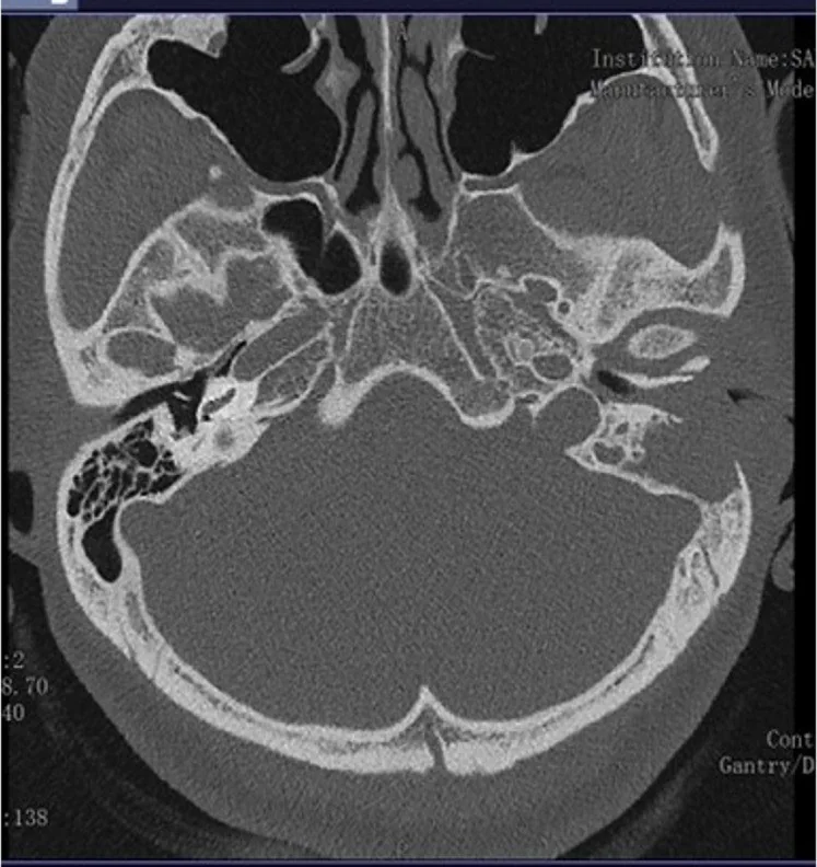
Temporal kemik inflamatuvar psödotümörü, orta kulak ve mastoidi tutabilen nadir, fibroinflamatuvar ve görüntülemede özgül olmayan kitle benzeri bir süreçtir. Kontrastsız yüksek çözünürlüklü BT kemik yıkımının yerini ve yayılımını gösterir, ancak görünüm tek başına histolojik tanı koydurmaz. Tegmen tympani veya mastoid kortekste kemik erozyonu ve komşu yumuşak doku kitlesi lezyonun yerel invazyonunu gösterir; manyetik rezonans görüntüleme dural kalınlaşma ve intrakraniyal uzanım değerlendirmesinde kullanılır. Agresif enfeksiyon ya da malign tümör sanılması tanısal gecikmeye; destrüksiyonun basit mastoid opasifikasyonu sayılması ise kafa tabanı yayılımının atlanmasına yol açabilir.
Faz ve pencere
- Kontrastsız tanısal
- İnce kesit kemik algoritmalı temporal kemik BT’de orta kulak/mastoid kitlesiyle tegmen veya mastoid korteks erozyonunun dağılımı incelenir; kontrastsız BT kemik sınırlarını gösterir, MR yumuşak doku ve dural/intrakraniyal yayılım değerlendirmesini tamamlar.
Önerilen pencereler: Kemik (G 1800 / M 400), Yumuşak doku (G 400 / M 40).
BT bulguları
- Orta kulak ve mastoid hava hücrelerini dolduran, sınırları belirgin olmayan yumuşak doku kitlesi
- Kafa tabanı kortikal erozyonu — tegmen tympani veya mastoid korteksin destrüksiyonu; komşu dura ve sigmoid sinüsle ilişkisi ayrıca değerlendirilir.
- Komşu kafa tabanı uzanımı — lezyon tegmen boyunca orta kafa çukuru/dura komşuluğuna ilerleyebilir; MR yumuşak doku ve intrakraniyal uzanımı daha iyi değerlendirmeyi sağlar.
- Mastoid korteks ve hava hücresi septalarında yıkım veya erozyon, bazen petroz kemik ve iç kulap yapısında da genişleme veya yıkım.
- Yumuşak doku kitlesinin orta kulak-mastoid sınırından komşu kafa tabanı kompartımanlarına devamlılığı.
- BT'de eşlik edebilen, ancak yayılım sınırını tek başına belirlemeyen dural komşuluk
Normalde
Normalde orta kulak ve mastoid hava hücreleri havalıdır; sigmoid plak, tegmen ve petroz kemik korteksi kesintisiz görünür. Kontrastsız ince kesitli temporal kemik BT'de orta kulak ve mastoiddeki yumuşak doku kitlesinin yayılımı ile tegmen veya mastoid kortekste kemik erozyonunun varlığı değerlendirilir.
Ayırıcı tanı
- Koalesan mastoidit
- mastoid septa ve kortekste destrüksiyon, klinik enfeksiyon bulguları veya subperiostal koleksiyonla birlikte olabilir.
- Temporal kemik skuamöz hücreli karsinomu
- dış kulak yolu ya da orta kulak merkezli infiltratif kitle ve düzensiz kemik yıkımı yapabilir.
- Kolesteatom
- epitimpanik merkez, scutum ve kemikçik erozyonu tipiktir; non-EPI difüzyon MR ayrımda yardımcı olur.
- Kafa tabanı osteomiyeliti
- kafa tabanı kortikal destrüksiyonu ve yaygın çevre yumuşak doku inflamasyonu, sıklıkla dış kulak yolu enfeksiyonu bağlamında görülür.
- Granülomatoz polianjiit
- orta kulak/mastoid opasifikasyonu yapabilir; eşlik eden sistemik hastalık ve diğer kulak bulguları tanısal bağlam sağlar.
Tuzaklar
- BT'de kemik erozyonunun bulunması inflamatuvar psödotümöre özgü değildir; skuamöz hücreli karsinom, osteomiyelit ve kolesteatom da destrüksiyon yapabilir.
- Mastoid opasifikasyonunu tek başına mastoidit diye adlandırma; septal yıkım, kortikal erozyon ve komşu yumuşak doku uzanımını ayrı ayrı ara.
- Dura ile temas tek başına invazyon anlamına gelmez; intrakraniyal yayılımın ve yumuşak doku tutulumunun değerlendirilmesinde MR gereklidir.
Kendini dene
Orta kulak-mastoid kitlesi tegmen ya da mastoid kortekste erozyon yapıyor. Bu BT görünümü için en doğru yaklaşım hangisidir?
Cevabı göster
Histoloji gerektiren agresif kitle. İnflamatuvar psödotümör dahil birden fazla agresif süreç kemik yıkımı yapar; BT yayılımı gösterir ama histolojik tanıyı tek başına koymaz. Kolesteatom için tipik merkez ve kemikçik/scutum bulguları gerekir; efüzyon kemik erozyonu yapmaz.
Opasifiye mastoidde septalar korunmuş, kortikal erozyon ve koleksiyon yok. Hangi tanı daha az desteklenir?
Cevabı göster
Koalesan mastoidit. Koalesan mastoiditte septal veya kortikal kemik yıkımı beklenir; korunmuş septalar bu tanıyı zayıflatır. Efüzyon, seröz otit ve GPA orta kulak-mastoid opasifikasyonu yapabilir ve BT'de erozyon olmadan görülebilir.
Kaynaklar
Bu sayfadaki 34 cümle ve 2 quiz sorusu aşağıdaki kaynaklardan alıntılarla tek tek karşılaştırıldı; 6 cümle bu karşılaştırmada düzeltildi (2026-10-08).
- Orbital pseudotumor and skull base infiltration as the sole manifestation of granulomatosis with polyangiitispmc.ncbi.nlm.nih.gov
- Tympanic Membrane Involvement of Multiple Myeloma: Report of a Casepmc.ncbi.nlm.nih.gov
- A case of an immunoglobulin G4 related inflammatory pseudotumor involving the clivus and the nasopharynx: A challenging case of a perplexing entity with an exhaustive overviewpmc.ncbi.nlm.nih.gov
- Proptosis Revealing a Neurological Form of IgG4-Related Disease: A Case Report and Literature Reviewpmc.ncbi.nlm.nih.gov
- Temporal bone plasmacytoma: An exceptional localization of solitary bone plasmacytoma highlighting the role of multimodal imaging and multidisciplinary managementpmc.ncbi.nlm.nih.gov
- Lateral skull base Inflammatory pseudotumor: A systematic review.pubmed.ncbi.nlm.nih.gov
- IgG4-Related Disease of Bilateral Temporal Bones.pubmed.ncbi.nlm.nih.gov
- Inflammatory Pseudotumor of the Temporal Bone: A Case Series.pubmed.ncbi.nlm.nih.gov
- Idiopathic Orbital Inflammatory Pseudotumor With Bone Erosion.pubmed.ncbi.nlm.nih.gov
- Inflammatory pseudotumor of the inner ear: are computed tomography changes pathognomonic?pubmed.ncbi.nlm.nih.gov
- Inflammatory pseudotumor (plasma cell granuloma) of the temporal bone.pubmed.ncbi.nlm.nih.gov
- Skull base infections, their complications, and management.pubmed.ncbi.nlm.nih.gov
- Inflammatory Pseudotumor of the Temporal Bone and Parapharyngeal Space: A Clinical Case.pubmed.ncbi.nlm.nih.gov
- Inflammatory Pseudotumor of the Temporal Bone Leading to Atypical Superior Semicircular Canal Dehiscence Syndrome: A Novel Case Report.pubmed.ncbi.nlm.nih.gov
- Inflammatory Pseudotumour of Temporal Bone in a Multiple-Comorbidity Patient.pubmed.ncbi.nlm.nih.gov
- Are there atypical sites of IgG4 related disease in head and neck region? Personal experience and literature review.pubmed.ncbi.nlm.nih.gov
- IgG4-Related Disease of Temporal Bone Presenting as Unilateral Mastoiditis and Cerebral Thrombosis in a 22-Year-Old Man.pubmed.ncbi.nlm.nih.gov
- Immunoglobulin G4-Related Disease Involving Various Head and Neck Regions: A Case Report.pubmed.ncbi.nlm.nih.gov
- Orbital pseudotumor and skull base infiltration as the sole manifestation of granulomatosis with polyangiitis.pubmed.ncbi.nlm.nih.gov
- Tympanic Membrane Involvement of Multiple Myeloma: Report of a Case.pubmed.ncbi.nlm.nih.gov
- A case of an immunoglobulin G4 related inflammatory pseudotumor involving the clivus and the nasopharynx: A challenging case of a perplexing entity with an exhaustive overview.pubmed.ncbi.nlm.nih.gov
- Imaging of Pathologies of the Temporal Bone and Middle Ear: Inflammatory Diseases, Their Mimics and Potential Complications-Pictorial Review.pubmed.ncbi.nlm.nih.gov
- Critical infections in the head and neck: A pictorial review of acute presentations and complications.pubmed.ncbi.nlm.nih.gov
- Diagnosis and Management of Middle Ear Neuroendocrine Tumour (MeNET).pubmed.ncbi.nlm.nih.gov
- Mastoid cortex osteoma associated with middle-ear and mastoid cholesteatoma: a case report.pubmed.ncbi.nlm.nih.gov
- Medication-related osteonecrosis of the external auditory canal and treatment with teriparatide.pubmed.ncbi.nlm.nih.gov
- Surgical Techniques and Materials Used in the Treatment of Complicated Otomastoiditis: A Systematic Review.pubmed.ncbi.nlm.nih.gov
- ESR Essentials: acute infections of the head and neck-practice recommendations by the European Society of Head and Neck Radiology.pubmed.ncbi.nlm.nih.gov
- External auditory canal osteoma with coexisting canal wall cholesteatoma: a case report and review of literature.pubmed.ncbi.nlm.nih.gov
- A 43-year-old woman presenting with subacute, bilateral, sequential facial nerve palsies, then developing pseudotumour cerebri.pubmed.ncbi.nlm.nih.gov
- Inflammatory pseudotumor of the temporal bone: a case series.pubmed.ncbi.nlm.nih.gov
- Middle Ear Teratoma: Clinical and Imaging Features.pubmed.ncbi.nlm.nih.gov
- Chronic nasopharyngeal and otitis media tuberculosis: diagnosis and management difficulties.pubmed.ncbi.nlm.nih.gov
- A multicorridor strategy including the infralabyrinthine route for extensive skull base chondrosarcoma: illustrative case.pubmed.ncbi.nlm.nih.gov
- Bilateral Cavernous Sinus Thrombosis and Clival Osteomyelitis Complicating Pediatric Otitis Media.pubmed.ncbi.nlm.nih.gov
- A Novel Descriptive Framework for Petroclival and Temporal Bone Meningioma Surgical Outcomes.pubmed.ncbi.nlm.nih.gov
- Primary glioblastoma with spontaneous transdural invasion and contiguous calvarial/mastoid osteolysis: a case report and literature review.pubmed.ncbi.nlm.nih.gov
- An intra-axial wolf in extra-axial clothing: A rare case of high-grade astrocytoma and literature review.pubmed.ncbi.nlm.nih.gov
- Orbital Fungating Mass as a Rare Manifestation of Retinoblastoma: A Case Study in Palliative Care.pubmed.ncbi.nlm.nih.gov
- Middle ear malformations combined with cerebrospinal fluid otorrhea: A case report.pubmed.ncbi.nlm.nih.gov
- [A retrospective study on anatomical morphological changes of pneumatized mastoid and sigmoid sinus bony wall in different types of sigmoid sinus dominance combined with high jugular bulb].pubmed.ncbi.nlm.nih.gov
- Multimodal imaging in conductive hearing loss: Optimising CT, MRI and CTA for accurate diagnosis and management.pubmed.ncbi.nlm.nih.gov
- Ruptured Posterior Inferior Cerebellar Artery Aneurysms: Integrating Microsurgical Expertise, Endovascular Challenges, and AI-Driven Risk Assessment.pubmed.ncbi.nlm.nih.gov
- Chronically Discharging Ears: Evalution with High Resolution Computed Tomography.pubmed.ncbi.nlm.nih.gov
- Granulomatosis with polyangiitis presenting as isolated ear involvement: a case series and literature review.pubmed.ncbi.nlm.nih.gov
- Detection of Coalescent Acute Mastoiditis on MRI in Comparison with CT.pubmed.ncbi.nlm.nih.gov
- Trichoepithelioma of the External Auditory Canal Mimicking Cholesteatoma.pubmed.ncbi.nlm.nih.gov
- Mucoepidermoid carcinoma of the middle ear: a case report.pubmed.ncbi.nlm.nih.gov
- Serous Otitis Media Secondary to a Middle Ear Neuroendocrine Tumor of Five-Year Duration.pubmed.ncbi.nlm.nih.gov
- Autoincudotomy as an uncommon etiology of conductive hearing loss: Case report and review of literature.pubmed.ncbi.nlm.nih.gov
- CT and MRI Features of Middle Ear Fibrous Hamartoma of Infancy: A Case Report.pubmed.ncbi.nlm.nih.gov
- Usefulness of <sup>99m</sup>Tc-HMPAO leukocyte scintigraphy in necrotizing external otitis/skull base osteomyelitis.pubmed.ncbi.nlm.nih.gov
- Intrapetrous Internal Carotid Artery Pseudoaneurysm Secondary to Malignant Otitis Externa: A Case Report.pubmed.ncbi.nlm.nih.gov
- Otologic Manifestations and Peripheral Facial Nerve Palsy As the Initial Presentation of Granulomatosis With Polyangiitis: A Case Report.pubmed.ncbi.nlm.nih.gov
- Granulomatosis with polyangiitis initially presenting as secretory otitis media: a case report.pubmed.ncbi.nlm.nih.gov
- Hearing Involvement in Active ANCA-Associated Vasculitis: The Role of High-Frequency Audiometry in Early Detection.pubmed.ncbi.nlm.nih.gov
- Danger of Delay: A Case Report of a Hidden, Extensive, Congenital External Auditory Canal Cholesteatoma in a Pediatric Patient.pubmed.ncbi.nlm.nih.gov
- Squamous Cell Carcinoma of the External Auditory Canal Presenting as a Persistent Ear Infection: A Case Report and Imaging Features.pubmed.ncbi.nlm.nih.gov
- External Auditory Canal Squamous Papilloma Presenting with Otitis Externa and Severe Bony Erosion.pubmed.ncbi.nlm.nih.gov
- Comprehensive Management of Cholesteatoma in Otitis Media: Diagnostic Challenges, Imaging Advances, and Surgical Outcome.pubmed.ncbi.nlm.nih.gov
- Emerging and re-emerging infectious disease in otorhinolaryngology.pubmed.ncbi.nlm.nih.gov
- Minimal Anterior and Posterior Combined Transpetrosal Approach for Posterior Clinoid Process Meningiomas.pubmed.ncbi.nlm.nih.gov
- Pediatric Cholesteatoma Presenting as Persistent Otorrhea: A Case of Delayed Diagnosis Across Multiple ENT Specialists.pubmed.ncbi.nlm.nih.gov
- The preoperative temporal bone CT: a mnemonic for reducing surgical risk.pubmed.ncbi.nlm.nih.gov
- A Comparative Analysis of Intraoperative Assessment Versus Preoperative Computed Tomography in Determining Aditus Ad Antrum Patency in Mucosal Chronic Otitis Media.pubmed.ncbi.nlm.nih.gov
- Microsurgical Reconstruction of the Ear and Temporal Region: Structural and Functional Considerations Including Hearing Rehabilitation-A Narrative Review.pubmed.ncbi.nlm.nih.gov
- Brazilian Society of Otology task force - cochlear implant ‒ recommendations based on strength of evidence.pubmed.ncbi.nlm.nih.gov
- Computed Tomography in Craniofacial Fibrous Dysplasia: A Case Series with Review of Literature and Classification Update.pubmed.ncbi.nlm.nih.gov
- Inner ear ossification and mineralization kinetics in human embryonic development - microtomographic and histomorphological study.pubmed.ncbi.nlm.nih.gov
- Extradural anterior transpetrosal approach: how I do it.pubmed.ncbi.nlm.nih.gov
- Key pre-operative CT parameters for cochlear implantation: a focused radiologic review with surgical relevance.pubmed.ncbi.nlm.nih.gov
- Metal implantation and high-grade osteosarcoma of the femur: a case report and review of the literature.pubmed.ncbi.nlm.nih.gov
- Collaborative Diagnostic Pathways for Unilateral Orbital Disease: A Clinicopathological Series.pubmed.ncbi.nlm.nih.gov
- Temporal bone plasmacytoma: An exceptional localization of solitary bone plasmacytoma highlighting the role of multimodal imaging and multidisciplinary management.pubmed.ncbi.nlm.nih.gov
- HRCT Scan Evaluation of Temporal Bone Cholesteatoma and its Correlation with Peroperative Findings.pubmed.ncbi.nlm.nih.gov
- External auditory canal cholesteatoma: staging and treatment strategies.pubmed.ncbi.nlm.nih.gov
- Imaging for Pediatric Head and Neck Emergencies: Critical Insights.pubmed.ncbi.nlm.nih.gov
- Beyond Otitis Media: When Mastoiditis Leads to Life-Threatening Complications in Children.pubmed.ncbi.nlm.nih.gov
- A Rare Case of Luc's Abscess in an Immunocompromised Female Patient.pubmed.ncbi.nlm.nih.gov
- MRI findings of granulomatosis with polyangiitis can mimic those of cholesteatoma: A case report.pubmed.ncbi.nlm.nih.gov
- Aural Manifestations of Antineutrophil Cytoplasmic Antibody (ANCA)-Associated Vasculitis-Diagnosis, Symptoms, Treatment.pubmed.ncbi.nlm.nih.gov
- A Case of Unilateral Otologic Symptoms as Initial Manifestations of Granulomatosis With Polyangiitis.pubmed.ncbi.nlm.nih.gov
- Proptosis Revealing a Neurological Form of IgG4-Related Disease: A Case Report and Literature Review.pubmed.ncbi.nlm.nih.gov
- Pulsatile Tinnitus: A Comprehensive Clinical Approach to Diagnosis and Management.pubmed.ncbi.nlm.nih.gov
Eğitim amaçlıdır; hasta başında tanı ve tedavi kararının yerine geçmez. İçerik yapay zekâ destekli bir süreçle güncel literatüre göre cümle cümle kaynaklarla karşılaştırıldı, ancak uzman radyolog incelemesinden geçmedi. Klinik kararlarda güncel kılavuzları, kurum protokollerini ve radyoloji raporunu esas alın.