Özet
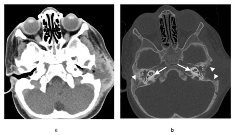
Çocukta otore, kulak çevresinde şişlik veya tedaviye rağmen düzelmeyen otomastoidit görünümünde temporal kemik Langerhans hücreli histiyositozu düşünülmelidir; erişkinde de görülebilir. Kontrastsız ince kesit BT, mastoid/skuamöz kemikte litik yıkımı ve komşu yumuşak doku kitlesini kemik sınırlarıyla birlikte gösterir; tanıyı kesinleştirmez ve MR intrakraniyal-yumuşak doku yayılımını daha iyi değerlendirir. Otomastoidit diye yorumlanan destrüktif lezyonun gecikmesi sistemik hastalık değerlendirmesini erteleyebilir. Tersine, her mastoid opasitesini Langerhans hastalığı saymak da enfeksiyonun klinik ve görüntüleme özelliklerini göz ardı ettirir.
Faz ve pencere
- Kontrastsız tanısal
- İnce kesit BT, mastoid ve skuamöz temporal kemikteki litik kemik yıkımını ve eşlik eden yumuşak doku kitlesini gösterir; kemik destrüksiyonunun değerlendirmesi için altın standarttır.
- Kemik algoritması tanısal
- BT, lezyonun mastoid, skuamöz temporal kemik, dış kulak yolu, orta kulak ve petroz apeks gibi alt bölgelere yayılımını gösterir; kemikçik zinciri çoğunlukla korunmakla birlikte otik kapsül tutulumu görülebilir.
Önerilen pencereler: Kemik (G 1800 / M 400), Yumuşak doku (G 400 / M 40), Beyin (G 80 / M 40).
BT bulguları
- Litik temporal kemik odağı — mastoid veya skuamöz bölümde sınırları belirgin olabilen kemik yıkımı.
- Eşlik eden yumuşak doku kitlesi — litik açıklıktan dış kulak yolu, mastoid veya orta kulak kompartımanına uzanabilir.
- Eğimli kenar bulgusu — iç ve dış korteksin eşit olmayan yıkımı litik lezyon kenarını eğimli gösterebilir; bulgu kalvaryal lezyonlarda tanımlanmıştır ve temporal kemik LCH’sine özgü değildir.
- Düğme sekestrumu (button sekestrum) — litik odak içinde rezidü kemik parçası görülebilir; LCH’ye özgü değildir ve osteomiyelitte de izlenebilir.
- Mastoid odaklı destrüksiyon — mastoid hücre duvarları ve korteks yıkılırken yumuşak doku komponenti eşlik edebilir.
- Kemikçik veya otik kapsül ilişkisi — yaygın kemik yıkımına karşın kemikçik zinciri ve labirent sıklıkla korunabilir; tutulmaları ayrıca raporlanır.
- Ekstratemporal uzanım — komşu kafa derisi, orta kafa çukuru veya kafa tabanına uzanım için yumuşak doku ve beyin pencereleri taranır.
Normalde
Normal aynı taraf temporal kemik BT'sinde mastoid septalar ve dış korteks süreklidir; otik kapsül yoğun, kesintisiz kemik halkası şeklinde, kemikçikler ise küçük kortike yapılar olarak izlenir. Karşı taraf mastoidiyle kıyaslarken yalnız hava hücrelerinin opaklaşmasına değil, septa/korteks yıkımına, litik odağın kenarına ve kemik kaybına eşlik eden yumuşak doku kitlesine odaklanın.
Normal BT ile kıyasla
Aynı bölgenin, kaynak veri setinde patoloji içermediği belirtilen bir BT incelemesini kesit kesit kaydırın; organ sınırlarını açarak anatomiyi hasta görüntüsüyle karşılaştırın. Küçük rastlantısal bulgular tümüyle dışlanmamıştır.
Kaynak: TotalSegmentator v2.01 veri seti (Wasserthal ve ark., Radiology: Artificial Intelligence 2023), CC BY 4.0, vaka s0103; organ sınırları veri setinin otomatik segmentasyonundan, birleştirilerek sadeleştirildi. Görüntüler 2,5 mm kesit aralığına yeniden örneklendi.
Ayırıcı tanı
- Akut koalesan mastoidit
- mastoid septa erozyonu ve yumuşak doku olabilir; klinik enfeksiyon ve kontrastlı incelemede apse eşliği tanıyı destekler.
- Rabdomiyosarkom
- dış kulak yolu veya orta kulakta yumuşak doku kitlesi ve agresif kemik yıkımı yapabilir; MR yayılımı gösterir, doku tanısı gerekir.
- Metastaz, özellikle nöroblastom
- çocukta litik temporal kemik lezyonu yapabilir; başka kemik veya sistemik odaklar ayırıcı tanıyı değiştirir.
- Kolesteatom
- keratin içeren orta kulak kitlesi scutum ve ossikülleri erode eder; izole mastoid litik kemik kitlesi görünümü daha az tipiktir.
- Osteomiyelit
- enfeksiyon zemininde kortikal destrüksiyon ve yumuşak doku inflamasyonu oluşturur; klinik ve eşlik eden apse bulguları ile ayrılır.
Tuzaklar
- Mastoid opasifikasyonu tek başına mastoidit değildir; septa kaybı ve destrüktif litik odak varsa Langerhans hücreli histiyositoz dahil kitle nedenlerini araştırın.
- Temporal kemik Langerhans odağında otik kapsülün korunması tanıyı destekleyebilir fakat şart değildir; yalnız bu işarete dayanarak hastalığı dışlamayın.
- BT’de yumuşak doku ve kemik yıkımı özgül değildir; eğimli kenar ve düğme sekestrumu da tanıyı kanıtlamaz. İntrakraniyal uzanım ve doku karakteri için MR, kesin tanı için histopatolojik korelasyon gerekir.
Kendini dene
Çocukta temporal kemikte, özellikle mastoid/skuamöz parçada litik destrüksiyon ve eşlik eden yumuşak doku kitlesi var; otik kapsül görece korunmuş. Ayırıcı tanıda özellikle hangi hastalık düşünülmelidir?
Cevabı göster
Langerhans hücreli histiyositoz. Temporal kemik LCH’sinde litik destrüksiyon ve yumuşak doku kitlesi görülebilir; kemik labirent görece korunabilir. Bu örüntü uyarıcıdır ama tek başına tanı koydurmaz; enfeksiyon ve diğer destrüktif kitlelerle klinik/histopatolojik korelasyon gerekir.
Temporal kemik LCH’sinde eğimli kenar ve düğme sekestrumu nasıl yorumlanmalıdır?
Cevabı göster
Destekleyici fakat özgül olmayan; eğimli kenar özellikle kalvaryal lezyonlarda tariflenmiştir. Eğimli kenar ve düğme sekestrumu LCH’de görülebilir ancak patognomonik değildir; düğme sekestrumu osteomiyelitte de olabilir. Eğimli kenar bulgusu esas olarak kalvaryal litik lezyonlar için tanımlanmıştır, bu nedenle temporal kemik LCH’sinde tek başına tanı aracı değildir.
İlgili konular
Kaynaklar
Bu sayfadaki 38 cümle ve 2 quiz sorusu aşağıdaki kaynaklardan alıntılarla tek tek karşılaştırıldı; 2 cümle bu karşılaştırmada düzeltildi (2026-10-08).
- Skeletal involvement in Langerhans cell histiocytosispmc.ncbi.nlm.nih.gov
- Cholesteatoma Masquerading as Recurrent Langerhans Cell Histiocytosispmc.ncbi.nlm.nih.gov
- Temporal Bone Langerhans Cell Histiocytosis: An Uncommon Bilateral Presentationpmc.ncbi.nlm.nih.gov
- Bilateral Temporal Bone Langerhans Cell Histiocytosis: Radiologic Pearlspmc.ncbi.nlm.nih.gov
- An Unusual Presentation of Multisystem Langerhans Cell Histiocytosis in a Child: A Case Reportpmc.ncbi.nlm.nih.gov
- [A case of adult temporal bone langerhans cell histiocytosis presenting as posterior canal dehiscence syndrome and literature review].pubmed.ncbi.nlm.nih.gov
- Bilateral temporal bone langerhans cell histiocytosis: radiologic pearls.pubmed.ncbi.nlm.nih.gov
- Temporal Bone Langerhans Cell Histiocytosis: An Uncommon Bilateral Presentation.pubmed.ncbi.nlm.nih.gov
- Cholesteatoma Masquerading as Recurrent Langerhans Cell Histiocytosis.pubmed.ncbi.nlm.nih.gov
- Secondary Acquired Cholesteatoma in Langerhans Cell Histiocytosis Patient.pubmed.ncbi.nlm.nih.gov
- Temporal bone Langerhans cell histiocytosis with uterine cervix involvement and cutaneous Erdheim-Chester disease.pubmed.ncbi.nlm.nih.gov
- Petrous apex lesions in the pediatric population.pubmed.ncbi.nlm.nih.gov
- Intracranial temporal bone angiomatoid fibrous histiocytoma: illustrative case.pubmed.ncbi.nlm.nih.gov
- Eosinophilic granuloma of the temporal bone in children.pubmed.ncbi.nlm.nih.gov
- Langerhans Cell Histiocytosis of the Temporal Bone.pubmed.ncbi.nlm.nih.gov
- Pediatric Langerhans cell histiocytosis of the lateral skull base.pubmed.ncbi.nlm.nih.gov
- Langerhans Cell Histiocytosis of the Temporal Bone.pubmed.ncbi.nlm.nih.gov
- Solitary juvenile xanthogranuloma of temporal bone: a case report.pubmed.ncbi.nlm.nih.gov
- Skull base Langerhans cell histiocytosis presenting as persistent ear discharge with granulation-like tissue in the external ear canal.pubmed.ncbi.nlm.nih.gov
- COVID-19-Associated Severe Aplastic Anemia in an 18-Month-Old Child: A Case Report.pubmed.ncbi.nlm.nih.gov
- Mastoiditis Now Affects Adults: A Case Report of an Occurrence of the Disease in an 82-Year-Old Male.pubmed.ncbi.nlm.nih.gov
- The Challenge of Differentiating Mixed Histiocytosis: A Case Report.pubmed.ncbi.nlm.nih.gov
- An Unusual Presentation of Multisystem Langerhans Cell Histiocytosis in a Child: A Case Report.pubmed.ncbi.nlm.nih.gov
- Langerhans Cell Histiocytosis in Adults: A Canadian Multicenter Case Series.pubmed.ncbi.nlm.nih.gov
- Skull Base Rhabdomyosarcoma Mimicking Osteomyelitis in a Pediatric Patient.pubmed.ncbi.nlm.nih.gov
- A case of bilateral sensorineural hearing loss from Langerhans cell histiocytosis.pubmed.ncbi.nlm.nih.gov
- Bilateral Cavernous Sinus Thrombosis and Clival Osteomyelitis Complicating Pediatric Otitis Media.pubmed.ncbi.nlm.nih.gov
- Orbital pseudotumor and skull base infiltration as the sole manifestation of granulomatosis with polyangiitis.pubmed.ncbi.nlm.nih.gov
- Decoding calvarial lesions: Contemporary neurosurgical management integrating radiological and histopathological features.pubmed.ncbi.nlm.nih.gov
- Xanthogranulomatous Osteomyelitis as a Bone Tumor Mimic: A Systematic Review of Pre-biopsy Diagnostic Clues and Management Decision Points.pubmed.ncbi.nlm.nih.gov
- HRCT imaging of acquired cholesteatoma: a pictorial review.pubmed.ncbi.nlm.nih.gov
- External and middle ear diseases: radiological diagnosis based on clinical signs and symptoms.pubmed.ncbi.nlm.nih.gov
- Rare Disorders of the Vestibular Labyrinth: of Zebras, Chameleons and Wolves in Sheep's Clothing.pubmed.ncbi.nlm.nih.gov
- Operative diagnosis of a meningioma-like intraosseous skull lesion revealing multiple myeloma: Case report and literature review.pubmed.ncbi.nlm.nih.gov
- Bilateral temporal skull xanthoma characterized as a Langerhans cell histiocytosis: a case report.pubmed.ncbi.nlm.nih.gov
- Pediatric Langerhans cell histiocytosis of the temporal bone: clinical and imaging studies of 27 cases.pubmed.ncbi.nlm.nih.gov
- An Unusual Skull With Bilateral Multiple Bony Lesions on the Parietal and Frontal Bones of the Superior Surface of the Calvaria: A Case Report.pubmed.ncbi.nlm.nih.gov
- Review of imaging modalities and radiological findings of calvarial lesions.pubmed.ncbi.nlm.nih.gov
- Multidisciplinary Exploration of Computed-Tomographic and Ancient-DNA Data of an Iron Age Skull From Latvia With Multiple Lytic Bone Lesions: Differential Diagnosis Between Metastatic Carcinoma, Multiple Myeloma and Skeletal Tuberculosis.pubmed.ncbi.nlm.nih.gov
- Case Report: A rare adult multisystem Langerhans cell histiocytosis involving the skin, thyroid, jaw-periodontal region, and bone: diagnostic challenges and complementary PET/CT-SPECT/CT findings.pubmed.ncbi.nlm.nih.gov
- Rare Diseases of the Middle Ear and Lateral Skull Base.pubmed.ncbi.nlm.nih.gov
- Lateral Skull Base Approaches in Pediatric Skull Base Surgery.pubmed.ncbi.nlm.nih.gov
- Disease extent, not lesion location, determines relapse risk in single-system skeletal Langerhans cell histiocytosis.pubmed.ncbi.nlm.nih.gov
- Clinician-Guided Deep Learning Segmentation of Skull Base Pneumatization on Computed Tomography Using 3D Slicer and MONAI Label.pubmed.ncbi.nlm.nih.gov
- Key pre-operative CT parameters for cochlear implantation: a focused radiologic review with surgical relevance.pubmed.ncbi.nlm.nih.gov
- Cerebellar abscess complicating chronic suppurative otitis media: An unusual route of intracranial spread.pubmed.ncbi.nlm.nih.gov
- Correlation analysis between CT values of cochlear bone labyrinth and auditory function and disease progression in patients with chronic suppurative otitis media.pubmed.ncbi.nlm.nih.gov
- Acquired external auditory canal stenosis: clinical characteristics, surgical strategies and prognostic analysis.pubmed.ncbi.nlm.nih.gov
- Subacute Onset of Perineural Rhino-Cerebral Mucormycosis Mimicking Giant Cell Arteritis: A Case Report.pubmed.ncbi.nlm.nih.gov
- Acute Bacterial Meningitis and Petrous Apicitis in a Child with Aplasia Cutis Congenita: A Case Report.pubmed.ncbi.nlm.nih.gov
- Rhabdomyosarcoma of the temporal bone in children - a systematic review.pubmed.ncbi.nlm.nih.gov
- An ultra-late maxillofacial cavity following multimodal treatment for adult head and neck rhabdomyosarcoma: a case report with a 16-year follow-up.pubmed.ncbi.nlm.nih.gov
- SMARCB1-deficient sinonasal carcinoma in a pediatric patient: Imaging findings and differential diagnosis of a rare entity.pubmed.ncbi.nlm.nih.gov
- Neuroblastoma Metastasis to the Mandible in Children: A Case Report and Focused Narrative Review of Reported Cases.pubmed.ncbi.nlm.nih.gov
- Artificial Intelligence for Diagnosis, Risk Stratification, and Prognosis of Neuroblastoma - A Systematic Review and Meta-Analysis.pubmed.ncbi.nlm.nih.gov
- Tislelizumab combined with Anti-GD2 antibody-based chemoimmunotherapy for pediatric relapsed/refractory high-risk neuroblastoma: a two-Case Report.pubmed.ncbi.nlm.nih.gov
- Applications of Artificial Intelligence in Temporal Bone Imaging: Advances and Future Challenges.pubmed.ncbi.nlm.nih.gov
- An in-depth discussion of cholesteatoma, middle ear Inflammation, and langerhans cell histiocytosis of the temporal bone, based on diagnostic results.pubmed.ncbi.nlm.nih.gov
- Case report: Langerhans cell histiocytosis of the temporal bone in children: Challenging diagnosis of a rare disease with some pitfalls.pubmed.ncbi.nlm.nih.gov
- Prevalence of oral lesions in Langerhans cell histiocytosis: a systematic review and meta-analysis.pubmed.ncbi.nlm.nih.gov
- Temporal bone manifestation of primary extranodal Rosai-Dorfman disease: a case report.pubmed.ncbi.nlm.nih.gov
- Imaging of Skull Base Tumors.pubmed.ncbi.nlm.nih.gov
- Calvarial Langerhans cell histiocytosis in an Adult: Typical imaging findings in an atypical age group.pubmed.ncbi.nlm.nih.gov
- Atypical fibrous histiocytoma of the skull: a case report of temporal bone involvement and comprehensive literature review.pubmed.ncbi.nlm.nih.gov
- Diagnosis and Management of Middle Ear Neuroendocrine Tumour (MeNET).pubmed.ncbi.nlm.nih.gov
- Nocardia Otomastoiditis in a Chemotherapy-Induced Neutropenic Patient With Diffuse Large B-cell Lymphoma: Successful Medical Management.pubmed.ncbi.nlm.nih.gov
- Bezold's Abscess Secondary to a Subclinical Mastoid Infection: Diagnostic Challenges and Literature Review.pubmed.ncbi.nlm.nih.gov
- Retinoblastoma and beyond: pediatric orbital mass lesions.pubmed.ncbi.nlm.nih.gov
- Multifocal Osseous Tuberculosis Mimicking Langerhans' Cell Histiocytosis: A Case Series.pubmed.ncbi.nlm.nih.gov
- Retinal vasculitis with Chronic Recurrent Multifocal Osteomyelitis: a case report and review of the literature.pubmed.ncbi.nlm.nih.gov
- Aural Polyp is not Always Due to Chronic Otitis Media (COM): Preoperative Computed Tomographic Scan is Good Pointer for Sinister Lesions.pubmed.ncbi.nlm.nih.gov
- Targeted Therapy in Erdheim-Chester Disease: A Case Report and Review of the Literature.pubmed.ncbi.nlm.nih.gov
- Pediatric non-infectious inflammatory lytic lesions of the skull: The pitfalls of diagnosis.pubmed.ncbi.nlm.nih.gov
- Contrast-enhanced sonography in the diagnosis of Langerhans cell hyperplasia in children with isolated liver involvement: A case report and review of the literature.pubmed.ncbi.nlm.nih.gov
- Posttraumatic intradiploic cyst-like lesion of the frontal bone without cortical disruption: A case report.pubmed.ncbi.nlm.nih.gov
- Brazilian Society of Otology task force - cochlear implant ‒ recommendations based on strength of evidence.pubmed.ncbi.nlm.nih.gov
- Chronically Discharging Ears: Evalution with High Resolution Computed Tomography.pubmed.ncbi.nlm.nih.gov
- Classification and Current Management of Inner Ear Malformations.pubmed.ncbi.nlm.nih.gov
- Imaging for Plasma Cell Dyscrasias: What, When, and How?pubmed.ncbi.nlm.nih.gov
- Pulsatile Tinnitus: Differential Diagnosis and Radiological Work-Up.pubmed.ncbi.nlm.nih.gov
- Emerging and re-emerging infectious disease in otorhinolaryngology.pubmed.ncbi.nlm.nih.gov
- Long-term safety and effectiveness of the "OptEase" vena cava filter.pubmed.ncbi.nlm.nih.gov
Eğitim amaçlıdır; hasta başında tanı ve tedavi kararının yerine geçmez. İçerik yapay zekâ destekli bir süreçle güncel literatüre göre cümle cümle kaynaklarla karşılaştırıldı, ancak uzman radyolog incelemesinden geçmedi. Klinik kararlarda güncel kılavuzları, kurum protokollerini ve radyoloji raporunu esas alın.