Özet
Çiğneme sırasında ağrı, krepitasyon veya hareket kısıtlılığı olan hastada TME'nin dejeneratif kemik yeniden şekillenmesi araştırılır. İnce kesit kontrastsız BT ve CBCT, kondil ile temporal eklem yüzündeki kortikal ve subkortikal kemik değişikliklerini diske ait yumuşak doku bulgularından daha iyi gösterir. Osteofit, erozyon, subkortikal kist veya yaygın skleroz tanısal dejeneratif değişikliklerdir; tek başına hafif düzleşme ya da sınırlı skleroz özgül değildir. Değişiklikleri aşırı yorumlamak normal yeniden şekillenmeyi hastalık saydırabilir; belirgin erozyon veya kondiler deformite ise ilerleyici eklem hasarını gözden kaçırmamak için tanımlanmalıdır.
Faz ve pencere
- Kontrastsız tanısal
- İnce kesit kemik algoritmasında kondil başı ve glenoid fossa/eminens korteksi incelenir; marjinal osteofit, kortikal yüzey kaybı, subkortikal kist ve yük taşıyan bölgede skleroz görünür. Kontrastlı faz gerekmez; eklem diski ve sinovyal yumuşak dokular için BT yeterli değildir.
Önerilen pencereler: Kemik (G 1800 / M 400).
BT bulguları
- Marjinal osteofit — eklem kenarında sklerotik sınırlı, dışa doğru açılan kemik çıkıntısı; kondil başında ön veya üst kenarda belirginleşebilir.
- Kortikal erozyon — normal kesintisiz eklem korteksinde düzensiz kayıp veya çökme vardır; yalnız yüzey düzleşmesiyle eş tutulmaz.
- Subkortikal kist — dejeneratif osteoartrit için tanısal BT ölçütlerinden biridir; eklem yüzü altında, normal trabeküler kemikten farklı yoğunlukta, iyi sınırlı kaviter odak olarak izlenir.
- Yaygın kondiler skleroz — kondil başının trabeküler ayrıntısı silinir ve yoğun kemik görünümü başın geniş bölümüne yayılır.
- Subkondral skleroz — yük taşıyan eklem yüzünün hemen altındaki kortikal plak komşu yük taşımayan kemikten daha kalın/yoğun izlenir.
- Yüzey düzleşmesi — kondilin yuvarlak üst konturu kaybolur; erozyon veya osteofit olmaksızın tek başına osteoartrit kanıtı değildir.
- Karşılıklı eklem remodelingi — temporal eklem yüzündeki skleroz, osteofit veya erozyon kondil bulgularıyla aynı tarafta kıyaslanır.
Ölçütler ve sınıflamalar
- Ahmad görüntüleme ölçütleri
- A: osteoartrit yok; kondil boyutu olağan, subkortikal skleroz ve yüzey düzleşmesi yok, deformasyon bulgusu yok. B: belirsiz; düzleşme ve/veya subkortikal skleroz olabilir ancak kist, erozyon, osteofit veya yaygın sklerozla deformasyon yoktur. C: osteoartrit; subkortikal kist, yüzey erozyonu, osteofit veya yaygın sklerozla deformasyon vardır.
Normalde
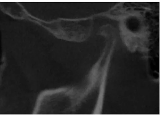
Normal kondil başı düzgün kortikal örtüye ve yuvarlak kontura sahiptir; glenoid fossa ile artiküler eminensin kemik yüzleri kesintisizdir ve subkortikal trabeküler yapı seçilir. İki tarafı aynı kemik penceresinde kıyaslayarak osteofit, kortikal erozyon veya subkortikal kistin gerçekten bulunduğunu arayın; yalnız düzleşme ve sınırlı skleroz dejeneratif hastalık tanısı koydurmaz.
Ayırıcı tanı
- Yaşa bağlı adaptif remodelingi
- düzgün sınırlı düzleşme veya hafif skleroz vardır; kortikal erozyon, belirgin osteofit ya da subkortikal kist yoktur.
- Romatoid/inflamatuvar artrit
- erozyonlar ve eklem hasarı osteoartritten farklı olarak sistemik inflamatuvar eklem bulguları ve klinik öyküyle birlikte olabilir.
- Septik artrit
- şiddetli ağrı, ateş, şişlik ve fonksiyon kaybı ile eklemi hızla bozabilir; kronik marjinal osteofit paterni beklenmez.
- Osteokondrom
- kondil yüzeyinden dışa doğru büyüyen kitle formasyonu gösterir; malign transformasyon çok nadirdir.
- Kondil osteonekrozu veya rezorpsiyonu
- kondil hacminde azalma ve yüzey çökmesi ön plandadır; karşı eklem yüzündeki osteofit/kist paterni olmayabilir.
Tuzaklar
- Kondil düzleşmesi ve sınırlı subkortikal skleroz tek başına osteoartrit değildir; osteofit, erozyon, kist veya yaygın sklerozla yapısal deformasyon arayın.
- Eklem diski kontrastsız BT'de güvenilir değerlendirilemez; klik veya disk yer değiştirmesi sorusunda kemik bulgularını disk tanısı gibi sunmayın.
- Tek taraflı düzleşme anatomik yüklenme veya eski remodelingi yansıtabilir; karşı TME ve temporal eklem yüzünü aynı planda kıyaslayın.
Kendini dene
Kondil başında düzgün yüzey düzleşmesi ve hafif subkortikal yoğunluk artışı var; osteofit, kist ya da erozyon yok. Ahmad ölçütlerine göre?
Cevabı göster
Belirsiz görüntü grubu. Sınırlı düzleşme ve subkortikal skleroz, deformasyon oluşturan kist/erozyon/osteofit/yaygın skleroz olmadan belirsiz gruptadır. Kesin osteoartritte deformasyon kriterlerinden biri gerekir; normal grupta bu değişiklikler bulunmaz; septik artrit bu kronik paternle tanınmaz.
Kondil kenarından çıkan sklerotik marjinal çıkıntı ve yüzey altında kaviter odak birlikte görülüyor. Hangi süreç daha olası?
Cevabı göster
Dejeneratif osteoartrit. Marjinal osteofit ile subkortikal kist birlikte dejeneratif eklem hastalığını destekler. Osteokondromlar eklem yüzünden dışa doğru büyüyen ve kortikomedüller süreklilik gösteren lezyonlardır; ankilozda eklem boşluğunda kemik köprü izlenir; kondiler çıkıkta kondil glenoid fossa dışına yer değiştirmiştir.
Kaynaklar
Bu sayfadaki 40 cümle ve 2 quiz sorusu aşağıdaki kaynaklardan alıntılarla tek tek karşılaştırıldı; 6 cümle bu karşılaştırmada düzeltildi (2026-10-08).
- Temporomandibular joint pathologies: pictorial reviewpmc.ncbi.nlm.nih.gov
- Temporomandibular Joint Osteoarthritis Diagnosis Employing Artificial Intelligence: Systematic Review and Meta-Analysis.pubmed.ncbi.nlm.nih.gov
- Association of bone marrow edema with temporomandibular joint (TMJ) osteoarthritis and internal derangements.pubmed.ncbi.nlm.nih.gov
- Current Applications of Deep Learning and Radiomics on CT and CBCT for Maxillofacial Diseases.pubmed.ncbi.nlm.nih.gov
- Equine temporomandibular joint diseases: A systematic review.pubmed.ncbi.nlm.nih.gov
- Understanding the Internal Derangement and Degenerative Joint Disease Process Through Radiographic Imaging.pubmed.ncbi.nlm.nih.gov
- Mapping cell diversity and dynamics in inflammatory temporomandibular joint osteoarthritis with pain at single-cell resolution.pubmed.ncbi.nlm.nih.gov
- Minimally Invasive Approach for Diagnosing TMJ Osteoarthritis.pubmed.ncbi.nlm.nih.gov
- 3D Auto-Segmentation of Mandibular Condyles.pubmed.ncbi.nlm.nih.gov
- Evolution of mirror-image pain in temporomandibular joint osteoarthritis mouse model.pubmed.ncbi.nlm.nih.gov
- Automated Detection of TMJ Osteoarthritis Based on Artificial Intelligence.pubmed.ncbi.nlm.nih.gov
- 3D superimposition and understanding temporomandibular joint arthritis.pubmed.ncbi.nlm.nih.gov
- Effectiveness of treatment with viscosupplementation in temporomandibular joints with or without effusion.pubmed.ncbi.nlm.nih.gov
- Lacc1-engineered extracellular vesicles reprogram mitochondrial metabolism to alleviate inflammation and cartilage degeneration in TMJ osteoarthritis.pubmed.ncbi.nlm.nih.gov
- Mechanical stress reduces secreted frizzled-related protein expression and promotes temporomandibular joint osteoarthritis via Wnt/β-catenin signaling.pubmed.ncbi.nlm.nih.gov
- Sleep Disorders and Their Relationship With Temporomandibular Joint Disorders Derived From a Psychoemotional Disorder.pubmed.ncbi.nlm.nih.gov
- Prevalence and characteristics of temporomandibular joint disc displacement based on Magnetic Resonance Imaging (MRI) in pre-clinical diagnosis: A retrospective study.pubmed.ncbi.nlm.nih.gov
- Complete Denture as Pivot Splint: A Better Approach for the Treatment of Temporomandibular Joint Osteoarthritis.pubmed.ncbi.nlm.nih.gov
- The Cervical-Hyoid Dynamics in Correlation to Craniomandibular Disorders: A Report of Two Cases.pubmed.ncbi.nlm.nih.gov
- Quantitative Imaging of the Mandibular Condyle: Radiomics and Microarchitectural Biomarkers.pubmed.ncbi.nlm.nih.gov
- Anterior Disc Displacement in a Young Patient With Stage II Osteoarthritis: Diagnostic and Therapeutic Approach. A Case Report.pubmed.ncbi.nlm.nih.gov
- Tooth Wear Severity and Temporomandibular Joint Degenerative Findings on Cone-Beam Computed Tomography: A Cross-Sectional Study in Adults.pubmed.ncbi.nlm.nih.gov
- Can Deep Learning Methods Differentiate Temporomandibular Joint Disorders From Healthy Joints? A 3D Artificial Intelligence Algorithm Study Based on CBCT Images.pubmed.ncbi.nlm.nih.gov
- Bone Changes in the Temporomandibular Joint: A Retrospective Cone Beam Computed Tomography Study.pubmed.ncbi.nlm.nih.gov
- Biomimetic osteochondral regeneration of the TMJ condyle: integrating BMP-2 osteoinduction, MSC-mediated fibrochondrogenesis, and gradient 3D-printed scaffolds.pubmed.ncbi.nlm.nih.gov
- Single-cell RNA sequencing of healthy and diseased rat temporomandibular joint condyle cartilage.pubmed.ncbi.nlm.nih.gov
- Identification of aging-related signature genes in temporomandibular joint degeneration based on bioinformatics.pubmed.ncbi.nlm.nih.gov
- U-HRCT and MRI-Based Indicators for Limited Mouth Opening in Patients With Temporomandibular Disorders.pubmed.ncbi.nlm.nih.gov
- Imaging Evaluation for Jaw Deformities: Diagnostic Workup and Pre-Treatment Imaging Checklist for Orthognathic Surgery.pubmed.ncbi.nlm.nih.gov
- Diagnostic Value of Panoramic Radiographs in the Assessment of Degenerative Joint Disease: A Retrospective Study.pubmed.ncbi.nlm.nih.gov
- A rare case of malignant transformation of condylar osteochondroma: Diagnosis and treatment.pubmed.ncbi.nlm.nih.gov
- Contrast-Enhanced Brain MRI for Ambulatory Evaluation of Headache: Comprehensive or Excessive?pubmed.ncbi.nlm.nih.gov
- Real-time morphological and dosimetric adaptation in nasopharyngeal carcinoma radiotherapy: insights from autosegmented fractional fan-beam CT.pubmed.ncbi.nlm.nih.gov
- Vision-Language Model as a 'Zero-Shot' Assistant for Evaluating Condylar Osseous Changes in Cone-beam Computed Tomography.pubmed.ncbi.nlm.nih.gov
- Radiographic evaluation of degenerative findings in the temporomandibular joint using CBCT: a prevalence-based study.pubmed.ncbi.nlm.nih.gov
- Case Report: Longitudinal temporomandibular joint remodeling in MMP2 associated nodulosis arthropathy osteolysis syndrome during serial panoramic follow up.pubmed.ncbi.nlm.nih.gov
- Accuracy of Computed Tomography in Diagnosing Temporomandibular Joint Osteoarthritis Relative to Histopathological Findings-An Ex Vivo Study of 41 Horses.pubmed.ncbi.nlm.nih.gov
- Comparative Evaluation of Osteoarthritic Changes in the Temporomandibular Joint Using Cone-Beam Computed Tomography Among Individuals With and Without Temporomandibular Disorders: A Comprehensive Review.pubmed.ncbi.nlm.nih.gov
- Functional Biomaterials and 3D Bioprinting Approaches for Temporomandibular Joint Reconstruction: A Narrative Review.pubmed.ncbi.nlm.nih.gov
- Cone-Beam CT-Based Analysis of Temporomandibular Joint Osseous Changes in Orthognathic Surgery Patients: A Retrospective Cross-Sectional Study.pubmed.ncbi.nlm.nih.gov
- Prevalence of Osteoarthritis Using Cone Beam in Patients at the University of Cuenca.pubmed.ncbi.nlm.nih.gov
- Computer-Aided Diagnosis of Equine Temporomandibular Joint Osteoarthritis Using Machine Learning Integrating Computed Tomography Findings and Synovial Fluid Biomarkers.pubmed.ncbi.nlm.nih.gov
- Mastication-Induced Electrical Stimulation Activates Prg4<sup>+</sup> Chondroprogenitors for Osteoarthritis Therapy.pubmed.ncbi.nlm.nih.gov
- Clinical and imaging outcomes following non-invasive management of temporomandibular joint osteoarthritis: An exploratory retrospective cohort study.pubmed.ncbi.nlm.nih.gov
- Condylar Morphology in Patients with Unilateral Maxillary Impacted Canines: A Panoramic Radiography Study.pubmed.ncbi.nlm.nih.gov
- Evaluation of TMJ bone microarchitecture in malocclusion and tooth loss using fractal dimension analysis.pubmed.ncbi.nlm.nih.gov
- Temporomandibular Joint Defects: Etiology and Reconstruction Strategy: A Narrative Review.pubmed.ncbi.nlm.nih.gov
- Goat temporomandibular joint osteochondral defects for future regenerative studies of osteoarthritis-like degeneration.pubmed.ncbi.nlm.nih.gov
- Preliminary Evaluation of Static and Kinematic Magnetic Resonance Imaging (MRI) of the Feline Temporomandibular Joint (TMJ): A Comparative Analysis of Joint Inclination Angles in Intact and Dislocated Conditions.pubmed.ncbi.nlm.nih.gov
- Evaluation of temporomandibular joint disc displacement via MRI and computerized axiography: a case report.pubmed.ncbi.nlm.nih.gov
- Association of articular eminence inclination and glenoid fossa roof thickness with sagittal skeletal classes: a CBCT study.pubmed.ncbi.nlm.nih.gov
- Correlation Between Osseous Degenerative Changes on Cone Beam Computed Tomography and Disc Displacement on Magnetic Resonance Imaging in Temporomandibular Joint Osteoarthritis.pubmed.ncbi.nlm.nih.gov
- Dynamic Mechanics and Longitudinal Changes in Temporomandibular Joint Structural Integrity.pubmed.ncbi.nlm.nih.gov
- Clinical and radiographic outcomes of a novel posterior occlusal splint in adults with temporomandibular joint osteoarthritis: a retrospective case series.pubmed.ncbi.nlm.nih.gov
- Correlation Between Condylar Shape and Malocclusion: CBCT Analysis.pubmed.ncbi.nlm.nih.gov
- Evaluation of the mandibular condylar bone microarchitecture in people living with HIV.pubmed.ncbi.nlm.nih.gov
- Quantitative Radiographic Progression of Joint Space Narrowing in Medial and Lateral Compartment Knee Osteoarthritis After Intraarticular Platelet-Rich Plasma Injection.pubmed.ncbi.nlm.nih.gov
- Recommendations for Imaging of the Temporomandibular Joint. Position Statement from the American Academy of Oral and Maxillofacial Radiology and the American Academy of Orofacial Pain.pubmed.ncbi.nlm.nih.gov
- A Comparative Study of Temporomandibular Joints in Adults with Definite Sleep Bruxism on Magnetic Resonance Imaging and Cone-Beam Computer Tomography Images.pubmed.ncbi.nlm.nih.gov
- Defining subcellular synovial responses in TMJ osteoarthritis onset via mechanical stress and articular disk derangement models.pubmed.ncbi.nlm.nih.gov
- Assessment of Temporomandibular Joint Degeneration in Rheumatoid Arthritis Patients Using Cone-Beam Computed Tomography and Musculoskeletal Ultrasound.pubmed.ncbi.nlm.nih.gov
- Relationship between severe tooth wear and temporomandibular disorders: a cross-sectional study using propensity score matching analysis.pubmed.ncbi.nlm.nih.gov
- A Jaw out of joint: Uncovering a rare infectious dislocation of temporo-mandibular junction: A case report.pubmed.ncbi.nlm.nih.gov
- Mandibular Condyle Osteomyelitis Following Third Molar Extraction: A Case Report and Literature Review.pubmed.ncbi.nlm.nih.gov
- Septic arthritis of the temporomandibular joint-a case report and review of the literature.pubmed.ncbi.nlm.nih.gov
- Association between systemic osteoporosis and mandibular condyle characteristics: A comparative imaging study.pubmed.ncbi.nlm.nih.gov
- Clinician-Recorded Temporomandibular Disorder at 12 Months After Digitally Planned Mandibular Fracture Repair: Associations with Condylar Injury and Early Functional Signs.pubmed.ncbi.nlm.nih.gov
- Arthrocentesis Plus Injectable Platelet-Rich Fibrin in Patients With Temporomandibular Joint Dysfunction: An Updated Meta-Analysis With Trial Sequential Analysis.pubmed.ncbi.nlm.nih.gov
- Magnetic resonance imaging in oral and maxillofacial surgery: a structured guide for trainees.pubmed.ncbi.nlm.nih.gov
- CBCT-based assessment of temporomandibular joint morphology in adults with unilateral cleft lip and palate: a comparative study.pubmed.ncbi.nlm.nih.gov
- Chondroid Tenosynovial Giant Cell Tumor of the Temporomandibular Joint: A Case Report.pubmed.ncbi.nlm.nih.gov
- Valgus Tibial Component Placement: A Novel Risk Factor for Tibial Fracture in Cementless Oxford® Partial Knee Arthroplasty.pubmed.ncbi.nlm.nih.gov
- Application of artificial intelligence in the diagnosis and management of temporomandibular joint osteoarthritis using cone-beam computed tomography: An evidence-based systematic review.pubmed.ncbi.nlm.nih.gov
- Capability of dental practitioners to identify condylar changes on orthopantomograms: a multicenter cross-sectional study in Pakistan.pubmed.ncbi.nlm.nih.gov
- Beyond Cartilage Regeneration: Targeting the Subchondral Bone with Extracorporeal Shock Wave Therapy and Regenerative Medicine in Knee Osteoarthritis.pubmed.ncbi.nlm.nih.gov
- Subchondral Bone Regeneration of the Porcine Knee Joint Using α-TCP Biocement Enriched with Manuka Honey-Qualitative Pilot Study.pubmed.ncbi.nlm.nih.gov
- Development of AI-based tools for assessing temporomandibular joint disorders using MRI images.pubmed.ncbi.nlm.nih.gov
- Ultrasonographic and Radiographic Evaluation of Osteoarthritic Changes in the Temporomandibular Joint.pubmed.ncbi.nlm.nih.gov
- Is Ultrasonography an Effective Method for Diagnosing Degenerative Changes in the Temporomandibular Joint?pubmed.ncbi.nlm.nih.gov
- Improving TMJ Diagnosis: A Deep Learning Approach for Detecting Mandibular Condyle Bone Changes.pubmed.ncbi.nlm.nih.gov
- Potential of ultra-high-resolution CT in detecting osseous changes of temporomandibular joint: experiences in temporomandibular disorders.pubmed.ncbi.nlm.nih.gov
- <i>In Vivo</i> comparative evaluation of early- and late-stage ACLT-hMnX rat models: a case study of avidin as a cartilage-penetrating nanocarrier for disease-modifying osteoarthritis drugs.pubmed.ncbi.nlm.nih.gov
- Imaging findings in the temporomandibular joint complex following radiation therapy for head and neck cancer: a review.pubmed.ncbi.nlm.nih.gov
- Demographic and structural indicators of temporomandibular joint osteoarthritis in asymptomatic adults: a cone-beam computed tomography-based multivariate analysis.pubmed.ncbi.nlm.nih.gov
Eğitim amaçlıdır; hasta başında tanı ve tedavi kararının yerine geçmez. İçerik yapay zekâ destekli bir süreçle güncel literatüre göre cümle cümle kaynaklarla karşılaştırıldı, ancak uzman radyolog incelemesinden geçmedi. Klinik kararlarda güncel kılavuzları, kurum protokollerini ve radyoloji raporunu esas alın.