Özet
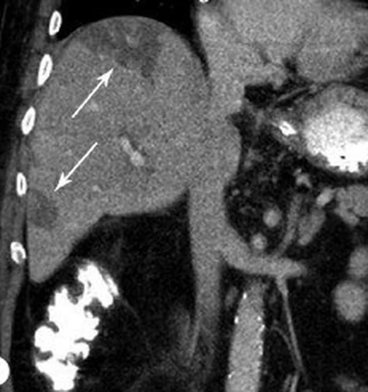
Fokal yağlanma ve fokal yağdan korunma, yaygın karaciğer yağlanmasının içinde veya dışında bölgesel atenüasyon farkı oluşturan kitle taklitçileridir. BT'de lezyon benzeri alanın şekli, tipik yerleşimi ve komşu damarlarla ilişkisi tanımayı sağlar; kontrastlı portal venöz görüntüler bu paternin parankim içindeki dağılımını gösterir. Kitle etkisinin olmaması ve damarların alan içinden doğal seyri, solid tümör olasılığını azaltır ancak atipik odakları tek başına dışlamaz. Yanlış kitle tanısı gereksiz ileri incelemeye, gerçek kitleyi yağ değişikliği sanmak ise gecikmiş tanıya neden olabilir.
Faz ve pencere
- Portal tanısal
- Steatoz BT’de karaciğer atenüasyonunu düşürür; portal venöz fazda safra kesesi fossası veya segment IV’ün arka-medialinde çevre parankime göre daha yüksek atenüasyon, steatozu destekleyebilir. Portal venöz fazda, safra kesesi fossası veya segment IV’ün arka-medialinde çevre parankime göre artmış atenüasyon görülebilir; bu bulgu steatozu destekleyebilir.
- Kontrastsız
- BT’de steatoz karaciğer atenüasyonunu düşürür; portal venöz fazda safra kesesi fossası veya segment IV’ün arka-medialindeki göreceli yüksek atenüasyon steatozu destekleyebilir. Portal venöz faz kontrastlı BT’de safra kesesi fossası veya segment IV’ün arka-medialinde çevre parankime göre artmış atenüasyon, steatozu destekleyebilir.
Önerilen pencereler: Karaciğer (G 150 / M 30), Batın (G 400 / M 50).
BT bulguları
- Coğrafi veya kama biçimli atenüasyon alanı; çoğu kez segmental ya da lobar dağılım gösterir.
- Fokal steatozun normal parankime göre hipoatenüasyonu veya yaygın steatoz içindeki korunmuş alanın göreceli hiperatenüasyonu.
- Safra kesesi fossası veya segment IV’ün arka-medialinde göreceli yüksek atenüasyon, kontrastlı BT’de steatozu destekleyebilir.
- Fokal yağlanma ve yağdan korunmuş alanlar gerçek karaciğer lezyonlarını taklit edebilir; tipik ve atipik görüntüleme özelliklerini tanımak yanlış sınıflandırmayı önlemeye yardımcı olur.
- Safra yollarında bası, segmental dilatasyon veya çevre dokuda kitle etkisinin bulunmaması.
- Portal venöz faz kontrastlı BT’de safra kesesi fossası veya segment IV’ün arka-medialinde çevre parankime göre artmış atenüasyon steatozu destekleyebilir.
- Üçüncü inflow oluşturan venlerin karaciğere drenajı fokal steatoz veya yağdan korunmuş alanlarla ilişkili olabilir.
Normalde
Şüpheli alanın içinden geçen portal/hepatik venlerin seyri, safra kanallarının kalibresi ve karaciğer konturunda dışa bombeleşme olup olmadığına aynı portal faz kesitlerinde bakın.
Normal BT ile kıyasla
Aynı bölgenin, kaynak veri setinde patoloji içermediği belirtilen bir BT incelemesini kesit kesit kaydırın; organ sınırlarını açarak anatomiyi hasta görüntüsüyle karşılaştırın. Küçük rastlantısal bulgular tümüyle dışlanmamıştır.
Kaynak: TotalSegmentator v2.01 veri seti (Wasserthal ve ark., Radiology: Artificial Intelligence 2023), CC BY 4.0, vaka s0541; organ sınırları veri setinin otomatik segmentasyonundan, birleştirilerek sadeleştirildi. Görüntüler 2,5 mm kesit aralığına yeniden örneklendi.
Ayırıcı tanı
- Metastaz
- fokal yağdan korunmuş alanlar, özellikle metastatik tümörleri taklit edebilir.
- Hepatoselüler karsinom
- sirotik zeminde arteriyel fazda belirgin kontrastlanma ve portal/geç fazda yıkanma gösterebilir.
- Geçici hepatik atenüasyon farkı
- arteriyel fazda kama biçimli hiperatenüasyon oluşturur ve portal fazda silikleşebilir.
- Hemangiom
- periferik nodüler kontrastlanma ve zamanla merkeze ilerleyen dolum paterni görülebilir.
Tuzaklar
- Hepatik psödolezyonların atipik görüntüleme özellikleri gerçek lezyonları taklit edebilir veya gizleyebilir; bu nedenle tipik ve atipik görünümler tanınmalıdır.
- Yağdan korunmuş alanlar yağlı karaciğerde kitleyi taklit edebilir; safra kesesi fossası veya segment IV’ün arka-medialindeki göreceli yüksek atenüasyon steatozu destekleyebilir.
- Arteriyel fazdaki perfüzyon farklılığı, yağlanma gibi hipodens ya da hiperdens alan oluşturabilir; fazlar arası görünüm değişimi değerlendirilir.
- Küçük veya belirsiz odaklarda manyetik rezonans görüntüleme, bilgisayarlı tomografi ile çelişen veya doğrulanması gereken fokal yağ değişimlerini teyit etmek için problem çözme aracı olarak kullanılabilir.
Kendini dene
Portal venöz fazlı BT’de safra kesesi fossası veya segment IV’ün arka-medialinde zemine göre artmış atenüasyon görülmesi en çok hangi durumu destekler?
Cevabı göster
Hepatik steatoz. Bu tipik bölgelerde zemine göre artmış atenüasyon, kontrastlı BT’de hepatik steatoz tanısını destekleyebilir; yöntemin duyarlılığı sınırlıdır.
Yaygın yağlı karaciğerde safra kesesi fossası veya segment IV’ün arka-medialinde çevre parankime göre artmış atenüasyon görülüyor. Bu alan neyi temsil edebilir?
Cevabı göster
Yağdan korunmuş parankim. Yağlı karaciğerde fokal yağdan korunma görülebilir; safra kesesi fossası veya segment IV’ün arka-medialindeki göreceli artmış atenüasyon kontrastlı BT’de steatozu destekleyebilir. Fokal yağlanma ve yağdan korunma solid karaciğer lezyonlarını taklit edebilir.
İlgili konular
Kaynaklar
Bu sayfadaki 35 cümle ve 2 quiz sorusu aşağıdaki kaynaklardan alıntılarla otomatik olarak karşılaştırıldı; 20 cümle bu karşılaştırmada düzeltildi (2026-10-09).
- Imaging of non alcoholic fatty liver disease: A road less travelledpmc.ncbi.nlm.nih.gov
- Hepatic Steatosis: Etiology, Patterns, and Quantification.pubmed.ncbi.nlm.nih.gov
- Hepatic pseudolesions caused by alterations in intrahepatic hemodynamics.pubmed.ncbi.nlm.nih.gov
- Unveiling the unreal: Comprehensive imaging review of hepatic pseudolesions.pubmed.ncbi.nlm.nih.gov
- The current status of contrast-enhanced ultrasound in China.pubmed.ncbi.nlm.nih.gov
- The cheating liver: imaging of focal steatosis and fatty sparing.pubmed.ncbi.nlm.nih.gov
- Hepatic incidentaloma: the rule of tens.pubmed.ncbi.nlm.nih.gov
- Imaging patterns and focal lesions in fatty liver: a pictorial review.pubmed.ncbi.nlm.nih.gov
- Detection of hepatic steatosis on contrast-enhanced CT images: diagnostic accuracy of identification of areas of presumed focal fatty sparing.pubmed.ncbi.nlm.nih.gov
- Nodular focal fat sparing of liver mimicking hepatocellular carcinoma in contrast-enhanced ultrasound: A case report.pubmed.ncbi.nlm.nih.gov
- Tc-99m Sulfur Colloid Scintigraphy in Differentiating Fat Sparing from Lymphomatous Involvement of Liver-Old Wine in New Bottle.pubmed.ncbi.nlm.nih.gov
- Fatty liver deposition and sparing: a pictorial review.pubmed.ncbi.nlm.nih.gov
- 68Ga-DOTATATE PET/CT in Focal Fatty Sparing of the Liver.pubmed.ncbi.nlm.nih.gov
- Findings and implications of focal fatty sparing of the liver at follow-up: a preliminary study based on sonography, computed tomography, and magnetic resonance imaging.pubmed.ncbi.nlm.nih.gov
- Comparison of contrast-enhanced ultrasonography with grey-scale ultrasonography and contrast-enhanced computed tomography in diagnosing focal fatty liver infiltrations and focal fatty sparing.pubmed.ncbi.nlm.nih.gov
- Multifocal fatty sparing areas in a steatotic liver mimicking hepatic lesions: a case report and review of the literature.pubmed.ncbi.nlm.nih.gov
- Focal Hepatic Steatosis Mimicking a Hilar Tumor With Rapid Resolution After Glycemic Control: A Case Report.pubmed.ncbi.nlm.nih.gov
- Focal fatty sparing areas of the pediatric steatotic liver: pseudolesions on hepatobiliary phase magnetic resonance images.pubmed.ncbi.nlm.nih.gov
- Multiparametric Ultrasound in Chronic Viral Hepatitis: From Fibrosis and Portal Hypertension to Steatosis and Focal Lesion Characterisation.pubmed.ncbi.nlm.nih.gov
- Multiparametric Abdominal Ultrasound as a Source of Imaging Biomarkers in Predictive and Personalized Medicine: A Narrative Review.pubmed.ncbi.nlm.nih.gov
- Giant Atypical Hepatocellular Adenoma of Unknown Etiology in a Young Male: A Case Report.pubmed.ncbi.nlm.nih.gov
- 불분명한 소아 복부 종괴: 영상 기반 진단 접근법.pubmed.ncbi.nlm.nih.gov
- Case report and literature review of IgG4-related autoimmune pancreatitis secondary to pancreatic involvement of IgG4-related sclerosing disease.pubmed.ncbi.nlm.nih.gov
- Fibrolamellar carcinoma: The role of imaging in diagnosis and management.pubmed.ncbi.nlm.nih.gov
- Transformer-enhanced deep ensemble for multi-class liver disease classification using computed tomography images.pubmed.ncbi.nlm.nih.gov
- Preventing false positive imaging diagnosis of HCC: differentiating HCC from mimickers and practical strategies.pubmed.ncbi.nlm.nih.gov
- Case Report: Regenerative hepatic pseudotumor induced by tislelizumab in a lung cancer patient.pubmed.ncbi.nlm.nih.gov
- Major liver resections, perioperative issues and posthepatectomy liver failure: A comprehensive update for the anesthesiologist.pubmed.ncbi.nlm.nih.gov
- Focal hepatic lesions in Gd-EOB-DTPA enhanced MRI: the atlas.pubmed.ncbi.nlm.nih.gov
- International Society of Paediatric Surgical Oncology (IPSO) Surgical Practice Guidelines.pubmed.ncbi.nlm.nih.gov
- The Gastric Vein Variants: An Evidence-Based Systematic Review of Prevalence and Clinical Considerations.pubmed.ncbi.nlm.nih.gov
- Imaging of the chemotherapy-induced hepatic damage: Yellow liver, blue liver, and pseudocirrhosis.pubmed.ncbi.nlm.nih.gov
- Neuroimaging Pearls from the MDS Congress Video Challenge. Part 1: Genetic Disorders.pubmed.ncbi.nlm.nih.gov
- Malignancy in mirror image anatomy reveals situs inversus totalis as an anatomic modifier of oncologic care.pubmed.ncbi.nlm.nih.gov
- Contrast-Enhanced Low-Mechanical-Index Endoscopic Ultrasound for the Evaluation of Focal Liver Lesions.pubmed.ncbi.nlm.nih.gov
- Imaging in scrub typhus - a pictorial review.pubmed.ncbi.nlm.nih.gov
- Thoracic Imaging Findings in Systemic Lupus Erythematosus.pubmed.ncbi.nlm.nih.gov
- A Stepwise Approach to Computed Tomography Imaging of Pulmonary Hypertension.pubmed.ncbi.nlm.nih.gov
- Paediatric liver ultrasound: a pictorial essay.pubmed.ncbi.nlm.nih.gov
- CT, MRI and PET imaging in peritoneal malignancy.pubmed.ncbi.nlm.nih.gov
- Hepatic perfusion disorders: etiopathogenesis and related diseases.pubmed.ncbi.nlm.nih.gov
- Ectopic liver tissue on the gallbladder wall with independent vascular supply: a case report.pubmed.ncbi.nlm.nih.gov
- Computed tomography of pediatric abdominal trauma: optimizing utilization and enhancing diagnostic interpretation.pubmed.ncbi.nlm.nih.gov
- Special Issue: EACR 2026 Congress: Innovative Cancer Science, 8-11 June 2026.pubmed.ncbi.nlm.nih.gov
- Hepatic and Pancreatobiliary Immune-Related Adverse Events in Patients Receiving Immune Checkpoint Inhibitors: A Multidisciplinary Approach.pubmed.ncbi.nlm.nih.gov
- International Multidisciplinary Consensus Report on Definitions, Diagnostic Criteria, and Management of Fatty Pancreas: A Joint Statement Endorsed by EPC, APA, EASD, EASL, ESGAR, ESGE, ESP, ESPCG, ESPEN, ESPGHAN, IAP, JPS, KPBA, LAPSG, and UEG.pubmed.ncbi.nlm.nih.gov
- Discontinuous peripheral enhancement of focal liver lesions on CT and MRI: outside the box of typical cavernous hemangioma.pubmed.ncbi.nlm.nih.gov
- Advances in Artificial Intelligence-Based Liver-Related Semantic Segmentation Techniques and Applications Using CT Imaging.pubmed.ncbi.nlm.nih.gov
- Best practices for pediatric liver MRI: guidelines from members of the Society for Pediatric Radiology Magnetic Resonance and Abdominal Imaging Committees.pubmed.ncbi.nlm.nih.gov
- Bibliometric Analysis of Gut Microbiota and Hepatocellular Carcinoma: Research Trends and Frontiers.pubmed.ncbi.nlm.nih.gov
- The pathogenesis of cerebral small vessel disease and vascular cognitive impairment.pubmed.ncbi.nlm.nih.gov
- EACR 2025 Congress: Innovative Cancer Science, 16-19 June 2025.pubmed.ncbi.nlm.nih.gov
- Ciliated Hepatic Foregut Cyst: Definitive Diagnosis Is Critical to the Optimal Treatment Pathway.pubmed.ncbi.nlm.nih.gov
- That liver lesion on MDCT in the oncology patient: is it important?pubmed.ncbi.nlm.nih.gov
- Lymphatic Vessel Network Structure and Physiology.pubmed.ncbi.nlm.nih.gov
- Rare Solid Pancreatic Lesions on Cross-Sectional Imaging.pubmed.ncbi.nlm.nih.gov
- Long-term safety and effectiveness of the "OptEase" vena cava filter.pubmed.ncbi.nlm.nih.gov
- Lymph Node Assessment with Multiparametric Ultrasound: Normal Values, Morphologic Patterns, and Diagnostic Algorithms.pubmed.ncbi.nlm.nih.gov
- Hepatocellular Carcinoma Precursor Lesions: From Pathological Basis to Risk Stratification and Precision Intervention.pubmed.ncbi.nlm.nih.gov
- Use of the Term "Atypical Hepatic Hemangioma" in Radiology Reports: An Analysis of Diagnostic Precision.pubmed.ncbi.nlm.nih.gov
- Assessment of the prevalence and consistency of microvascular flow imaging patterns in focal nodular hyperplasia.pubmed.ncbi.nlm.nih.gov
- Imaging of liver cancer.pubmed.ncbi.nlm.nih.gov
- Radiological Assessment After Pancreaticoduodenectomy for a Precision Approach to Managing Complications: A Narrative Review.pubmed.ncbi.nlm.nih.gov
- Infarcts and ischemia in the abdomen: an imaging perspective with an emphasis on cross-sectional imaging findings.pubmed.ncbi.nlm.nih.gov
- Role of whole body MRI in paraneoplastic/autoimmune syndromes: An MRPET study to standardize protocols, pattern interpretation, and establish yield of MRI.pubmed.ncbi.nlm.nih.gov
- WFUMB Review Paper. Incidental Findings in Otherwise Healthy Subjects, How to Manage: Liver.pubmed.ncbi.nlm.nih.gov
- Practical Considerations in Abdominal MRI: Sequences, Patient Preparation, and Clinical Applications.pubmed.ncbi.nlm.nih.gov
- Unexpected hosts: imaging parasitic diseases.pubmed.ncbi.nlm.nih.gov
- Medical hyperspectral imaging: an updated review of technology advancements and biomedical applications.pubmed.ncbi.nlm.nih.gov
- 2025 Heart Disease and Stroke Statistics: A Report of US and Global Data From the American Heart Association.pubmed.ncbi.nlm.nih.gov
- 2024 Heart Disease and Stroke Statistics: A Report of US and Global Data From the American Heart Association.pubmed.ncbi.nlm.nih.gov
- Deep learning-based high-resolution united compressed sensing for gadoxetic acid-enhanced liver magnetic resonance imaging in the detection of colorectal liver metastases.pubmed.ncbi.nlm.nih.gov
- Understanding Photon-Counting CT: Physics, Detector Technology, and Image Reconstructions.pubmed.ncbi.nlm.nih.gov
- Clinical applications of 4D flow magnetic resonance imaging in hepatic vascular disorders: practical biomarkers, protocol design, and translational implementation.pubmed.ncbi.nlm.nih.gov
- Incidental finding of hepatic pseudolesion from aberrant right gastric vein in a breast cancer patient.pubmed.ncbi.nlm.nih.gov
- Preoperative imaging of colorectal liver metastases: what the radiologist and the multidisciplinary team need to know.pubmed.ncbi.nlm.nih.gov
- Imaging of aberrant left gastric vein and associated pseudolesions of segments II and III of the liver and mimickers.pubmed.ncbi.nlm.nih.gov
- Hepatic Hemangioma: Review of Imaging and Therapeutic Strategies.pubmed.ncbi.nlm.nih.gov
- Joint Consensus Statement of the Indian National Association for Study of the Liver and Indian Radiological and Imaging Association for the Diagnosis and Imaging of Hepatocellular Carcinoma Incorporating Liver Imaging Reporting and Data System.pubmed.ncbi.nlm.nih.gov
- Commentary: an eye on PET quantification.pubmed.ncbi.nlm.nih.gov
- Lower Abdominal and Periumbilical Pain in a Patient With Focal Nodular Hyperplasia of the Liver: A Case Report and Literature Review.pubmed.ncbi.nlm.nih.gov
- The role of ceus in the characterization of indeterminate focal liver lesions at second-level imaging methods (INFOLIL STUDY).pubmed.ncbi.nlm.nih.gov
- Knowledge mapping of ⁹⁰Y transarterial radioembolization combined with immunotherapy in hepatocellular carcinoma: a bibliometric analysis.pubmed.ncbi.nlm.nih.gov
- Current Imaging Diagnosis of Hepatocellular Carcinoma.pubmed.ncbi.nlm.nih.gov
- 18 F-FDG PET/CT Finding of Hepatocellular Carcinoma Mimicking Focal Fat Sparing in Severe Fatty Liver.pubmed.ncbi.nlm.nih.gov
- Common and Uncommon Splenic Lesions: A Review.pubmed.ncbi.nlm.nih.gov
- Magnetic Resonance Imaging Features of Hepatic Hydatid Disease: A Pictorial Review with Emphasis on Atypical Presentations and Differential Diagnosis.pubmed.ncbi.nlm.nih.gov
- Management of pneumonia by the emergency radiologist.pubmed.ncbi.nlm.nih.gov
- The role of number-of-excitations (NEX) for the usability of free-breathing acquired diffusion-derived 'vessel density' (DDVD) and slow diffusion coefficient (SDC) maps in separating liver cysts, hemangiomas, and solid masses with a focus on small lesions.pubmed.ncbi.nlm.nih.gov
- Giant Cavernous Hepatic Hemangioma Masking a Separate Pleural Empyema: A Diagnostic Pitfall and Anchoring Bias.pubmed.ncbi.nlm.nih.gov
- The Abscess That Was Cancer: Pyogenic Liver Abscess as the Initial Manifestation of Hepatocellular Carcinoma in a Noncirrhotic Patient.pubmed.ncbi.nlm.nih.gov
- Non-viral hepatocellular carcinoma masquerading as a liver abscess: a case report highlighting a diagnostic challenge.pubmed.ncbi.nlm.nih.gov
- Immunoglobulin-G4 Related Disease: A Rare Entity with Many Clinical Faces - Literature Review and Case Illustration.pubmed.ncbi.nlm.nih.gov
- Accuracy of Non-Invasive Imaging Techniques for the Diagnosis of MASH in Patients With MASLD: A Systematic Review.pubmed.ncbi.nlm.nih.gov
- Embolization in Pediatric Patients: A Comprehensive Review of Indications, Procedures, and Clinical Outcomes.pubmed.ncbi.nlm.nih.gov
- Contrast-Enhanced CT Finding of Hot Spot Sign Secondary to Supra Hilar Mass Causing Superior Vena Cava Syndrome: A Case Report.pubmed.ncbi.nlm.nih.gov
- Inflammatory mass or malignancy? The radiology of fish bone phlegmon: a review of 3 cases.pubmed.ncbi.nlm.nih.gov
- Hemorrhagic hepatocellular carcinoma managed with urgent transarterial particle embolization: A case report.pubmed.ncbi.nlm.nih.gov
- Diffusion-Weighted Imaging in the Musculoskeletal System: Evolving Role in Modern Imaging Practice.pubmed.ncbi.nlm.nih.gov
- MRI for Lung Cancer Management: Any Closer to Clinical Application?pubmed.ncbi.nlm.nih.gov
- Resveratrol and Curcumin in Stroke Therapy: From Experimental Evidence to Clinical Perspectives.pubmed.ncbi.nlm.nih.gov
- Automated Deep Learning Detection of Hepatic Steatosis on Noncontrast Computed Tomography Scans and Discrepancy With Scan Reports.pubmed.ncbi.nlm.nih.gov
- Case Report: Elastography-negative acute fatty liver of pregnancy diagnosed by histopathology and electron microscopy.pubmed.ncbi.nlm.nih.gov
- Imaging features and management of focal liver lesions.pubmed.ncbi.nlm.nih.gov
- CT Attenuation Thresholds for Grading Hepatic Steatosis in Adults with Overweight or Obesity and Metabolic Dysfunction-Associated Steatotic Liver Disease.pubmed.ncbi.nlm.nih.gov
- Virtual non-contrast images from dual-layer spectral CT: comparison with true non-contrast across abdominal structures.pubmed.ncbi.nlm.nih.gov
- Erector spinae muscle characteristics predict 90-day survival in elderly pneumonia patients.pubmed.ncbi.nlm.nih.gov
- Giant solid pseudopapillary neoplasm of the pancreas in an adolescent girl: a case report with narrative review.pubmed.ncbi.nlm.nih.gov
- Lumbar Plexus Disorders Presenting as Low Back Pain: A Narrative Review of a Practical Diagnostic Imaging Approach.pubmed.ncbi.nlm.nih.gov
- Comparative analysis of natural language processing methodologies for classifying computed tomography enterography reports in Crohn's disease patients.pubmed.ncbi.nlm.nih.gov
- Pseudolesion in the right parafissural liver parenchyma on CT: The base is found in embryology and collagen content.pubmed.ncbi.nlm.nih.gov
Eğitim amaçlıdır; hasta başında tanı ve tedavi kararının yerine geçmez, tıbbi tavsiye değildir. Metinler yapay zekâ desteğiyle yazıldı ve otomatik bir adımda kaynak alıntılarıyla karşılaştırıldı; bu bir tıbbi doğrulama değildir ve içerik uzman radyolog incelemesinden geçmedi. Klinik kararlarda güncel kılavuzları, kurum protokollerini ve radyoloji raporunu esas alın.