Özet
Hepatik venlerin veya intrahepatik vena kava inferiorun trombozu, karaciğerden venöz dönüşü bozarak Budd–Chiari spektrumunda konjesyon ve portal hipertansiyona yol açabilir. Çok fazlı kontrastlı BT, trombüsün hangi hepatik venleri ya da IVC segmentini tuttuğunu ve karaciğer parankimindeki ikincil değişiklikleri birlikte gösterir. Damarın yalnızca seçilememesi yeterli değildir; akım zamanlaması ve kollateral damarlar görünümü etkileyebilir. Tıkanıklığı atlamak ilerleyen karaciğer hasarını, tümör trombüsünü veya müdahale planını değiştirecek anatomiyi gözden kaçırabilir.
Faz ve pencere
- Portal tanısal
- Hepatik ven veya IVC lümeninde çevresel kontrastlanan kanla çevrili düşük atenüasyonlu dolum defekti, lümen daralması ya da opaklaşmama aranır; hepatik konjesyon periferde yamalı hipoenhansman ve kaudat lobda göreli belirgin kontrastlanma yapabilir.
- Arteriyel tanısal
- Şüpheli intraluminal dokunun damar duvarıyla birlikte genişleyip genişlemediği ve trombüs içinde arteriyel kontrastlanma olup olmadığı değerlendirilir; kontrastlanan yumuşak doku tümör invazyonunu destekler.
- Gecikmiş
- Yavaş akımlı veya dar segmentteki opaklaşmanın devam edip etmediği ve karaciğer parankimindeki gecikmiş periferik kontrastlanma farkı izlenir; bu faz tek başına trombüs tanısı koydurmaz.
Önerilen pencereler: Batın (G 400 / M 50), Karaciğer (G 150 / M 30), Anjiyo (G 600 / M 150).
BT bulguları
- Hepatik ven lümeni boyunca uzanan düşük atenüasyonlu dolum defekti; damar genişlemesi ve trombüs içi kontrastlanma tümör trombüsünü destekler.
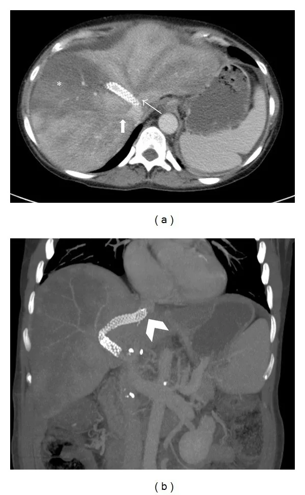
- Hepatik venin ya da intrahepatik IVC’nin segmental opaklaşmaması, kısa kesilmesi veya membranöz daralması
- Birden fazla hepatik ven tutulumu ve hepatik venöz kollateral yolların belirginleşmesi
- Akut konjesyonda hepatomegali ve periferik yamalı hipoenhansman; kaudat lob daha iyi kontrastlanabilir
- Kronik çıkış tıkanıklığında kaudat lob hipertrofisi, intrahepatik kollateraller ve asit
- Tümör trombüsünde damar genişlemesi, trombüs içinde kontrastlanma ve komşu kitleyle süreklilik
Ölçütler ve sınıflamalar
- Budd–Chiari anatomik tutulum örüntüsü
- Görüntüleme raporunda hepatik ven tutulumu, IVC tutulumu veya her ikisinin birlikte tutulumu ayrı ayrı tarif edilir; tıkalı damar ve kollateral drenaj yolları belirtilir.
Normalde
Aynı venlerin beklenen birleşme noktalarını ardışık aksiyel ve koronal kesitlerde izleyerek gerçek dolum defektini, akım karışımından veya parsiyel volüm etkisinden ayır.
Normal BT ile kıyasla
Aynı bölgenin, kaynak veri setinde patoloji içermediği belirtilen bir BT incelemesini kesit kesit kaydırın; organ sınırlarını açarak anatomiyi hasta görüntüsüyle karşılaştırın. Küçük rastlantısal bulgular tümüyle dışlanmamıştır.
Kaynak: TotalSegmentator v2.01 veri seti (Wasserthal ve ark., Radiology: Artificial Intelligence 2023), CC BY 4.0, vaka s0541; organ sınırları veri setinin otomatik segmentasyonundan, birleştirilerek sadeleştirildi. Görüntüler 2,5 mm kesit aralığına yeniden örneklendi.
Ayırıcı tanı
- Tümör trombüsü
- damar genişlemesi ve trombüs dokusunda kontrastlanma, özellikle komşu karaciğer kitlesiyle devamlılık varsa lehinedir.
- Akım karışım artefaktı
- IVC’de tabakalı opaklaşma olabilir; daha kaudal ve kraniyal kesitlerde lümen homojenleşir, sabit damar duvarı genişlemesi yoktur.
- Ekstrinsik IVC basısı, benign veya malign komşu oluşumların IVC'ye dıştan basısıyla gelişebilir.
- Siroza bağlı hepatik venlerin zor seçilmesi
- damar lümeninde doğrudan trombüs kanıtı olmayabilir; tek başına görünmeme tromboz tanısı değildir.
Tuzaklar
- Hepatik venlerin BT’de seçilememesi yalancı pozitif sonuç verebilir; her veni IVC birleşimine kadar izleyip doğrudan dolum defekti arayın.
- IVC’de karışık kontrast bolusu geçici yalancı dolum defekti yaratabilir; kraniokaudal süreklilik ve ek faz görüntüsüyle sabit trombüsten ayırın.
- Kontrastlanan damar içi yumuşak doku tümör trombüsünü destekler; damar genişlemesi ve komşu primer tümörle süreklilik de bu yorumu destekleyen bulgulardır.
- Budd–Chiari şüphesinde ilk görüntüleme genellikle Doppler ultrasonografidir; BT veya MR tanıyı doğrulamak ve yayılımı belirlemek için kullanılır.
Kendini dene
Venöz dolum defektinde tümör trombüsünü bland trombüsten ayırmada en özgül bulgu hangisidir?
Cevabı göster
Trombüsün primer tümörle benzer kontrastlanma göstermesi. Trombüsün primer tümörle benzer kontrastlanması tümör trombüsü için en özgül bulgudur. Kontrastlanma olmaması bland trombüsle uyumludur; difüzyon kısıtlanması her iki trombüs tipinde görülebilir ve dolum defekti tek başına ayırt ettirmez.
IVC'de kontrastlanan ve komşu karaciğer kitlesiyle kesintisiz devam eden intraluminal doku için en uygun yorum hangisidir?
Cevabı göster
Tümör trombüsü. Trombüsün kontrastlanma örüntüsünün komşu primer tümörle benzer olması tümör trombüsünü güçlü biçimde destekler. Kaynak, kontrastlanmanın tümör trombüsünü bland trombüsten ayırmada yardımcı olduğunu ve tümör trombüsünün komşu organla süreklilik gösterebildiğini bildiriyor.
İlgili konular
Kaynaklar
Bu sayfadaki 35 cümle ve 2 quiz sorusu aşağıdaki kaynaklardan alıntılarla otomatik olarak karşılaştırıldı; 5 cümle bu karşılaştırmada düzeltildi (2026-10-09).
- Budd-Chiari Syndrome Imaging Diagnosis: State of the Art and Future Perspectivespmc.ncbi.nlm.nih.gov
- Tumor in the veins: an abdominal perspective with an emphasis on CT and MR imagingpmc.ncbi.nlm.nih.gov
- Congestive hepatopathy.pubmed.ncbi.nlm.nih.gov
- Budd-Chiari Syndrome.pubmed.ncbi.nlm.nih.gov
- Management of portal hypertension and ascites in polycystic liver disease.pubmed.ncbi.nlm.nih.gov
- The Budd-Chiari syndrome.pubmed.ncbi.nlm.nih.gov
- Liver vascular anatomy: a refresher.pubmed.ncbi.nlm.nih.gov
- Living-donor liver transplantation: Right versus left.pubmed.ncbi.nlm.nih.gov
- Role of contrast-enhanced ultrasound in the evaluation of post-liver transplant vasculature.pubmed.ncbi.nlm.nih.gov
- An essential accessory.pubmed.ncbi.nlm.nih.gov
- Current role of intraoperative ultrasonography in hepatectomy.pubmed.ncbi.nlm.nih.gov
- Post-operative imaging in liver transplantation: state-of-the-art and future perspectives.pubmed.ncbi.nlm.nih.gov
- Combined biembolization induces higher hypertrophy than portal vein embolization before major liver resection.pubmed.ncbi.nlm.nih.gov
- Improving Image Quality and Reducing Radiation Dose for Pediatric CT by Using Deep Learning Reconstruction.pubmed.ncbi.nlm.nih.gov
- CT angiography of the hepatic and pancreatic circulation.pubmed.ncbi.nlm.nih.gov
- Should transjugular intrahepatic portosystemic shunt stent grafts be underdilated?pubmed.ncbi.nlm.nih.gov
- Liver and Spleen Stiffness as Non-Invasive Tools Assessing Liver Congestion in Patients With Budd-Chiari Syndrome.pubmed.ncbi.nlm.nih.gov
- JAK2-Positive Myeloproliferative Neoplasm: Polycythaemia Vera Presenting As Budd-Chiari Syndrome.pubmed.ncbi.nlm.nih.gov
- Multiparametric Ultrasound in Budd-Chiari Syndrome: From Venous Outflow Obstruction to Microcirculatory Dysfunction.pubmed.ncbi.nlm.nih.gov
- Contrast-Enhanced CT Finding of Hot Spot Sign Secondary to Supra Hilar Mass Causing Superior Vena Cava Syndrome: A Case Report.pubmed.ncbi.nlm.nih.gov
- Application of "parenchyma-first" approach in laparoscopic living donor left lateral sectionectomy.pubmed.ncbi.nlm.nih.gov
- Imaging features, diagnosis, and clinical management of hepatic involvement in hereditary hemorrhagic telangiectasia: a case series report and literature review.pubmed.ncbi.nlm.nih.gov
- Acute portal vein thrombosis postimage-guided liver biopsy in common variable immunodeficiency.pubmed.ncbi.nlm.nih.gov
- Renal vein thrombosis with inferior vena cava extension in PLA2R-positive membranous nephropathy: A radiological mimic of acute pyelonephritis.pubmed.ncbi.nlm.nih.gov
- Case Report: Acute superior mesenteric vein and portal vein thrombosis complicated by small intestinal necrosis: a case of successful endovascular treatment.pubmed.ncbi.nlm.nih.gov
- Cross-sectional imaging in an inferior vena cava agenesis associated with antiphospholipid syndrome, lower limb, and portomesenteric deep vein thrombosis and renal insufficiency in a young patient: A rare presentation.pubmed.ncbi.nlm.nih.gov
- Thickness Reduction Versus Conventional Left Lateral Sector Strategies in Children Below 8 kg Undergoing Living Donor Liver Transplantation.pubmed.ncbi.nlm.nih.gov
- Interrupted Inferior Vena Cava and Duplicated Superior Vena Cavae Identified During Catheter Ablation.pubmed.ncbi.nlm.nih.gov
- Transcatheter closure of patent ductus venosus in an adolescent using a KONAR-Multifunctional Occluder device.pubmed.ncbi.nlm.nih.gov
- Portal Vein Invasion Secondary to Hepatic Hydatid Disease: A Rare Case Report.pubmed.ncbi.nlm.nih.gov
- Endoscopic ultrasound-guided cyanoacrylate therapy for gastric variceal bleeding secondary to sinistral portal hypertension: a retrospective case series.pubmed.ncbi.nlm.nih.gov
- Multimodal imaging features to diagnose abdominal complications of sickle cell disease.pubmed.ncbi.nlm.nih.gov
- Whole lung lavage in the setting of exogenous lipoid pneumonia.pubmed.ncbi.nlm.nih.gov
- A rare pediatric case: Budd-Chiari Syndrome and upper gastrointestinal bleeding in a 5-year-old boy.pubmed.ncbi.nlm.nih.gov
- Dissemination along the hepatocaval ligament in a caudate-origin intrahepatic cholangiocarcinoma: a case report with video.pubmed.ncbi.nlm.nih.gov
- Ultrasound Characteristics of Budd-Chiari Syndrome: A Multicenter Analysis.pubmed.ncbi.nlm.nih.gov
- Giant Clear Cell Renal Cell Carcinoma With Continuous Tumor Thrombus Extension Into the Left Renal, Gonadal, Ascending Lumbar, and Hemiazygos Veins: A Case Report.pubmed.ncbi.nlm.nih.gov
- HAIC-Based Combination Therapy for Advanced Hepatocellular Carcinoma with Vp4 Portal Vein Tumor Thrombus and Child-Pugh B Liver Function: A Case Report.pubmed.ncbi.nlm.nih.gov
- Multimodal ultrasound in the diagnosis and treatment of caudate lobe Klebsiella pneumoniae liver abscess complicated by rare gas-containing inferior vena cava thrombosis: a case report.pubmed.ncbi.nlm.nih.gov
- Iatrogenic portobiliary fistula treated with covered stent following percutaneous transhepatic biliary drainage.pubmed.ncbi.nlm.nih.gov
- Clinical applications of 4D flow magnetic resonance imaging in hepatic vascular disorders: practical biomarkers, protocol design, and translational implementation.pubmed.ncbi.nlm.nih.gov
- When Cancer Clots: An Extensive Radiologic Analysis of Cancer-Associated Thromboembolism.pubmed.ncbi.nlm.nih.gov
- Open Surgical Management of Renal Cell Carcinoma with Infradiaphragmatic Venous Tumor Thrombus (Mayo Levels 0-III): The Epitome of Surgical Self-Reliance in Urology.pubmed.ncbi.nlm.nih.gov
- Natural History of Treated and Untreated Bland Portal Vein Thrombosis in Patients with Hepatocellular Carcinoma.pubmed.ncbi.nlm.nih.gov
- Establishment and validation of a predictive scoring system for titanium clip retention at six months after endoscopic submucosal dissection for gastric lesions.pubmed.ncbi.nlm.nih.gov
- Artificial Intelligence in Pulmonary Hypertension: Current State and Future Prospects.pubmed.ncbi.nlm.nih.gov
- Technical advances and application strategies in anatomical hepatic resection based on Laennec's capsule.pubmed.ncbi.nlm.nih.gov
- Radiographic overestimation of inferior vena cava involvement in pediatric neuroblastoma: a case report.pubmed.ncbi.nlm.nih.gov
- Case Report: Primary inferior vena cava leiomyosarcoma with hepatic metastases initially misinterpreted as primary hepatic leiomyosarcoma - ultrasonographic features and diagnostic insights.pubmed.ncbi.nlm.nih.gov
- Extensive Iliofemoral Deep Vein Thrombosis Secondary to Uterine Leiomyoma-Induced Iliac Vein Compression: A Case Report.pubmed.ncbi.nlm.nih.gov
- Case Report: Primary myelofibrosis presenting with non-cirrhotic portal hypertension and initially misdiagnosed as cirrhosis.pubmed.ncbi.nlm.nih.gov
- Overview of Non-Cirrhotic Portal Hypertension in Pediatric Patients.pubmed.ncbi.nlm.nih.gov
- Perioperative Point-of-Care Ultrasound Utilization in Abdominal Organ Transplantation. Part II: Postoperative Care.pubmed.ncbi.nlm.nih.gov
- Radiologic Interventions for Gastrointestinal Bleeding.pubmed.ncbi.nlm.nih.gov
- Society for Cardiovascular Magnetic Resonance 2024 Cases of SCMR case series.pubmed.ncbi.nlm.nih.gov
- Computed tomography and magnetic resonance imaging for pulmonary embolus evaluation in children: up-to-date review on practical imaging protocols.pubmed.ncbi.nlm.nih.gov
- Technical Recommendations for Computed Tomography Guidance of Intervention in the Right Ventricular Outflow Tract: Native RVOT, Conduits and Bioprosthetic Valves: A White Paper of the Society of Cardiovascular Computed Tomography (SCCT), Congenital Heart Surgeons' Society (CHSS), and Society for Cardiovascular Angiography & Interventions (SCAI).pubmed.ncbi.nlm.nih.gov
- Preoperative MRI Model for Predicting Inferior Vena Cava Wall Invasion in Renal Cell Carcinoma with Tumor Thrombus.pubmed.ncbi.nlm.nih.gov
- Endovenous reconstruction of congenital iliocaval anomalies.pubmed.ncbi.nlm.nih.gov
- Pediatric scimitar syndrome with an intrahepatic venous-venous shunt: A rare imaging case report.pubmed.ncbi.nlm.nih.gov
- Liver transplantation for Type I Abernethy malformation with liver tumors in children: a report of two cases with contrasting portal vein reconstruction strategies.pubmed.ncbi.nlm.nih.gov
- Efficacy and safety of combined TACE-HAIC and targeted immunotherapy in primary liver cancer with portal vein tumor thrombus.pubmed.ncbi.nlm.nih.gov
- Magnetic Resonance-Guided Radiotherapy for Unresectable Hepatocellular Carcinoma with Bile Duct Tumor Thrombus: A Case Series and Review of Treatment Options.pubmed.ncbi.nlm.nih.gov
- Hereditary hemorrhagic telangiectasia in a 72-year-old woman presenting with recurrent epistaxis and liver cirrhosis: a case report.pubmed.ncbi.nlm.nih.gov
- Imaging findings in Fontan-associated liver disease: Results of a single center study.pubmed.ncbi.nlm.nih.gov
- Laparoscopic unroofing for hepatic cysts compressing the left portal vein, partial TIPE ALPPS, and hepatic vein deprivation to overcome a small future liver remnant before right hemihepatectomy.pubmed.ncbi.nlm.nih.gov
- Transhepatic venous access as a vessel-preserving salvage strategy for temporary hemodialysis in venous access exhaustion.pubmed.ncbi.nlm.nih.gov
- Primary Renal Ewing Sarcoma Presenting With Divergent Radiological Phenotypes: A Radiology-Focused Case Series Featuring the Mimicking of Renal Cell Carcinoma and Urinary Tract Urothelial Carcinoma.pubmed.ncbi.nlm.nih.gov
- IL-32 drives PR3-dependent NET formation to promote portal vein tumor thrombosis and radioresistance in HCC.pubmed.ncbi.nlm.nih.gov
- Peribiliary Inflammatory Phlegmon Masquerading as Cholangiocarcinoma in Janus Kinase 2 V617F-positive Primary Myelofibrosis: A Multimodality Imaging Diagnosis.pubmed.ncbi.nlm.nih.gov
- Pathophysiological management of gastric varices: From hemodynamics to targeted therapies.pubmed.ncbi.nlm.nih.gov
- Managing High-Risk Liver Transplantation: A Case of Budd-Chiari Syndrome With Preemptive Venovenous Extracorporeal Membrane Oxygenation (ECMO).pubmed.ncbi.nlm.nih.gov
- The crucial role of multimodality ultrasound in the postoperative evaluation of pediatric ischemic diseases.pubmed.ncbi.nlm.nih.gov
- Atypically presented flank pain in a patient with pulmonary embolism; a case report and literature review of a diagnostic dilemma.pubmed.ncbi.nlm.nih.gov
- Pancreatic Pseudocyst Masquerading as Right Atrial Thrombus: An Unusual Intracardiac Extension.pubmed.ncbi.nlm.nih.gov
- En Bloc Multivisceral Resection and Portal Vein Thrombectomy for Metastatic Solid Pseudopapillary Neoplasm of the Pancreas: A Case Report.pubmed.ncbi.nlm.nih.gov
- Balloon-occluded hepatic arterial infusion for unresectable hepatocellular carcinoma: a phase II trial interim analysis.pubmed.ncbi.nlm.nih.gov
- Alpha-Fetoprotein Is a Potential Biomarker for Pancreatic Ductal Adenocarcinoma (PDAC): A Case Report.pubmed.ncbi.nlm.nih.gov
- Balloon-assisted coil embolization for high-flow renal arteriovenous fistula with inferior vena cava dilatation: A report of two cases.pubmed.ncbi.nlm.nih.gov
- An unusual superior mesenteric arteriovenous malformation causing obscure gastrointestinal bleeding: a case report.pubmed.ncbi.nlm.nih.gov
- From congenital variants to critical clues: A radiologic review of inferior vena cava pathologies.pubmed.ncbi.nlm.nih.gov
- Diagnosis and Surgical Management for Advanced Pancreatic Cancer Requiring Vascular Resection.pubmed.ncbi.nlm.nih.gov
- Prevalence and Risk Factors for Portal Cavernoma in Adult Patients with Portal Vein Thrombosis.pubmed.ncbi.nlm.nih.gov
- Primary extraskeletal osteosarcoma of the post-hepatic caudal vena cava in a dog-Case report.pubmed.ncbi.nlm.nih.gov
- Subcapsular Pancreatic Pseudocyst of the Right Hepatic Lobe: A Rare Case Report and Literature Review.pubmed.ncbi.nlm.nih.gov
- Early and late outcomes of patients treated with graft replacement of the inferior vena cava for malignant disease: A single-center experience over three decades.pubmed.ncbi.nlm.nih.gov
- Can Artificial Intelligence Really Help? A Practicing Radiologist's Simplified Guide to AI, with a Critical Appraisal of the Use of AI in Cancer-Associated Thromboembolism.pubmed.ncbi.nlm.nih.gov
- Photon-counting detector CT with iodine quantification: improved distinction between bland and neoplastic portal vein thrombosis.pubmed.ncbi.nlm.nih.gov
- The role of advanced imaging in the diagnosis and management of scimitar syndrome in pediatric patients.pubmed.ncbi.nlm.nih.gov
- Liver Imaging Reporting and Data System (LI-RADS): A Comprehensive Review.pubmed.ncbi.nlm.nih.gov
- Portal vein aneurysm mimicking a pancreatic mass: A case report with an emphasis on imaging pitfalls.pubmed.ncbi.nlm.nih.gov
- Gastric varices: a contemporary review of vascular anatomy and therapeutic strategies.pubmed.ncbi.nlm.nih.gov
- Adult Hepatic Infarction for Internists: A Practical Diagnostic and Management Pathway to Avoid Misdiagnosis, Unnecessary Drainage, and Delayed Vascular Recognition.pubmed.ncbi.nlm.nih.gov
- MRI findings of intrahepatic cholangiocarcinoma with sarcomatoid differentiation: a retrospective case series.pubmed.ncbi.nlm.nih.gov
- Primary leiomyosarcoma of the portal vein: A progressively enhancing fusiform mass without biliary dilatation as a rare differential for hilar tumors.pubmed.ncbi.nlm.nih.gov
- Case Report: Leiomyosarcoma of the inferior vena cava presenting as a right atrial mass: the value of multimodality imaging.pubmed.ncbi.nlm.nih.gov
- Right adrenal metastasis from left apical non-small cell lung carcinoma with contiguous tumor thrombus extending through the inferior vena cava into the right atrium.pubmed.ncbi.nlm.nih.gov
- Incidental Detection of Hepatocellular Carcinoma by Echocardiographic Imaging During Acute Coronary Syndrome Evaluation.pubmed.ncbi.nlm.nih.gov
Eğitim amaçlıdır; hasta başında tanı ve tedavi kararının yerine geçmez, tıbbi tavsiye değildir. Metinler yapay zekâ desteğiyle yazıldı ve otomatik bir adımda kaynak alıntılarıyla karşılaştırıldı; bu bir tıbbi doğrulama değildir ve içerik uzman radyolog incelemesinden geçmedi. Klinik kararlarda güncel kılavuzları, kurum protokollerini ve radyoloji raporunu esas alın.