
Özet
Amip karaciğer absesi, Entamoeba histolytica'nın bağırsaktan portal dolaşımla karaciğere ulaşmasıyla gelişen, çoğu kez tek odaklı enfeksiyondur. Kontrastlı BT’de rüptürle ilişkili toraks uzanımı ve hepatik ven ya da inferior vena cava trombozu gösterilebilir; tip I morfoloji ağır klinik seyir ve daha yüksek rüptür sıklığıyla ilişkilidir. BT’de eksik duvarlı, düzensiz ve kesintili kontrastlanan tip I; tam duvar kontrastlanması ve hipodens halo gösteren tip II ya da kontrastlanmayan duvarlı tip III morfoloji görülebilir; görüntüleme bulguları piyojenik apseyle örtüşebilir. BT bulguları amip ve piyojenik apseyi güvenilir biçimde ayırt ettirmez; serolojik ve moleküler testler etken tanısına katkı sağlar, BT ise rüptür ve damar trombozu gibi komplikasyonları gösterebilir.
Faz ve pencere
- Portal tanısal
- Tip I morfolojide duvar yok ya da eksik olabilir; kenarlar düzensiz ve kontrastlanma kesintilidir. Tip II’de tam duvar kontrastlanması ve çevresel hipodens halo, tip III’te ise kontrastlanmayan duvar görülür. Bu görünümler piyojenik apseyle örtüşebilir.
- Kontrastsız
- Merkezdeki sıvılaşmış nekroz ile çevre parankim arasında orta atenüasyonlu geçiş kuşağı veya kanama/gaz seçilebilir; duvarın kontrastlanma davranışı bu fazda değerlendirilemez.
Önerilen pencereler: Karaciğer (G 150 / M 30), Batın (G 400 / M 50), Mediasten (G 350 / M 50).
BT bulguları
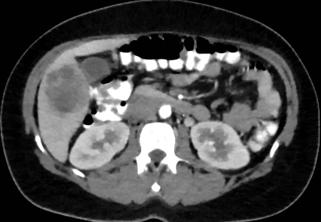
- Genellikle sağ lobda, tek ve yuvarlak-oval düşük atenüasyonlu kavite; sol lob odağı da mümkündür
- Erken tip I morfoloji — eksik/olmayan duvar, düzensiz kenar, kesintili kontrastlanma ve periferik ince septalar
- Tip II morfoloji — düzgün tam duvar kontrastlanması ve duvar çevresinde hipodens halo; çift hedef düzeni oluşabilir
- Tip III morfoloji — düzgün ve tam duvarda kontrastlanma olmaması; gecikmiş ve hafif bulgulu seyirle ilişkilidir.
- Kavite çevresinde hipodens halo bulunabilir.
- Rüptürde diyafram altı veya intratoraks uzantısı, komşu plevral veya peritoneal boşluklarda yeni sıvı izlenir.
- Amip karaciğer absesinde hepatik ven ve inferior vena cava trombozu bildirilmiştir.
- Rüptürde diyaframı aşarak toraksa uzanım ve plevral koleksiyon görülebilir; amip karaciğer absesinin abdominotorasik yapılara rüptürü de bildirilmiştir.
Ölçütler ve sınıflamalar
- BT morfolojik amip absesi tipleri
- BT morfolojisine göre: Tip I’de duvar yok/eksik, kenar düzensiz ve kontrastlanma kesintili olabilir; Tip II’de düzgün tam duvar kontrastlanır ve çevresinde hipodens halo bulunabilir; Tip III’te düzgün duvar kontrastlanmaz. Bu BT morfoloji grupları klinik özelliklerle ilişkilidir; ancak görüntüleme tek başına amip ve piyojenik apseyi güvenilir biçimde ayırt ettirmez.
Normalde
Aynı seviyede kavitenin kenar düzenini, kapsüle mesafesini, çevresindeki kama biçimli perfüzyon alanını ve plevral/peritoneal boşluklarda yeni sıvı olup olmadığını karşılaştırın.
Normal BT ile kıyasla
Aynı bölgenin, kaynak veri setinde patoloji içermediği belirtilen bir BT incelemesini kesit kesit kaydırın; organ sınırlarını açarak anatomiyi hasta görüntüsüyle karşılaştırın. Küçük rastlantısal bulgular tümüyle dışlanmamıştır.
Kaynak: TotalSegmentator v2.01 veri seti (Wasserthal ve ark., Radiology: Artificial Intelligence 2023), CC BY 4.0, vaka s0541; organ sınırları veri setinin otomatik segmentasyonundan, birleştirilerek sadeleştirildi. Görüntüler 2,5 mm kesit aralığına yeniden örneklendi.
Ayırıcı tanı
- Piyojenik karaciğer absesi
- çoklu veya biliyer dağılımlı odaklar ve küme işareti bakteriyel apsede daha sık olsa da duvar/halo görünümü örtüşebilir.
- Nekrotik metastaz
- düzensiz nodüler canlı kenar, başka metastazlar veya bilinen primer tümör malignite lehinedir.
- Basit karaciğer kisti
- homojen sıvı atenüasyonu ve kontrastlanmayan ince duvar vardır; inflamatuvar halo beklenmez.
- Enfekte hidatik kist
- hidatik kist, karaciğer apsesinin ayırıcı tanısında düşünülmelidir.
Tuzaklar
- Tam halka biçimli kontrastlanma amip için özgül değildir; piyojenik apse aynı çift hedef görünümünü oluşturabilir, bu nedenle BT ile etken atamayın.
- Erken tip I lezyonda tam kapsül görülmeyebilir; düzensiz sınır, çevre parankim perfüzyonu ve olası rüptür alanını birlikte izleyin.
- Tip III morfolojide duvar bulunur ancak kontrastlanmaz.
- Rüptür değerlendirmesinde kapsül defektini ve komşu boşluklara uzanımı arayın.
Kendini dene
Ateş ve sağ üst kadran ağrısında BT'de eksik duvarlı, kesintili kontrastlanan ve düzensiz kenarlı kavite izleniyor. Hangi morfoloji tanımı uygundur?
Cevabı göster
Tip I amip morfolojisi. Tip I görünümde duvar eksik olabilir ve sınır düzensiz olup kontrastlanma düzensiz ya da kesintilidir. Tip II’de duvarın tamamı kontrastlanır; Tip III düzgün fakat kontrastlanmayan duvar gösterir. CE3a ayrılmış membranlı hidatik kist evresidir.
Duvarı tam halka biçiminde kontrastlanan tek karaciğer kavitesi, amip ve piyojenik apse arasında etkeni kesin ayırıyor mu?
Cevabı göster
Hayır, örtüşen görünüm. Amip ve piyojenik apseler halka biçimli kontrastlanma ve çevresel halo gösterebilir; BT tek başına güvenilir etken ayrımı sağlamaz. Seroloji, kültür ve klinik bağlam gerekir. Tek başına halka metastazı da kesinleştirmez.
İlgili konular
Kaynaklar
Bu sayfadaki 42 cümle ve 2 quiz sorusu aşağıdaki kaynaklardan alıntılarla otomatik olarak karşılaştırıldı; 12 cümle bu karşılaştırmada düzeltildi (2026-10-09).
- Entamoeba histolytica and amoebic liver abscess in northern Sri Lanka: a public health problempmc.ncbi.nlm.nih.gov
- Liver abscess: diagnostic and management issues found in the low resource settingpmc.ncbi.nlm.nih.gov
- Ruptured Amoebic Liver Abscess With Empyema, Venous Thromboembolism and Bronchopleural Fistula: A Case Reportpmc.ncbi.nlm.nih.gov
- Amoebic liver abscess: diagnosis and management of a single case in Guangdong, Chinapmc.ncbi.nlm.nih.gov
- Accidental cannulation of amoebic liver abscess during pericardiocentesis: a case reportpmc.ncbi.nlm.nih.gov
- Three cases of amoebic liver abscess causing inferior vena cava obstruction, with a review of the literature.pubmed.ncbi.nlm.nih.gov
- [A case of amoebic liver abscess complicated by a gastric fistula cured with oral medication only].pubmed.ncbi.nlm.nih.gov
- Case report: Huge amoebic liver abscesses in both lobes.pubmed.ncbi.nlm.nih.gov
- Amoebic Liver Abscess: A disease native to Oman?pubmed.ncbi.nlm.nih.gov
- An uncommon case of hepatopulmonary amoebiasis.pubmed.ncbi.nlm.nih.gov
- Fatal case of amoebic liver abscess in a child.pubmed.ncbi.nlm.nih.gov
- Tubercular liver abscess: a distinct entity not to be missed.pubmed.ncbi.nlm.nih.gov
- Bacterial Superinfection of Amoebic Liver Abscess.pubmed.ncbi.nlm.nih.gov
- A review of amoebic liver abscess for clinicians in a nonendemic setting.pubmed.ncbi.nlm.nih.gov
- [Amoebic liver abscess in a patient initially diagnosed with pneumonia: case report and discussion of relevant literature].pubmed.ncbi.nlm.nih.gov
- Neurological complications during treatment of liver abscess: think of metronidazole toxicity.pubmed.ncbi.nlm.nih.gov
- Enhanced Drainage Protocol in Large Amoebic Liver Abscess.pubmed.ncbi.nlm.nih.gov
- Recent advances in the diagnosis and management of amoebiasis.pubmed.ncbi.nlm.nih.gov
- <i>Entamoeba histolytica</i>-induced NETs are highly cytotoxic on hepatic and colonic cells due to serine proteases and myeloperoxidase activities.pubmed.ncbi.nlm.nih.gov
- Diversity and Plasticity of Virulent Characteristics of Entamoeba histolytica .pubmed.ncbi.nlm.nih.gov
- Amebic Liver Abscess: An Unusual Cause of Budd-Chiari Syndrome.pubmed.ncbi.nlm.nih.gov
- A rare case report of pyogenic hepatic abscess secondary to sigmoid diverticulitis.pubmed.ncbi.nlm.nih.gov
- Ruptured Amoebic Liver Abscess With Empyema, Venous Thromboembolism and Bronchopleural Fistula: A Case Report.pubmed.ncbi.nlm.nih.gov
- <i>Klebsiella pneumoniae</i> Liver Abscess in a Filipino Patient With Previously Undiagnosed Diabetes: Successful Conservative Management.pubmed.ncbi.nlm.nih.gov
- Complications following surgical management of a fish bone-related abscess in the liver.pubmed.ncbi.nlm.nih.gov
- Exploration of Various Diagnostic Modalities for Detection of Amoebic Liver Abscess and Co-Occurrence of Other Infective Aetiology, Eastern India.pubmed.ncbi.nlm.nih.gov
- Liver abscesses in Southern Europe: clinical and microbiological characterization of a retrospective cohort.pubmed.ncbi.nlm.nih.gov
- Amebiasis as a neglected tropical disease: Current knowledge gaps and future directions.pubmed.ncbi.nlm.nih.gov
- Case Report: Massive subcapsular hepatic hematoma after spontaneous rupture of pyogenic liver abscess in a healthy young woman.pubmed.ncbi.nlm.nih.gov
- An Imported Case of Gigantic Amoebic Liver Abscess in a 24-Year-Old Woman in Singapore.pubmed.ncbi.nlm.nih.gov
- Case Report: A case of intrahepatic papillary neoplasm of the bile duct with invasive carcinoma in a young patient and review of the literature.pubmed.ncbi.nlm.nih.gov
- Adult Hepatic Infarction for Internists: A Practical Diagnostic and Management Pathway to Avoid Misdiagnosis, Unnecessary Drainage, and Delayed Vascular Recognition.pubmed.ncbi.nlm.nih.gov
- Case Report: Two rare cases of aggressive fibromatosis originating in the intestinal tract.pubmed.ncbi.nlm.nih.gov
- Common and Uncommon Splenic Lesions: A Review.pubmed.ncbi.nlm.nih.gov
- Edwardsiella tarda infections in humans: a systematic review of reported cases and clinical outcomes.pubmed.ncbi.nlm.nih.gov
- Amoebic liver abscess: diagnosis and management of a single case in Guangdong, China.pubmed.ncbi.nlm.nih.gov
- Gastrointestinal Tract Mycobacterial Infection in People Living With Human Immunodeficiency Virus (HIV)/Acquired Immunodeficiency Syndrome (AIDS) (PLWHA): Case Reports and Patterns of Endoscopic Involvement.pubmed.ncbi.nlm.nih.gov
- CT of amebic liver abscess: different morphological types with different clinical features.pubmed.ncbi.nlm.nih.gov
- Complex cystic liver lesions: classification, diagnosis, and management.pubmed.ncbi.nlm.nih.gov
- Amebic liver abscess: An update.pubmed.ncbi.nlm.nih.gov
- Amebic liver abscess: Clinico-radiological findings and interventional management.pubmed.ncbi.nlm.nih.gov
- Culture-negative cryptogenic splenic abscess in an immunocompetent host with recurrent misdiagnosis as pleural effusion: a case report with a review of the literature.pubmed.ncbi.nlm.nih.gov
- Imaging insights into pediatric liver masses: A comprehensive minireview for hepatology practice.pubmed.ncbi.nlm.nih.gov
- Contrast-Enhanced CT Finding of Hot Spot Sign Secondary to Supra Hilar Mass Causing Superior Vena Cava Syndrome: A Case Report.pubmed.ncbi.nlm.nih.gov
- Case Report: Indocyanine green fluorescence imaging in complex focal nodular hyperplasia resection: report of two cases.pubmed.ncbi.nlm.nih.gov
- Radiation segmentectomy for hepatocellular carcinoma: redefining the limits of curative-intent therapy.pubmed.ncbi.nlm.nih.gov
- TACE combined with DynaCT-guided MWA in the treatment of high-risk location liver cancers with ≤5 cm tumor size.pubmed.ncbi.nlm.nih.gov
- Ablation manual for liver cancer.pubmed.ncbi.nlm.nih.gov
- A pictorial essay of thoracic wall diseases: multiple pathologies in the same anatomical site.pubmed.ncbi.nlm.nih.gov
- Evolving management of pyogenic liver abscess: A narrative review of laparoscopic and open drainage approaches with contextual insights from Pakistan.pubmed.ncbi.nlm.nih.gov
- Ultrasound-Based Staging and Its Impact on Clinical Management of Hepatic Hydatid Cysts in an Endemic Setting: A Cross-Sectional Study in Eastern Afghanistan.pubmed.ncbi.nlm.nih.gov
- Phenotype and molecular characterization of K1-ST700 hypervirulent Klebsiella pneumoniae isolated from bone marrow aspirate of a patient with multi-system infection.pubmed.ncbi.nlm.nih.gov
- Controversies in contrast-enhanced ultrasound (CEUS): pregnancy, paediatric, abdominal trauma, complex renal cysts, and endovascular aortic repair follow-up.pubmed.ncbi.nlm.nih.gov
- Magnetic Resonance Imaging Features of Hepatic Hydatid Disease: A Pictorial Review with Emphasis on Atypical Presentations and Differential Diagnosis.pubmed.ncbi.nlm.nih.gov
- Computed Tomographic Findings in Dogs with Hepatic Bacterial Parenchymal Infection and Abscessation.pubmed.ncbi.nlm.nih.gov
- The Infectious Clue: Linking Bacterial Infections to Underlying Malignancies.pubmed.ncbi.nlm.nih.gov
- Case Report: Primary gastric extraskeletal osteosarcoma and literature review.pubmed.ncbi.nlm.nih.gov
- Case Report: A hepatic lesion is not always metastasis: synchronous rectal adenocarcinoma and primary hepatocellular carcinoma followed by a benign necrotic liver nodule.pubmed.ncbi.nlm.nih.gov
- Advanced Uterine Carcinosarcoma With Fatal Suspected Pulmonary Embolism in a Resource-Limited Setting: A Rare Case Report.pubmed.ncbi.nlm.nih.gov
- A case of left adrenal echinococcosis misdiagnosed as a left renal cyst: Case report.pubmed.ncbi.nlm.nih.gov
- Case Report: Retroperitoneal bronchogenic cyst mimicking adrenal mass: Clinical implications and comprehensive literature analysis.pubmed.ncbi.nlm.nih.gov
- Breast cystic echinococcosis with ultrasonographic "cerebral gyrus sign": a case report.pubmed.ncbi.nlm.nih.gov
- Agenesis of the left pulmonary artery and cardiac hydatid cysts: A dual rarity.pubmed.ncbi.nlm.nih.gov
- Common and Rare Radiological Findings of Pulmonary and Thoracic Hydatid Cysts.pubmed.ncbi.nlm.nih.gov
- Complications and expected imaging findings after endoscopic retrograde cholangiopancreatography.pubmed.ncbi.nlm.nih.gov
- Duodenal imaging on the spotlight: from A to Z.pubmed.ncbi.nlm.nih.gov
- Gastrointestinal perforation: clinical and MDCT clues for identification of aetiology.pubmed.ncbi.nlm.nih.gov
- Liver-gallbladder anatomical adjacency-related severe infection: a case report of liver abscess rupturing into the gallbladder inducing septic shock and literature review.pubmed.ncbi.nlm.nih.gov
- Percutaneous <i>vs</i> surgical management of World Health Organization cystic echinococcosis 1 and 3a liver hydatid cysts.pubmed.ncbi.nlm.nih.gov
- Cross-sectional imaging after pancreatic surgery: The dialogue between the radiologist and the surgeon.pubmed.ncbi.nlm.nih.gov
- Biliary Mucinous Cystadenoma: A Review of the Literature.pubmed.ncbi.nlm.nih.gov
- International Society of Paediatric Surgical Oncology (IPSO) Surgical Practice Guidelines.pubmed.ncbi.nlm.nih.gov
- Pediatric emergency medicine point-of-care ultrasound: summary of the evidence.pubmed.ncbi.nlm.nih.gov
- The anatomical compartments and their connections as demonstrated by ectopic air.pubmed.ncbi.nlm.nih.gov
- "First Reported Case of Rapidly Progressive Pyogenic Liver Abscess with Isolation of <i>Priestia megaterium</i>: Case Report and Literature Review".pubmed.ncbi.nlm.nih.gov
- Practical Guidance for Clinical Microbiology Laboratories: Laboratory Diagnosis of Parasites from the Gastrointestinal Tractpubmed.ncbi.nlm.nih.gov
- Epidemiology, pathology, diagnosis, and management of amoebic liver abscesses in Sub-Saharan Africa: a retrospective cross-sectional study.pubmed.ncbi.nlm.nih.gov
- The Abscess That Was Cancer: Pyogenic Liver Abscess as the Initial Manifestation of Hepatocellular Carcinoma in a Noncirrhotic Patient.pubmed.ncbi.nlm.nih.gov
- A Comprehensive Review of SURGICEL®: Efficacy, Applications, and Radiological Insights in Surgery.pubmed.ncbi.nlm.nih.gov
- Breaking Barriers with Sound: The Implementation of Histotripsy in Cancer.pubmed.ncbi.nlm.nih.gov
- Imaging of Acute Abdominopelvic Pain in Pregnancy and Puerperium-Part II: Non-Obstetric Complications.pubmed.ncbi.nlm.nih.gov
- Case Report: Isolated splenic hydatid cyst with negative serology managed by laparoscopic splenectomy.pubmed.ncbi.nlm.nih.gov
- Role of interventional radiology in the management of hepatic hydatid disease.pubmed.ncbi.nlm.nih.gov
- Long-term safety and effectiveness of the "OptEase" vena cava filter.pubmed.ncbi.nlm.nih.gov
- Multimodal ultrasound in the diagnosis and treatment of caudate lobe Klebsiella pneumoniae liver abscess complicated by rare gas-containing inferior vena cava thrombosis: a case report.pubmed.ncbi.nlm.nih.gov
- Amebic liver abscess rupture into inferior vena cava with pulmonary thrombo embolism: a case report.pubmed.ncbi.nlm.nih.gov
- Observational Study of Clinical Profiles and Management of Liver Abscess in Hospitalized Patients: A North Indian Tertiary Care Perspective.pubmed.ncbi.nlm.nih.gov
- Necrotic Liver Metastasis From Anorectal Mucosal Melanoma Mimicking a Pyogenic Liver Abscess: A Case Report.pubmed.ncbi.nlm.nih.gov
- Guidelines for Antibiotics Prescription in Critically Ill Patients.pubmed.ncbi.nlm.nih.gov
- Cytomorphological Spectrum of Hepatic Lesions on Ultrasonography (USG) or CT-Guided Fine-Needle Aspiration Cytology (FNAC) in a Tertiary Care Centre, Rajendra Institute of Medical Sciences (RIMS), Ranchi, Jharkhand, India.pubmed.ncbi.nlm.nih.gov
- Culture-Negative Pyogenic Liver Abscess Presenting as Lung Abscess and Empyema Through Transdiaphragmatic Spread.pubmed.ncbi.nlm.nih.gov
- Coexisting Pyogenic and Tuberculous Liver Abscess in Uncontrolled Diabetes: A Diagnostic Challenge.pubmed.ncbi.nlm.nih.gov
- Washout on Contrast-Enhanced Ultrasound of Benign Focal Liver Lesions-A Review on Its Frequency and Possible Causes.pubmed.ncbi.nlm.nih.gov
- Major liver resections, perioperative issues and posthepatectomy liver failure: A comprehensive update for the anesthesiologist.pubmed.ncbi.nlm.nih.gov
- Imaging findings of splenic emergencies: a pictorial review.pubmed.ncbi.nlm.nih.gov
- Amoebic Liver Abscesses in Returning Travelers: Lessons to Keep in Mind From a Nonendemic Area.pubmed.ncbi.nlm.nih.gov
- Case Report: Rare spontaneous hepatic rupture hemorrhage complicated by autoimmune liver disease.pubmed.ncbi.nlm.nih.gov
- Spontaneous subcapsular hepatic hematoma with hepatic rupture and hemorrhage in the postpartum period: a case report and literature review.pubmed.ncbi.nlm.nih.gov
- Contrast-enhanced ultrasound (CEUS) in urgent and emergent pediatric conditions: a comprehensive review.pubmed.ncbi.nlm.nih.gov
- Recurrent Liver Abscess Caused by Mucoid-Type Klebsiella pneumoniae Successfully Treated With Hepatic Artery Antibiotic Infusion Therapy: A Case Report.pubmed.ncbi.nlm.nih.gov
- Neuroparasitic infections: cestodes, trematodes, and protozoans.pubmed.ncbi.nlm.nih.gov
- Liver Imaging Reporting and Data System (LI-RADS): A Comprehensive Review.pubmed.ncbi.nlm.nih.gov
- Dual-modality imaging with CEUS and MRI for early recurrence assessment in hepatocellular carcinoma following combined radiofrequency ablation and transarterial chemoembolization.pubmed.ncbi.nlm.nih.gov
- Giant Hepatobiliary Cystadenoma Requiring Biliary Reconstruction in a Cat: A Case Report.pubmed.ncbi.nlm.nih.gov
- Benign and malignant focal liver lesions displaying rim arterial phase hyperenhancement on CT and MRI.pubmed.ncbi.nlm.nih.gov
- Non-infectious Hepatic Cystic Lesions: A Narrative Review.pubmed.ncbi.nlm.nih.gov
Eğitim amaçlıdır; hasta başında tanı ve tedavi kararının yerine geçmez, tıbbi tavsiye değildir. Metinler yapay zekâ desteğiyle yazıldı ve otomatik bir adımda kaynak alıntılarıyla karşılaştırıldı; bu bir tıbbi doğrulama değildir ve içerik uzman radyolog incelemesinden geçmedi. Klinik kararlarda güncel kılavuzları, kurum protokollerini ve radyoloji raporunu esas alın.