Özet
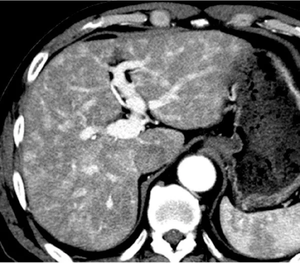
THAD, bölgesel portal akım azalması veya arteriyel katkı artışının dinamik kontrastlı BT'de oluşturduğu geçici parankim görünümüdür. Arteriyel fazda kitle gibi parlak seçilebildiği için özellikle sirotik karaciğerde hepatoselüler karsinomla karıştırılabilir. Fazlar arası kaybolması, kama biçimi ve içinden geçen normal damarlar BT'nin başlıca ayırt ettirici katkısıdır. Gerçek nodül sanılması gereksiz ileri incelemeye; gerçek bir tümör çevresindeki perfüzyon değişikliğinin lezyon sınırına katılması ise boyutun hatalı değerlendirilmesine yol açabilir.
Faz ve pencere
- Arteriyel tanısal
- Arteriyel fazda çoğunlukla periferik, düz kenarlı kama veya üçgen biçimli hiperatenüöz parankim görülür; alan gerçek bir solid kitle gibi yuvarlak kontur kazanmaz ve içinden normal damar dalları geçebilir.
- Portal tanısal
- Portal venöz fazda bölge çevre parankimle izodens hale gelir; kalıcı nodüler kontrastlanma veya kitle etkisi oluşturan kontur değişikliği gözlenmez.
- Gecikmiş tanısal
- Gecikmiş faz tek başına THAD tanısı koydurmaz. Tipik perfüzyon psödolezyonu arteriyel faz dışındaki serilerde silinir ve kitle etkisi oluşturmaz; başka fazlarda süren yuvarlak, kitle biçimli görünüm gerçek fokal lezyon olasılığını artırır.
Önerilen pencereler: Karaciğer (G 150 / M 30), Batın (G 400 / M 50).
BT bulguları
- Arteriyel fazda kama, üçgen veya koni biçimli bölgesel hiperatenüasyon; çoğu kez subkapsüler yerleşim.
- Alan kenarının düz ve segmental perfüzyon sınırına uyan görünümü; yuvarlak, ekspansil bir kitle konturu oluşturmaması.
- Portal ve hepatik ven dallarının parankim odağından doğal kalibrasyon ve doğrultuyla geçmesi.
- Portal fazda çevre karaciğerle atenüasyonun eşitlenmesi; kalıcı nodüler kontrastlanma kusurunun bulunmaması.
- THAD alanı geçici parankimal kontrastlanma farkıdır; normal damarların bu alandan geçmesi gerçek kitlelerden ayrımına yardımcı olur.
- Portal ven trombozu, kompresyonu ya da tümör çevresi gibi yerel akım değişikliğini açıklayabilecek nedenin eşlik edebilmesi.
Normalde
THAD değerlendirmesinde, arteriyel fazdaki bölgesel kontrastlanmanın portal venöz fazda çevre parankimle normal ya da normale yakın atenüasyona dönüp dönmediğini ve normal damarların alan içinden geçip geçmediğini karşılaştır. Kıyaslamada kuşkulu alanın aynı anatomik düzeydeki komşu segmentlerden yalnız arteriyel fazda ayrışıp ayrışmadığını ve portal ven dalının içinden itilmeden geçip geçmediğini kontrol et.
Normal BT ile kıyasla
Aynı bölgenin, kaynak veri setinde patoloji içermediği belirtilen bir BT incelemesini kesit kesit kaydırın; organ sınırlarını açarak anatomiyi hasta görüntüsüyle karşılaştırın. Küçük rastlantısal bulgular tümüyle dışlanmamıştır.
Kaynak: TotalSegmentator v2.01 veri seti (Wasserthal ve ark., Radiology: Artificial Intelligence 2023), CC BY 4.0, vaka s0541; organ sınırları veri setinin otomatik segmentasyonundan, birleştirilerek sadeleştirildi. Görüntüler 2,5 mm kesit aralığına yeniden örneklendi.
Ayırıcı tanı
- HCC arteriyel faz hiperatenüansı ve portal ya da gecikmiş faz yıkanması gösterebilir; THAD ise kama biçimi, alan içinden geçen normal damarlar ve portal fazda normale yakın atenüasyona dönmesiyle ayırt edilebilir.
- Fokal nodüler hiperplazi, arteriyel kontrastlanma ve merkezî skar gösterebilen bir karaciğer lezyonudur.
- Hemangiom
- periferik kesintili nodüler kontrastlanma ve sonraki fazlarda içe doğru dolum eğilimi gösterir.
- Hepatik infarkt gerçek parankimal iskemi/infarktı ifade eder; fokal portal venöz obstrüksiyona eşlik eden heterojen karaciğer kontrastlanması ise THAD olarak görülebilir ve gerçek parankimal infarktla karıştırılmamalıdır.
Tuzaklar
- Arteriyel fazın yanlış zamanlanması kontrastlanmayı ve atenüasyon ölçümlerini etkileyebilir; değerlendirmede aortadaki atenüasyonu referans almak yardımcı olabilir.
- THAD genellikle lezyonun periferinde bulunur ve lezyonla komşudur; hastanın takibinde THAD alanını lezyon çapına ekleyerek hastalığın boyutunu abartmamak gerekir.
- Tipik THAD portal venöz fazda çevre parankimle normal ya da normale yakın atenüasyona döner; diğer fazlarda süren bir kontrastlanma farkı tipik geçici görünümle uyumlu değildir.
Kendini dene
THAD olarak tanımlanan arteriyel fazdaki kama biçimli alan, portal venöz fazda tipik olarak nasıl değişir?
Cevabı göster
Çevre karaciğer parankimiyle aynı ya da yakın atenüasyona döner. THAD alanı portal venöz fazda normal ya da normale yakın atenüasyona döner.
Arteriyel seride kama biçimli karaciğer hiperatenüasyonu görülüyor. Hangi bulgu, THAD'ın beklenen fazlar arası görünümüne uymaz?
Cevabı göster
Portal fazda kalıcı, yuvarlak ve kitle biçimli hipoatenüan odağın kalması. THAD tipik olarak arteriyel fazda kama biçimli görünür, normal damarları içerir ve portal/venöz dönemde çevre parankimle izodens hale gelir. Kalıcı, yuvarlak ve kitle biçimli odak yalnız geçici perfüzyon farkıyla açıklanmaz; gerçek fokal lezyon açısından değerlendirilmelidir.
Bu konunun yer aldığı turlar
İlgili konular
Kaynaklar
Bu sayfadaki 35 cümle ve 2 quiz sorusu aşağıdaki kaynaklardan alıntılarla otomatik olarak karşılaştırıldı; 9 cümle bu karşılaştırmada düzeltildi (2026-10-09).
- Acute portal vein thrombosis postimage-guided liver biopsy in common variable immunodeficiencypmc.ncbi.nlm.nih.gov
- Comparing the computed tomography radiologic features of cytokeratin 19-positive hepatocellular carcinoma to those of conventional hepatocellular carcinoma and intrahepatic cholangiocarcinomapmc.ncbi.nlm.nih.gov
- Hepatic pseudolesions caused by alterations in intrahepatic hemodynamicspmc.ncbi.nlm.nih.gov
- Hepatic sclerosed hemangioma and sclerosing cavernous hemangioma: a radiological studypmc.ncbi.nlm.nih.gov
- The Impact of Transient Hepatic Attenuation Differences in the Diagnosis of Pseudoaneurysm and Arteriovenous Fistula on Follow-Up CT Scans after Blunt Liver Traumapmc.ncbi.nlm.nih.gov
- Ongoing challenges in the diagnosis of hepatocellular carcinoma.pubmed.ncbi.nlm.nih.gov
- Transient hepatic attenuation difference or fat sparing? Aberrant right gastric vein determining a pseudolesion at the border of the IInd/IIIrd liver segments. Review of developmental concepts.pubmed.ncbi.nlm.nih.gov
- Hepatic pseudolesions caused by alterations in intrahepatic hemodynamics.pubmed.ncbi.nlm.nih.gov
- Hepatic sclerosed hemangioma and sclerosing cavernous hemangioma: a radiological study.pubmed.ncbi.nlm.nih.gov
- A Comprehensive Approach to Hepatic Vascular Disease.pubmed.ncbi.nlm.nih.gov
- Diagnostic performance of multidetector CT for acute cholangitis: evaluation of a CT scoring method.pubmed.ncbi.nlm.nih.gov
- The Impact of Transient Hepatic Attenuation Differences in the Diagnosis of Pseudoaneurysm and Arteriovenous Fistula on Follow-Up CT Scans after Blunt Liver Trauma.pubmed.ncbi.nlm.nih.gov
- Xanthogranulomatous cholecystitis: diagnostic performance of US, CT, and MRI for differentiation from gallbladder carcinoma.pubmed.ncbi.nlm.nih.gov
- Differentiation between biliary cystic neoplasms and simple cysts of the liver: accuracy of CT.pubmed.ncbi.nlm.nih.gov
- Transient Hepatic Attenuation Differences in Computed Tomography from Extrahepatic Portal Vein Compression.pubmed.ncbi.nlm.nih.gov
- Identification of MRI features associated with injury type, severity, and prognosis in drug-induced liver injury.pubmed.ncbi.nlm.nih.gov
- Septic thrombophlebitis of the portal venous system: clinical and imaging findings in thirty-three patients.pubmed.ncbi.nlm.nih.gov
- Multislice Computed Tomographic Manifestation of Transient Hepatic Attenuation Difference in the Left Lobe of the Liver: A Retrospective Study.pubmed.ncbi.nlm.nih.gov
- Classification of hypervascular liver lesions based on hepatic artery and portal vein blood supply coefficients calculated from triphasic CT scans.pubmed.ncbi.nlm.nih.gov
- Case Report: Multimodality imaging features and serial metabolic changes on <sup>18</sup> F-FDG PET/CT in a dog with granulomatous hepatitis.pubmed.ncbi.nlm.nih.gov
- Computed Tomographic Findings and Clinical Manifestations of Acute Severe Hypersensitivity to a Non-Ionic Iodinated Contrast Agent in a Castrated Male Dog: A Case Report.pubmed.ncbi.nlm.nih.gov
- Low concentration isotonic iodixanol on hepatic and portal veins imaging quality and renal function: A 320-detector CT dual-energy study.pubmed.ncbi.nlm.nih.gov
- Emerging Perspectives in the Diagnosis and Management of Metabolic Dysfunction-Associated Steatotic Liver Disease (MASLD): A Narrative Review.pubmed.ncbi.nlm.nih.gov
- Human biomarker navigator.pubmed.ncbi.nlm.nih.gov
- That liver lesion on MDCT in the oncology patient: is it important?pubmed.ncbi.nlm.nih.gov
- Post-operative imaging in liver transplantation: state-of-the-art and future perspectives.pubmed.ncbi.nlm.nih.gov
- The TSANZ Practical Guide for Clinicians in the Management of Screen- and Incidentally-Detected Nodules.pubmed.ncbi.nlm.nih.gov
- EACR 2025 Congress: Innovative Cancer Science, 16-19 June 2025.pubmed.ncbi.nlm.nih.gov
- Imaging findings of mimickers of hepatocellular carcinoma.pubmed.ncbi.nlm.nih.gov
- Non-invasive ultrasound assessment of chronic liver disease: current position and future directions for a "one-stop" liver ultrasound approach.pubmed.ncbi.nlm.nih.gov
- Hypoxic pulmonary vasoconstriction.pubmed.ncbi.nlm.nih.gov
- Acute portal vein thrombosis postimage-guided liver biopsy in common variable immunodeficiency.pubmed.ncbi.nlm.nih.gov
- Assessing Pancreatic Enhancement Variability in ICU Patients with CT Hypoperfusion Complex: A Retrospective Single-Center Study.pubmed.ncbi.nlm.nih.gov
- Patterns of injury and violence in Yaoundé Cameroon: an analysis of hospital data.pubmed.ncbi.nlm.nih.gov
- Non-invasive diagnosis of focal liver lesions: an individualized approach.pubmed.ncbi.nlm.nih.gov
- Hepatic perfusion disorders: etiopathogenesis and related diseases.pubmed.ncbi.nlm.nih.gov
- Long-term safety and effectiveness of the "OptEase" vena cava filter.pubmed.ncbi.nlm.nih.gov
- Interventional, surgical, and clinical management of Abernethy malformations: A case series and novel anatomic variant.pubmed.ncbi.nlm.nih.gov
- Guidelines on the Use of Therapeutic Apheresis in Clinical Practice-Evidence-Based Approach From the Writing Committee of the American Society for Apheresis: The Tenth Special Issue.pubmed.ncbi.nlm.nih.gov
- Short- and long-term outcome and quality of life assessment in dogs undergoing transvenous coil embolization of congenital intrahepatic portosystemic shunts.pubmed.ncbi.nlm.nih.gov
- Low-dose l-carnitine supplementation attenuates hepatic lipid accumulation in chow-fed mice: Insights from integrated multi-omics analysis.pubmed.ncbi.nlm.nih.gov
- A Diagnostic Dilemma in Hepatic Cystic Lesions: Radiological Mimicry of Biliary Cystadenoma and Hydatid Cyst in a Histologically Confirmed Simple Hepatic Cyst.pubmed.ncbi.nlm.nih.gov
- Comparison of the clinical characteristics and imaging findings of acute cholangitis with and without biliary dilatation.pubmed.ncbi.nlm.nih.gov
- Cross-sectional imaging of congenital and acquired abnormalities of the portal venous system.pubmed.ncbi.nlm.nih.gov
- Portal Vein Thrombosis: State-of-the-Art Review.pubmed.ncbi.nlm.nih.gov
- Gut-Liver Axis Dysregulation in Portal Hypertension: Emerging Frontiers.pubmed.ncbi.nlm.nih.gov
- Transformative biomechanics and mechanobiology breakthroughs shaping the future of health and medicine.pubmed.ncbi.nlm.nih.gov
- TACE-HAIC Plus Targeted Therapy and Immunotherapy for HCC with Tumor Thrombus and Arteriovenous Fistula.pubmed.ncbi.nlm.nih.gov
- Exploratory investigation of dose calculation and tumor-feeding artery identification using dual CBCT in selective internal radiation therapy with Yttrium-90 microspheres.pubmed.ncbi.nlm.nih.gov
- A contrast‑enhanced CT histogram‑driven nomogram for predicting post‑radiotherapy liver regeneration in hepatocellular carcinoma.pubmed.ncbi.nlm.nih.gov
- Clinical value of contrast-enhanced ultrasound in early diagnosis of hepatocellular carcinoma.pubmed.ncbi.nlm.nih.gov
- Three Types of Collateral Arterial Supply to the Spleen After Spleen-Preserving Distal Pancreatectomies with Splenic Vessels Resection-How to Use This Knowledge for Organ(s) Preservation in Locally Advanced and Borderline Resectable Pancreatic Head Cancers Surgery-Hemodynamic, Surgical and Oncological Outcomes of 134 Spleen-Preserving Pancreatectomies.pubmed.ncbi.nlm.nih.gov
- Clinicopathological insights and management of liver metastases: Current advances and future perspectives.pubmed.ncbi.nlm.nih.gov
- Comparing the computed tomography radiologic features of cytokeratin 19-positive hepatocellular carcinoma to those of conventional hepatocellular carcinoma and intrahepatic cholangiocarcinoma.pubmed.ncbi.nlm.nih.gov
- Long-Term Care in Fontan Circulation: Surveillance and Management of Fontan-Associated Liver Disease and Protein-Losing Enteropathy.pubmed.ncbi.nlm.nih.gov
- Metabolic Dysfunction-Associated Steatotic Liver Disease (MASLD): New Perspectives on an Evolving Epidemic.pubmed.ncbi.nlm.nih.gov
- Imaging insights into pediatric liver masses: A comprehensive minireview for hepatology practice.pubmed.ncbi.nlm.nih.gov
- Imaging at the nexus: how state of the art imaging techniques can enhance our understanding of cancer and fibrosis.pubmed.ncbi.nlm.nih.gov
- Bubbles in the barely born-contrast-enhanced ultrasound in neonates: a single-center experience.pubmed.ncbi.nlm.nih.gov
- Liver transplantation for pediatric liver tumors: a case report of hepatic angiosarcoma with literature review.pubmed.ncbi.nlm.nih.gov
- Use of the Term "Atypical Hepatic Hemangioma" in Radiology Reports: An Analysis of Diagnostic Precision.pubmed.ncbi.nlm.nih.gov
- Comprehensive review of pulmonary embolism imaging: past, present and future innovations in computed tomography (CT) and other diagnostic techniques.pubmed.ncbi.nlm.nih.gov
- Current concepts in the management of non-cirrhotic non-malignant portal vein thrombosis.pubmed.ncbi.nlm.nih.gov
- Diagnostic and therapeutic recommendations in pancreatic ductal adenocarcinoma. Recommendations of the Working Group of the Polish Pancreatic Club.pubmed.ncbi.nlm.nih.gov
- Advances in Biomaterials and Technologies for Vascular Embolization.pubmed.ncbi.nlm.nih.gov
- Preoperative imaging of colorectal liver metastases: what the radiologist and the multidisciplinary team need to know.pubmed.ncbi.nlm.nih.gov
- MR Imaging of the Perihepatic Space.pubmed.ncbi.nlm.nih.gov
- Evaluating the Effectiveness of Yttrium-90 Glass Microspheres in the Treatment of Hepatocellular Carcinoma, Intrahepatic Cholangiocarcinoma, and Metastatic Colorectal Cancer in Practice: Protocol for the Prospective PROACTIF Phase IV Registry Study in France.pubmed.ncbi.nlm.nih.gov
- Longitudinal Inpatient Trajectories After Splenic Artery Embolization in Cirrhosis: Real-World Evidence from Kazakhstan.pubmed.ncbi.nlm.nih.gov
- Ultrasound Evaluation of Fontan-Associated Liver Disease: A State-of-the-Art Review.pubmed.ncbi.nlm.nih.gov
- Perioperative nursing coordination and complication management in Yttrium-90 radioactive microsphere therapy for hepatocellular carcinoma.pubmed.ncbi.nlm.nih.gov
- Hepatobiliary phase imaging in HCC: revisiting diagnostic algorithms and clinical utility of gadoxetic acid-enhanced MRI across international guidelines.pubmed.ncbi.nlm.nih.gov
- Liver Imaging Reporting and Data System (LI-RADS): A Comprehensive Review.pubmed.ncbi.nlm.nih.gov
- Magnetic Resonance-Guided Radiotherapy for Unresectable Hepatocellular Carcinoma with Bile Duct Tumor Thrombus: A Case Series and Review of Treatment Options.pubmed.ncbi.nlm.nih.gov
- Non-contrast microvascular flow imaging in abdominal ultrasound: a pictorial essay.pubmed.ncbi.nlm.nih.gov
- Pylorus-Preserving Pancreatoduodenectomy with Arterial Reconstruction and Right Gastric Vessel Preservation for Pancreatic Head Cancer with Atherosclerotic Celiac Axis Stenosis and Common Hepatic Artery Occlusion: A Case Report.pubmed.ncbi.nlm.nih.gov
- Venous Thromboembolism in Neonates, Children, and Adolescents: A Comprehensive Narrative Review of Risk Factors, Diagnosis, Treatment, and Prevention.pubmed.ncbi.nlm.nih.gov
- EAT Thickness and BAT-Related Thermogenesis: Dual Imaging Phenotypes Associated with Hepatic Steatosis Severity in MASLD.pubmed.ncbi.nlm.nih.gov
- The Association of Neutrophil-To-Lymphocyte and Platelet-To-Lymphocyte Ratios With Distant Metastasis in Vietnamese Gastric Cancer Patients.pubmed.ncbi.nlm.nih.gov
- Ultrasound Radiomics in Pediatric Imaging: Current Applications, Challenges, and Future Directions Toward Clinical Implementation.pubmed.ncbi.nlm.nih.gov
- Application of immunoPET imaging to enhance head and neck squamous cell carcinoma clinical management.pubmed.ncbi.nlm.nih.gov
- Comparison of abdominal computed tomography to ultrasonography in the diagnosis of biliary disease in dogs with acute abdominal signs.pubmed.ncbi.nlm.nih.gov
- Alveolar Type 2 Cell Dysfunction Is Associated with Bile Acid Alterations in Experimental Hepatopulmonary Syndrome.pubmed.ncbi.nlm.nih.gov
- Vendor-agnostic versus vendor-specific boost methods in low-concentration contrast-enhanced computed tomography of the abdomen.pubmed.ncbi.nlm.nih.gov
- Study on the combined application of selected virtual monoenergetic imaging, virtual non-contrast imaging, and adaptive statistical iterative reconstruction-V in head and neck CT angiography using 256-slice wide-body CT.pubmed.ncbi.nlm.nih.gov
- "WBC-HBP Dissociation" Unveils Occult Xanthogranulomatous Inflammation Mimicking Hepatocellular Carcinoma in Diabetic Patients.pubmed.ncbi.nlm.nih.gov
- Focal nodular hyperplasia: a rare benign liver tumour in early childhood without predisposing factors.pubmed.ncbi.nlm.nih.gov
- Contrast-Enhanced CT Finding of Hot Spot Sign Secondary to Supra Hilar Mass Causing Superior Vena Cava Syndrome: A Case Report.pubmed.ncbi.nlm.nih.gov
- Focal Nodular Hyperplasia Presenting With Biliary Obstruction and Intrahepatic Gallstones: A Case Report and Literature Review.pubmed.ncbi.nlm.nih.gov
- Associations Among CTA Collateral Scores, Multimodal MRI Lesion Volumes, and Clinical Severity in Acute Middle Cerebral Artery Infarction.pubmed.ncbi.nlm.nih.gov
- Recent Progress in PET and SPECT Myocardial Perfusion Imaging: From Radiopharmaceuticals to Imaging Innovations.pubmed.ncbi.nlm.nih.gov
- Arterial Thrombosis in Severe Ulcerative Colitis: A Case-Based Narrative Review of Current Evidence.pubmed.ncbi.nlm.nih.gov
- Multicontrast Multiphase Intestinal Imaging Using Photon-Counting CT: A Feasibility Study in Rats.pubmed.ncbi.nlm.nih.gov
- Phase identification for dynamic CT enhancements with generative adversarial network.pubmed.ncbi.nlm.nih.gov
- Systemic pharmacological treatments for chronic plaque psoriasis: a network meta-analysis.pubmed.ncbi.nlm.nih.gov
- Computed Tomographic Findings of Liver Cirrhosis in Dogs: Comparison between Compensated and Decompensated Cirrhosis.pubmed.ncbi.nlm.nih.gov
- Transarterial embolization with bleomycin-lipiodol for symptomatic multifocal hepatic hemangiomas: Concurrent nontarget lesion devascularization via permitted reflux.pubmed.ncbi.nlm.nih.gov
- Role of SIRT in Patients With Hepatocellular Carcinoma: Current Clinical Indications and Future Perspectives.pubmed.ncbi.nlm.nih.gov
- Application value of dual-phase cone-beam computed tomography during hepatic arteriography in the detection of ≤2 cm hypervascular hepatocellular carcinoma and its feeding arteries.pubmed.ncbi.nlm.nih.gov
- Ultrasound innovations in abdominal radiology: techniques and clinical applications in pediatric imaging.pubmed.ncbi.nlm.nih.gov
- Obstructive Sleep Apnea: Epidemiology, Pathophysiology, Complications, Diagnosis, Management, and Emerging Fibrosis-Linked Remodeling.pubmed.ncbi.nlm.nih.gov
- Commentary: an eye on PET quantification.pubmed.ncbi.nlm.nih.gov
- Ultrasound elastography in pediatric care: bridging innovation and noninvasive diagnostics.pubmed.ncbi.nlm.nih.gov
- A case of hepatic segmental atrophy mimicking spontaneously necrotized hepatocellular carcinoma.pubmed.ncbi.nlm.nih.gov
- Atypical Appearance of Hepatocellular Carcinoma and Its Mimickers: How to Solve Challenging Cases Using Gadoxetic Acid-Enhanced Liver Magnetic Resonance Imaging.pubmed.ncbi.nlm.nih.gov
- Recent Advances in the Imaging Diagnosis of Hepatocellular Carcinoma: Value of Gadoxetic Acid-Enhanced MRI.pubmed.ncbi.nlm.nih.gov
- Practical approach to diagnose and manage benign liver masses.pubmed.ncbi.nlm.nih.gov
- Glomerulonephritis through the lens of ultrasound and radiology.pubmed.ncbi.nlm.nih.gov
- Washout on Contrast-Enhanced Ultrasound of Benign Focal Liver Lesions-A Review on Its Frequency and Possible Causes.pubmed.ncbi.nlm.nih.gov
- Acute paraumbilical vein recanalization: an unusual complication of acute pancreatitis.pubmed.ncbi.nlm.nih.gov
- Current Management of Infective Endocarditis: A Narrative Review Focused on Unmet Clinical Needs and the Multidisciplinary Approach.pubmed.ncbi.nlm.nih.gov
- Nanoparticle Contrast Agents for Photon-Counting Computed Tomography: Recent Developments and Future Opportunities.pubmed.ncbi.nlm.nih.gov
- Computed Tomography Techniques, Protocols, Advancements, and Future Directions in Liver Diseases.pubmed.ncbi.nlm.nih.gov
- Hepatobiliary phases in magnetic resonance imaging using liver-specific contrast for focal lesions in clinical practice.pubmed.ncbi.nlm.nih.gov
- The AFSUMB Consensus Statements and Recommendations for the Clinical Practice of Contrast-Enhanced Ultrasound using Sonazoid.pubmed.ncbi.nlm.nih.gov
Eğitim amaçlıdır; hasta başında tanı ve tedavi kararının yerine geçmez, tıbbi tavsiye değildir. Metinler yapay zekâ desteğiyle yazıldı ve otomatik bir adımda kaynak alıntılarıyla karşılaştırıldı; bu bir tıbbi doğrulama değildir ve içerik uzman radyolog incelemesinden geçmedi. Klinik kararlarda güncel kılavuzları, kurum protokollerini ve radyoloji raporunu esas alın.