Özet
Karaciğer kist hidatiği, Echinococcus larva kistinin karaciğerde büyümesiyle oluşur ve Türkiye gibi endemik bölgelerde kistik karaciğer lezyonlarının önemli bir paraziter nedenidir. BT, kistin segmental yerini, kalsifiye duvarını, kitle etkisini ve komşu safra yolları/damarlarla ilişkisini gösterir; kist iç mimarisinin ve canlılığın evrelenmesinde ultrason temel yöntemdir. Kız veziküller veya ayrılmış membran paraziter kökeni kuvvetle desteklerken yalnız tek odacıklı sıvı kist BT'de özgül olmayabilir. Rüptür veya biliyer iletişimin gözden kaçması enfeksiyon, obstrüksiyon ve peritoneal yayılım riskini saklayabilir.
Faz ve pencere
- Kontrastsız tanısal
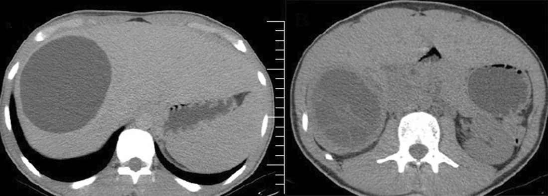
- Suya yakın düşük atenüasyonlu kist içeriği, kalsifiye perikist duvarı, intrakistik membran/debris ve kız veziküller değerlendirilebilir; duvar kalsifikasyonu bu fazda en iyi seçilir.
- Portal tanısal
- Kontrastlı BT’de perikist ince, kontrastlanan bir halka olarak görülebilir; kız veziküller ana kist içinde sıvı dolu odacıklar oluşturur. BT’de safra yolu genişlemesi ve damar basısı gibi komşu yapı etkileri değerlendirilebilir.
Önerilen pencereler: Karaciğer (G 150 / M 30), Batın (G 400 / M 50).
BT bulguları
- İyi sınırlı, homojen düşük atenüasyonlu tek odacıklı kist BT’de basit kistten güvenilir biçimde ayırt edilemeyebilir; CE1 evresi ultrason morfolojisine dayanır.
- Kız veziküller — ana kist içinde çok sayıda yuvarlak, daha küçük kistik odacık; bal peteği veya rozet düzeni
- Ayrılmış endokist membranı — hidatik kiste özgü 'su lili işareti' olarak bilinen, sıvı içinde dalgalanan ince parazitik tabaka görünümüdür.
- Kist duvarında kavisli, halka biçimli veya parçalı kalsifikasyon; kontrastsız seride belirgin
- Kist içi detaylar ve iç yapı, BT'ye göre ultrasonun morfolojik evrelemede öncelikli olduğu akılda tutularak değerlendirilmelidir.
- Komşu portal veya hepatik venlerde, vena kava inferiorda ya da safra kanallarında kitle etkisine bağlı bası ve obstrüksiyon bulgularını değerlendirin.
- İntrahepatik safra yolu genişlemesi kistobiliyer iletişime eşlik edebilir; biliyer iletişimin değerlendirilmesinde MR avantajlıdır.
Ölçütler ve sınıflamalar
- WHO-IWGE CE ultrason evrelemesi
- WHO-IWGE evrelemesine göre CE1 tek odacıklı sıvı dolu kistir; CE2 kız veziküller içeren ve septumlarla bölünmüş aktif kistir; CE3a sıvı içinde avasküler, ince ve düzenli dalgalanan ayrılmış parazitik tabakası olan kistir; CE3b avasküler matriks içinde kız veziküller içeren karmaşık içeriğe sahip kistir; CE4 ve CE5 ise dejeneratif ve inaktif kist evreleridir. Sınıflama ultrason morfolojisine dayanır; BT’den tek başına kesin CE evresi verilmemelidir.
Normalde
Kistin yerleştiği segmentte komşu portal ven, hepatik ven ve safra yollarının anatomik seyrini ve kitle etkisiyle oluşabilecek bası/yer değiştirmeyi değerlendirin.
Normal BT ile kıyasla
Aynı bölgenin, kaynak veri setinde patoloji içermediği belirtilen bir BT incelemesini kesit kesit kaydırın; organ sınırlarını açarak anatomiyi hasta görüntüsüyle karşılaştırın. Küçük rastlantısal bulgular tümüyle dışlanmamıştır.
Kaynak: TotalSegmentator v2.01 veri seti (Wasserthal ve ark., Radiology: Artificial Intelligence 2023), CC BY 4.0, vaka s0541; organ sınırları veri setinin otomatik segmentasyonundan, birleştirilerek sadeleştirildi. Görüntüler 2,5 mm kesit aralığına yeniden örneklendi.
Ayırıcı tanı
- Basit karaciğer kisti
- homojen su atenüasyonu ve ince düzgün duvar gösterir; kız vezikül, ayrılmış membran veya kalsifiye lameller duvar yoktur.
- Müsinöz kistik neoplazi
- görüntüleme bulguları diğer kistik karaciğer lezyonlarıyla sıkça örtüşür; kesin ayırım için histopatoloji gerekebilir.
- Piyojenik karaciğer absesi BT’de cluster sign veya double target sign gösterebilir; hidatik kistte kız veziküller ve periferik kalsifikasyonlar tanıya yardımcı olabilir.
- Amip karaciğer absesi tek, sağ subkapsüler yerleşimli bir lezyon şeklinde görülebilir.
- Alveoler ekinokokkoz
- düzgün sınırlı büyük kistten çok infiltratif, düzensiz ve kalsifik odaklı kitle benzeri büyüme gösterebilir.
Tuzaklar
- Basit tek odacıklı CE1 kisti basit kistten BT'de ayırt edilemeyebilir; endemik maruziyet ve ultrason iç yapısı tanısal güveni değiştirir.
- Duvar kalsifikasyonu tek başına kistin canlı ya da inaktif olduğunu kanıtlamaz; WHO-IWGE evresi US morfolojisiyle belirlenir.
- Kist içindeki kız veziküllerini değerlendirin; CE2 evresinde bunlar sıvı dolu kist içinde septalı görünüm oluşturur.
- Safra yolu ile yakın temas iletişim kanıtı değildir; kanal içi membran/debris veya belirgin biliyer obstrüksiyon bulgusu arayın.
Kendini dene
Endemik bölgede yaşayan hastada karaciğer kisti içinde çok sayıda yuvarlak küçük sıvı odacığı görülüyor. En olası tanı nedir?
Cevabı göster
Kist hidatik. Ana kavite içindeki kız veziküller kistik ekinokokkoz lehine karakteristik mimaridir. Ana kavitedeki kız veziküller (satellite cysts/honeycomb appearance) hidatik kisti destekler. Kız veziküllerin görülmesi bu olguda kistik ekinokokkoz tanısını destekler.
Bir karaciğer kisti çevresindeki belirgin duvar kalsifikasyonu WHO-IWGE evresini BT üzerinden tek başına belirler mi?
Cevabı göster
Hayır, sınıflama US temellidir. WHO-IWGE evreleri ultrason morfolojisine göre tanımlanmıştır. WHO-IWGE evreleri kistin morfolojik özelliklerine dayanır; duvar kalsifikasyonu tek başına evreyi belirlemez.
İlgili konular
Kaynaklar
Bu sayfadaki 40 cümle ve 2 quiz sorusu aşağıdaki kaynaklardan alıntılarla otomatik olarak karşılaştırıldı; 20 cümle bu karşılaştırmada düzeltildi (2026-10-09).
- Treatment of uncomplicated hepatic cystic echinococcosis (hydatid disease)pmc.ncbi.nlm.nih.gov
- Standardized endocystectomy technique for surgical treatment of uncomplicated hepatic cystic echinococcosispmc.ncbi.nlm.nih.gov
- Medical treatment versus “Watch and Wait” in the clinical management of CE3b echinococcal cysts of the liverpmc.ncbi.nlm.nih.gov
- Albendazole Monotherapy and Spleen Preservation in an Adolescent with Isolated Primary Splenic Cystic Echinococcosis: A Case Reportpmc.ncbi.nlm.nih.gov
- Alveolar echinococcosispmc.ncbi.nlm.nih.gov
- Standardized endocystectomy technique for surgical treatment of uncomplicated hepatic cystic echinococcosis.pubmed.ncbi.nlm.nih.gov
- Peritoneal Hydatid Cysts in Children: A Case Series of Rare Echinococcosis Localization.pubmed.ncbi.nlm.nih.gov
- Intrabiliary Rupture of a Hepatic Hydatid Cyst: A Case Report.pubmed.ncbi.nlm.nih.gov
- Long-term sonographic and serological follow-up of inactive echinococcal cysts of the liver: hints for a "watch-and-wait" approach.pubmed.ncbi.nlm.nih.gov
- Assessment of the accuracy of preoperative imaging methods in the diagnosis of hepatic single-chamber echinococcosis.pubmed.ncbi.nlm.nih.gov
- Surgical decision-making in splenic hydatid cyst management via successful total splenectomy: a case report.pubmed.ncbi.nlm.nih.gov
- Abdominal effusion revealing an exophytic hydatid cyst of the liver has developed under mesocolic.pubmed.ncbi.nlm.nih.gov
- Abdominal hydatid disease: role of imaging in diagnosis, complications, and management.pubmed.ncbi.nlm.nih.gov
- Hydatid cyst of the liver causing inferior vena caval obstruction.pubmed.ncbi.nlm.nih.gov
- Pleural migration of biliary stent 10 years following treatment of hepatic hydatic echinococcosis: A case report.pubmed.ncbi.nlm.nih.gov
- Case Report: Isolated splenic hydatid cyst with negative serology managed by laparoscopic splenectomy.pubmed.ncbi.nlm.nih.gov
- Primary diaphragmatic hydatid cyst: A rare location of echinococcosis.pubmed.ncbi.nlm.nih.gov
- Isolated renal hydatic cyst in a 14-year-old girl: A case report.pubmed.ncbi.nlm.nih.gov
- Ultrasound-Based Staging and Its Impact on Clinical Management of Hepatic Hydatid Cysts in an Endemic Setting: A Cross-Sectional Study in Eastern Afghanistan.pubmed.ncbi.nlm.nih.gov
- Albendazole Monotherapy and Spleen Preservation in an Adolescent with Isolated Primary Splenic Cystic Echinococcosis: A Case Report.pubmed.ncbi.nlm.nih.gov
- Hepatic Echinococcosis as an Incidental Finding in a Patient With a Perforated Gastric Ulcer: A Case Report.pubmed.ncbi.nlm.nih.gov
- Common and Rare Radiological Findings of Pulmonary and Thoracic Hydatid Cysts.pubmed.ncbi.nlm.nih.gov
- Laparoscopic management of an infected hepatic hydatid cyst with cystobiliary fistula in a child: a case report.pubmed.ncbi.nlm.nih.gov
- Hydatid Cyst in Pregnancy-A Diagnostic and Therapeutic Dilemma: Study Case Report.pubmed.ncbi.nlm.nih.gov
- Magnetic Resonance Imaging Features of Hepatic Hydatid Disease: A Pictorial Review with Emphasis on Atypical Presentations and Differential Diagnosis.pubmed.ncbi.nlm.nih.gov
- Role of interventional radiology in the management of hepatic hydatid disease.pubmed.ncbi.nlm.nih.gov
- A Rare Case of Extensive Relapsing Disseminated Hydatid Disease with Multi-Organ Involvement: A Case Report.pubmed.ncbi.nlm.nih.gov
- Breast cystic echinococcosis with ultrasonographic "cerebral gyrus sign": a case report.pubmed.ncbi.nlm.nih.gov
- Rare anatomical localizations of hydatid cysts (2020-2025): Narrative review of presentation, imaging, and management.pubmed.ncbi.nlm.nih.gov
- An Unusual Case of Anterior Mediastinal Cystic Echinococcosis Successfully Resolved with Multidisciplinary Approach.pubmed.ncbi.nlm.nih.gov
- Mucinous cystic neoplasms of the liver: current insights into epidemiology, diagnosis, and treatment.pubmed.ncbi.nlm.nih.gov
- Hepatic Alveolar Echinococcosis Mimicking Perihilar Cholangiocarcinoma: A Case Report.pubmed.ncbi.nlm.nih.gov
- Hydatid thrombosis of the portal vein: a rare case report.pubmed.ncbi.nlm.nih.gov
- Common and Uncommon Splenic Lesions: A Review.pubmed.ncbi.nlm.nih.gov
- Splenic pathologies: A pictorial review of common, wandering, twisting and rare presentations.pubmed.ncbi.nlm.nih.gov
- Gallbladder mucinous cystic neoplasm: Diagnostic challenges and multimodality imaging correlation.pubmed.ncbi.nlm.nih.gov
- Encysted Odyssey: A Clinical and Pictorial Analysis of Hydatid Cysts From Head to Toe.pubmed.ncbi.nlm.nih.gov
- Imaging Spectrum of Hydatid Disease: Usual and Unusual Locations.pubmed.ncbi.nlm.nih.gov
- Hydatid cyst of spleen: a diagnostic challenge.pubmed.ncbi.nlm.nih.gov
- A challenging imaging feature of mature teratoma in uterus.pubmed.ncbi.nlm.nih.gov
- Imaging insights into pediatric liver masses: A comprehensive minireview for hepatology practice.pubmed.ncbi.nlm.nih.gov
- Gossypiboma mimicking hepatic hydatid cyst after 22 years: a case report.pubmed.ncbi.nlm.nih.gov
- Primary Posterior Neck Hydatid Cyst: A Case Report and Review of the Literature.pubmed.ncbi.nlm.nih.gov
- A rare case of primary hydatid cyst of the adrenal gland: Case report and review of the literature.pubmed.ncbi.nlm.nih.gov
- Massive Inferior Vena Cava Thrombosis and Pulmonary Embolism Secondary to Hepatic Hydatid Cyst: A Fatal Case.pubmed.ncbi.nlm.nih.gov
- Laparoscopic management of a ruptured hepatic hydatid cyst with a cystobiliary fistula in an adolescent: a case report.pubmed.ncbi.nlm.nih.gov
- Predictors of cystobiliary communication in patients undergoing surgery for hepatic hydatid cysts.pubmed.ncbi.nlm.nih.gov
- Percutaneous <i>vs</i> surgical management of World Health Organization cystic echinococcosis 1 and 3a liver hydatid cysts.pubmed.ncbi.nlm.nih.gov
- When size misleads: CT attenuation and enhancement characteristics predict benignity in a giant ovarian mucinous cystadenoma.pubmed.ncbi.nlm.nih.gov
- Giant Hepatobiliary Cystadenoma Requiring Biliary Reconstruction in a Cat: A Case Report.pubmed.ncbi.nlm.nih.gov
- Contrast-Enhanced CT Finding of Hot Spot Sign Secondary to Supra Hilar Mass Causing Superior Vena Cava Syndrome: A Case Report.pubmed.ncbi.nlm.nih.gov
- A Silent Mimic: Biopsy-Proven Focal Nodular Hyperplasia Masquerading as Chronic Intrahepatic Biliary Obstruction.pubmed.ncbi.nlm.nih.gov
- Case Report: Late repeat pancreatic irreversible electroporation in the long-term multidisciplinary management of unresectable pancreatic ductal adenocarcinoma.pubmed.ncbi.nlm.nih.gov
- Prevalence of Liver Cystic Echinococcosis in Türkiye: A Systematic Review of Population-Based Ultrasound Imaging Surveys.pubmed.ncbi.nlm.nih.gov
- Iliopsoas hydatid cyst mimicking an ovarian tumor: a diagnostic pitfall in an endemic setting-a case report.pubmed.ncbi.nlm.nih.gov
- Practical approach to diagnose and manage benign liver masses.pubmed.ncbi.nlm.nih.gov
- Imaging features and management of focal liver lesions.pubmed.ncbi.nlm.nih.gov
- Diagnostic reliability of O-RADS score based on non-dynamic contrast-enhanced MRI and apparent diffusion coefficient in characterization of adnexal masses.pubmed.ncbi.nlm.nih.gov
- 불분명한 소아 복부 종괴: 영상 기반 진단 접근법.pubmed.ncbi.nlm.nih.gov
- Evolving management of pyogenic liver abscess: A narrative review of laparoscopic and open drainage approaches with contextual insights from Pakistan.pubmed.ncbi.nlm.nih.gov
- Amebic Liver Abscess: An Unusual Cause of Budd-Chiari Syndrome.pubmed.ncbi.nlm.nih.gov
- Alveolar echinococcosis.pubmed.ncbi.nlm.nih.gov
- Locally Aggressive Hepatic Echinococcosis With Colonic Involvement Requiring a Right Hemicolectomy: A Case Report and Focused Literature Review.pubmed.ncbi.nlm.nih.gov
- Computed tomography densitometry for discriminating between type 1 hydatid and simple hepatic cysts.pubmed.ncbi.nlm.nih.gov
- Medical, Surgical, and Combined Approaches in Pediatric Hydatid Liver Disease: A Systematic Review.pubmed.ncbi.nlm.nih.gov
- Clinical profile, imaging characteristics, and operative outcomes of pediatric cystic echinococcosis in Northern Jordan: a single-center retrospective study.pubmed.ncbi.nlm.nih.gov
- Pictorial review of hepatic echinococcosis: Ultrasound imaging and differential diagnosis.pubmed.ncbi.nlm.nih.gov
- Non-infectious Hepatic Cystic Lesions: A Narrative Review.pubmed.ncbi.nlm.nih.gov
- Hepatic Hydatid Cyst Complicated by Biliary Obstruction: A Case Report.pubmed.ncbi.nlm.nih.gov
- Cystic liver lesions: a pictorial review.pubmed.ncbi.nlm.nih.gov
- Unexpected hosts: imaging parasitic diseases.pubmed.ncbi.nlm.nih.gov
- Rare complex recurrent cystic echinococcosis with multi-organ involvement after inadequate postoperative therapy: a case report.pubmed.ncbi.nlm.nih.gov
- Fifty-five cases of hepatic alveolar echinococcosis combined with lymph node metastasis: A retrospective study.pubmed.ncbi.nlm.nih.gov
- Concomitant Hydatid Disease of the Lower Extremity and Retroperitoneum: A Case Report of Two Rare Locations.pubmed.ncbi.nlm.nih.gov
- Multisystem involvement in hydatid disease: a case report of disseminated echinococcosis.pubmed.ncbi.nlm.nih.gov
- Iconographies of rare causes of medullary compression due to primary vertebro-medullary and epidural hydatidosis: Contribution of imaging.pubmed.ncbi.nlm.nih.gov
- The wandering mimic: Meckel's diverticulum with enterolith simulating an intraperitoneal hydatid cyst.pubmed.ncbi.nlm.nih.gov
- Surgical management of cystic echinococcosis-A 20-year case series and outcome analysis.pubmed.ncbi.nlm.nih.gov
- Bacterial Infection of an Alveolar Echinococcus Cyst from C. perfringens Septicemia: A Case Report and Review of the Literature.pubmed.ncbi.nlm.nih.gov
- Application of Multi-Slice Computed Tomography for the Preoperative Diagnosis and Classification of Pulmonary Cystic Echinococcosis.pubmed.ncbi.nlm.nih.gov
- Disseminated Pulmonary and Hepatic Cystic Echinococcosis Presenting as Suspected Pulmonary Embolism and Mimicking Metastatic Malignancy.pubmed.ncbi.nlm.nih.gov
- Large Mesenchymal Hepatic Hamartoma in Pediatric Age: A Case Report.pubmed.ncbi.nlm.nih.gov
- Hydatid Disease: A Radiological Pictorial Review of a Great Neoplasms Mimicker.pubmed.ncbi.nlm.nih.gov
- Hydatid Disease: A Pictorial Review of Uncommon Locations.pubmed.ncbi.nlm.nih.gov
- Common cystic pancreatic neoplasms: A comprehensive magnetic resonance imaging-based review of typical and atypical imaging features.pubmed.ncbi.nlm.nih.gov
- Radiologic-Pathologic Correlation in Biliary Cystic Neoplasms: Insights From a Single-Centre Experience.pubmed.ncbi.nlm.nih.gov
- Amebiasis as a neglected tropical disease: Current knowledge gaps and future directions.pubmed.ncbi.nlm.nih.gov
- Typical imaging finding of hepatic infections: a pictorial essay.pubmed.ncbi.nlm.nih.gov
- The Abscess That Was Cancer: Pyogenic Liver Abscess as the Initial Manifestation of Hepatocellular Carcinoma in a Noncirrhotic Patient.pubmed.ncbi.nlm.nih.gov
- Concomitant Borreliosis and Invasive Amoebiasis: A Rare Case with Suspected Sexual Acquisition.pubmed.ncbi.nlm.nih.gov
- Ruptured Community-acquired Methicillin-Resistant <i>Staphylococcus aureus</i> (MRSA) Hepatic Abscess in an Immunocompetent Child Treated Successfully with Culture-guided Antibiotic Therapy: A Case Report.pubmed.ncbi.nlm.nih.gov
- Hepatic abscess on CT: pearls, pitfalls and diagnostic challenges.pubmed.ncbi.nlm.nih.gov
- Necrotic Liver Metastasis From Anorectal Mucosal Melanoma Mimicking a Pyogenic Liver Abscess: A Case Report.pubmed.ncbi.nlm.nih.gov
- Ciliated hepatic foregut cyst: a case report and review of literature on a rare hepatic cystic lesion.pubmed.ncbi.nlm.nih.gov
- Hepatic cysts: a survival guide.pubmed.ncbi.nlm.nih.gov
- Benign focal liver lesions: The role of magnetic resonance imaging.pubmed.ncbi.nlm.nih.gov
- MRI insights into endometrial stromal tumors.pubmed.ncbi.nlm.nih.gov
- Rapid Interval Growth of a Presumed Simple Hepatic Cyst Revealing a Mucinous Cystic Neoplasm in a Rare Male Presentation.pubmed.ncbi.nlm.nih.gov
- [Mucinous cystic neoplasm of the liver].pubmed.ncbi.nlm.nih.gov
- Incidental cystic pancreatic neuroendocrine neoplasm and differential diagnosis.pubmed.ncbi.nlm.nih.gov
- Simultaneous surgical treatment of multiple abdominal echinococcosis and benign pelvic organ tumours: a case report.pubmed.ncbi.nlm.nih.gov
- A Novel Approach to Recurrent Hepatic Hydatid Cyst Using EUS-Guided Aspiration and Ethanol Injection: A Case Report and Focused Literature Review.pubmed.ncbi.nlm.nih.gov
- Anesthetic Challenges in the Management of a Rare Giant Biliary Mucinous Cystadenoma With Major Vascular Compression.pubmed.ncbi.nlm.nih.gov
- Different scenarios and management of complicated hepatic cystic echinococcosis: a case series.pubmed.ncbi.nlm.nih.gov
- Acute Pancreatitis as a Complication of a Hydatid Liver Cyst.pubmed.ncbi.nlm.nih.gov
- Thoracobiliary Fistulae: Diagnostic Challenges and Surgical Solutions in Two Cases.pubmed.ncbi.nlm.nih.gov
- Characterization of a single liver cystic mass: a metastasis of squamous cell carcinoma of the nasopharynx.pubmed.ncbi.nlm.nih.gov
- Unmasking the mimic: vertebral alveolar echinococcosis diagnosed by metagenomic next-generation sequencing.pubmed.ncbi.nlm.nih.gov
- Cross-sectional imaging findings of splenic infections: is differential diagnosis possible?pubmed.ncbi.nlm.nih.gov
- Adrenohepatic Adhesion/Fusion Mimicking Hepatic Invasion: Two Cases of Benign Right Adrenal Lesions and a Review of the Literature.pubmed.ncbi.nlm.nih.gov
- Clinical and CT/MRI features of hepatic AL amyloidosis: preliminary experience in 10 cases.pubmed.ncbi.nlm.nih.gov
- Contrast-Enhanced CT of the Coelom in a Female Bald Eagle (<i>Haliaeetus leucocephalus</i>): Development of a Preliminary CT-Based Anatomical Segmentation Workflow.pubmed.ncbi.nlm.nih.gov
Eğitim amaçlıdır; hasta başında tanı ve tedavi kararının yerine geçmez, tıbbi tavsiye değildir. Metinler yapay zekâ desteğiyle yazıldı ve otomatik bir adımda kaynak alıntılarıyla karşılaştırıldı; bu bir tıbbi doğrulama değildir ve içerik uzman radyolog incelemesinden geçmedi. Klinik kararlarda güncel kılavuzları, kurum protokollerini ve radyoloji raporunu esas alın.