Özet
İntralüminal duodenal divertikül, çoğunlukla D2-D3 içinde uzanan ve lümeni iki kanallı gösterebilen nadir doğumsal bir membran/kese yapısıdır. BT'de kese içindeki sıvı veya oral kontrast, çevresinde kalan gerçek duodenal lümenle birlikte windsock ya da halo görünümü oluşturabilir. Görüntüleme, obstrüksiyon, kanama veya ampulla yakınında pankreatobiliyer etkilenme gibi komplikasyonları gösterir; düzlem ve oral kontrast içeriği tanınmayı kolaylaştırır. Kese polip veya kitle sanılırsa gereksiz tümör değerlendirmesi yapılabilir, gerçek lümen gözden kaçarsa obstrüksiyonun anatomisi yanlış anlaşılabilir.
Okuma sırası
- D2-D3 lümeni. Koronal planda D2'den D3'e inen duodenum lümenini izleyin; ana kanalın içinde ikinci bir uzamış kese olup olmadığını arayın.
- Kese duvarı ve halo. Kesenin çevresinde kalan ince çizgiyi takip edin; bu halo, kese duvarı ile gerçek duodenal lümeni birbirinden ayırır.
- Ana duodenal lümen. Dıştaki gerçek lümenin proksimal ve distal yönde devamını doğrulayın; yalnızca kıvrım içindeki sıvı kesesini divertikül kabul etmeyin.
- Papilla ve proksimal duodenum. Keseyi ampulla düzeyine kadar izleyin ve mide-duodenumda yukarı akım genişlemesi, iç içe geçme veya çevresel inflamasyon bulgusu arayın.
Faz ve pencere
- Portal tanısal
- Koronal/sagittal rekonstrüksiyonda duodenum lümeni içinde uzayan düşük atenüasyonlu kese; oral kontrast verilmişse kese ve gerçek lümen kontrastla dolup aradaki ince duvar/halo seçilebilir. İntralüminal divertikül kısmi veya tam duodenal obstrüksiyona yol açabileceğinden, duodenum obstrüksiyon açısından değerlendirilir.
Önerilen pencereler G genişlik, M merkez; değerler Hounsfield birimi (HU)
- BatınG 400 / M +50
- Yumuşak dokuG 400 / M +40
BT bulguları
- Windsock kesesi — D2 veya D3 lümeni boyunca uzanan, ince duvarla sınırlı uzamış kese.
- Halo işareti — kese ile dıştaki gerçek duodenal lümen arasında ince düşük atenüasyonlu çizgi.
- Çift lümen görünümü — kese iç boşluğu ve çevresinde devam eden ana duodenal kanal birlikte izlenir.
- Kese-duvar bağlantısı — İntralüminal divertikül, duodenal lümeni çaprazlayan ve gerçek lümene ayrı bir açıklık bırakan bir diyaframdan oluşur; kollabe kese BT'de intralüminal kitle veya polip gibi görünebilir.
- Lümen genişlemesi — İntralüminal divertikül, küçük açıklıklı doğumsal bir membran aracılığıyla kısmi veya tam duodenal obstrüksiyona yol açabilir.
- Ampulla yakınlığı — D2'de papilla komşuluğundaki kese pankreatikobiliyer akımı etkileyebilir.
- İnvajinasyon veya inflamasyon — duodeno-duodenal iç içe geçme, çevresel ödem ya da çevre yağda çizgilenme komplikasyon düşündürür.
- İntralüminal duodenal divertikülde oral kontrastla dolu kesenin duvarı BT'de düşük atenüasyonlu bir flep olarak seçilebilir; üst gastrointestinal incelemede kese çevresindeki radyolüsen halo windsock işaretini oluşturur.
Normalde
Papilla, genellikle D2'nin medial duvarında yer alır ve normalde bu bölgede ekstra lümensel kese veya kitlesel deformite bulunmaz. Kıyaslamada kese dışındaki gerçek lümenin devam edip etmediğine ve kesenin duvarla boyun üzerinden bağlantısına bakın.
Normal BT ile kıyasla
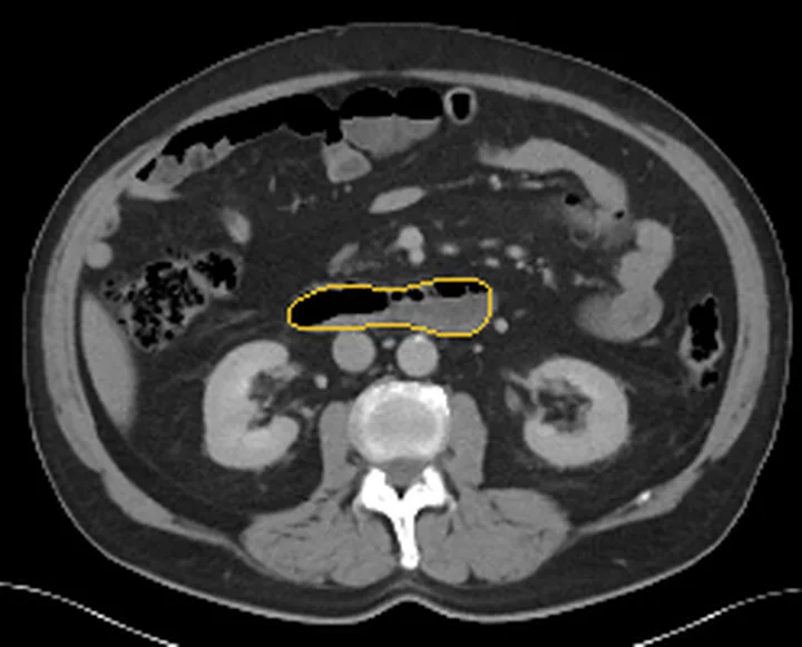
Aynı bölgenin, kaynak veri setinde patoloji içermediği belirtilen bir BT incelemesini kesit kesit kaydırın; organ sınırlarını açarak anatomiyi hasta görüntüsüyle karşılaştırın. Küçük rastlantısal bulgular tümüyle dışlanmamıştır.
Kaynak: TotalSegmentator v2.01 veri seti (Wasserthal ve ark., Radiology: Artificial Intelligence 2023), CC BY 4.0, vaka s0541; organ sınırları veri setinin otomatik segmentasyonundan, birleştirilerek sadeleştirildi. Görüntüler 2,5 mm kesit aralığına yeniden örneklendi.
Ayırıcı tanı
- Duodenal polip
- lümene uzanan solid yumuşak doku odağıdır; kese içinde sıvı/oral kontrast ve çevresel halo beklenmez.
- Duodenal duplikasyon kisti
- duvar komşuluğunda kistik yapı yapar ancak çoğunlukla ana lümenle iletişim göstermez.
- Koledokosel
- distal koledok/ampulla merkezli kistik genişlemedir; safra yolu ile ilişkisi izlenir.
- Periampuller kistik lezyon
- duodenum dışında yer alıp duvarı içeri iter; divertikülde kese lümen içinde bulunur.
- Duodeno-duodenal invajinasyon
- iç içe geçmiş duvar katları ve mezenterik yağ/ damarların taşınmasıyla tanınır.
Tuzaklar
- Kollabe kese BT'de polipoid duvar kalınlaşmasını taklit edebilir; koronal-sagittal planda kese boynunu ve lümen içindeki uzanımı arayın.
- Oral kontrast kese ve ana lümeni eşit doldurduğunda ince duvar silikleşebilir; çevreleyen gerçek lümen/halo sürekliliğini takip edin.
- Papillaya yakın kistik görünümü koledokosel sanmayın; kistik yapının safra yoluna mı duodenal lümene mi devam ettiğini izleyin.
Kendini dene
D2 içinde kontrastla dolu kese, çevresinde ince halo ve devam eden dış lümen izleniyor. Tanı nedir?
Cevabı göster
İntralüminal divertikül. Kese içi ve çevresindeki gerçek lümenin birlikte görülmesi windsock divertikülünü destekler. Polip solid çıkıntıdır; koledokosel safra yolu kökenlidir; duplikasyon kisti çoğunlukla ana lümene açılmaz.
Duodenal duplikasyon kistinin BT’de tipik görünümü hangisidir?
Cevabı göster
Ana lümenle genellikle iletişim göstermeyen, sıvı atenüasyonlu, iyi sınırlı kistik kitle. Duodenal duplikasyon kistleri BT’de tipik olarak sıvı atenüasyonlu, iyi sınırlı kistik kitle görünümündedir ve genellikle duodenum lümeniyle iletişim göstermez.
Kaynaklar
Bu sayfadaki 35 cümle ve 2 quiz sorusu aşağıdaki kaynaklardan alıntılarla otomatik olarak karşılaştırıldı; 5 cümle bu karşılaştırmada düzeltildi (2026-10-09).
- Duodenal imaging on the spotlight: from A to Zpmc.ncbi.nlm.nih.gov
- Imaging of non-neoplastic duodenal diseases. A pictorial review with emphasis on MDCTpmc.ncbi.nlm.nih.gov
- Intraluminal Duodenal (“Windsock”) Diverticulum: A Rare Cause of Gastrointestinal Bleedingpmc.ncbi.nlm.nih.gov
- Gone With the Windsock Diverticulum: An Incidentally Discovered Intraluminal Duodenal Diverticulumpmc.ncbi.nlm.nih.gov
- ENDOSCOPIC TREATMENT OF INTRALUMINAL DUODENAL DIVERTICULUM: A CASE REPORTpmc.ncbi.nlm.nih.gov
- Small Bowel Diverticulosis: Pathogenesis, Clinical Management, and New Concepts.pubmed.ncbi.nlm.nih.gov
- Duodenal diverticula: potential complications and common imaging pitfalls.pubmed.ncbi.nlm.nih.gov
- Recurrent pancreatitis caused by pancreatobiliary anomalies in children with annular pancreas.pubmed.ncbi.nlm.nih.gov
- Intraluminal duodenal diverticula: collective review with report of a laparoscopic excision.pubmed.ncbi.nlm.nih.gov
- Intraluminal Duodenal Diverticulum: A Rare Cause of Chronic Epigastric Pain.pubmed.ncbi.nlm.nih.gov
- Imaging Findings of an Intraluminal Duodenal Diverticulum Associated with Adult Duodeno-Duodenal Intussusception and Recurrent Pancreatitis: A Case Report.pubmed.ncbi.nlm.nih.gov
- Pancreatic and biliary obstruction years after retention of a swallowed coin in a duodenal diverticulum: a case report.pubmed.ncbi.nlm.nih.gov
- An Uncommon Case of Intraluminal Duodenal Diverticulum Complicated by Recurrent Gastrointestinal Bleeding and Small Bowel Obstruction.pubmed.ncbi.nlm.nih.gov
- Jejuno-ileal diverticulitis: Etiopathogenicity, diagnosis and management.pubmed.ncbi.nlm.nih.gov
- An unusual cause of small bowel obstruction: duodenal bezoar-induced diverticulitis.pubmed.ncbi.nlm.nih.gov
- Periampullary duodenal diverticulum causing obstructive jaundice: a case of Lemmel syndrome and review of the literature.pubmed.ncbi.nlm.nih.gov
- Gastroduodenal Emphysema with Portal Venous Air due to Congenital Duodenal Web in a Child: A Case Report and Review of Literature.pubmed.ncbi.nlm.nih.gov
- Exploring the neglected segment of the intestine: the duodenum and its pathologies.pubmed.ncbi.nlm.nih.gov
- Duodenal imaging on the spotlight: from A to Z.pubmed.ncbi.nlm.nih.gov
- Imaging of non-neoplastic duodenal diseases. A pictorial review with emphasis on MDCT.pubmed.ncbi.nlm.nih.gov
- MRI Findings of Intrinsic and Extrinsic Duodenal Abnormalities and Variations.pubmed.ncbi.nlm.nih.gov
- Gallbladder perforation due to the obstructing periampullary duodenal diverticulum (Lemmel's syndrome): a case report.pubmed.ncbi.nlm.nih.gov
- Major surgical conditions of childhood and their lifelong implications: comprehensive review.pubmed.ncbi.nlm.nih.gov
- Recurrent ascending cholangitis with acute pancreatitis and pancreatic atrophy caused by a juxtapapillary duodenal diverticulum: A case report and literature review.pubmed.ncbi.nlm.nih.gov
- Enterolithiasis.pubmed.ncbi.nlm.nih.gov
- Long-term safety and effectiveness of the "OptEase" vena cava filter.pubmed.ncbi.nlm.nih.gov
- Nonproliferative and Proliferative Lesions of the Gastrointestinal Tract, Pancreas and Salivary Glands of the Rat and Mouse.pubmed.ncbi.nlm.nih.gov
- Gastrointestinal Fistulas-What Gastroenterologists Need to Know in 2025.pubmed.ncbi.nlm.nih.gov
- Practical approach to linear endoscopic ultrasound examination of the gallbladder.pubmed.ncbi.nlm.nih.gov
- Neonatal gastrointestinal emergencies: a radiological review.pubmed.ncbi.nlm.nih.gov
- Urethral multiplicity in boys: systematic review of case reports and case series from the last 15 years.pubmed.ncbi.nlm.nih.gov
- Hepatic incidentaloma: An asymptomatic ectopic thyroid tissue.pubmed.ncbi.nlm.nih.gov
- International Harmonization of Nomenclature and Diagnostic Criteria (INHAND): Nonproliferative and Proliferative Lesions of the Rabbit.pubmed.ncbi.nlm.nih.gov
- Simultaneous heterotopia of the gastric mucosa and pancreatic tissue leads to perforation of the small intestine: case report and literature review.pubmed.ncbi.nlm.nih.gov
- Bouveret syndrome in a young patient: A case report and review of literature.pubmed.ncbi.nlm.nih.gov
- Gastrointestinal complications in intensive care.pubmed.ncbi.nlm.nih.gov
- Endovascular Treatment of Hepatic Artery Pseudoaneurysm after Pancreaticoduodenectomy: A Literature Review.pubmed.ncbi.nlm.nih.gov
- Patterns of injury and violence in Yaoundé Cameroon: an analysis of hospital data.pubmed.ncbi.nlm.nih.gov
- Surgical Rescue for Refractory Post-sphincterotomy Hemorrhage: Preserving a Critical, Vanishing Competency for the Next Generation of Surgeons.pubmed.ncbi.nlm.nih.gov
- Severe Dumping and Persistent Obesity in a Post-Roux-en-Y Gastric Bypass Patient With a Non-discernible Gastric Pouch and Symptomatic Improvement on Tirzepatide: A Case Report.pubmed.ncbi.nlm.nih.gov
- Third-Redo Laparoscopic Roux-en-Y Hepaticojejunostomy for Recurrent Anastomotic Stones: A Video-Based Case Report.pubmed.ncbi.nlm.nih.gov
- Approach to anaemia in gastrointestinal disease: A position paper by the ESPGHAN Gastroenterology Committee.pubmed.ncbi.nlm.nih.gov
- Ectopic pancreas in the upper gastrointestinal tract: Is endosonographic diagnosis reliable? Data from the German Endoscopic Ultrasound Registry and review of the literature.pubmed.ncbi.nlm.nih.gov
- Non-perforated peptic ulcer disease: multidetector CT findings, complications, and differential diagnosis.pubmed.ncbi.nlm.nih.gov
- Prenatal Ultrasound Progression and Postnatal Retrospective Analysis of a Case of Small Intestinal Atresia with Volvulus.pubmed.ncbi.nlm.nih.gov
- Fetal MRI: abdominal cystic lesions.pubmed.ncbi.nlm.nih.gov
- Surgical management of gastric duplication cyst with respiratory epithelium in an asymptomatic child.pubmed.ncbi.nlm.nih.gov
- Double gallbladder malformation with duodenal ulcer stricture leading to common bile duct dilation: a case report.pubmed.ncbi.nlm.nih.gov
- A large type IV-A choledochal cyst mimicking hydatid cyst of the liver: A case report.pubmed.ncbi.nlm.nih.gov
- Treatment of Wirsungocele Induced Chronic Pancreatitis by Endoscopic Retrograde Cholangiopancreatography and Stent Placement: A Case Report.pubmed.ncbi.nlm.nih.gov
- Multifocal small bowel gastrointestinal stromal tumor causing perforation peritonitis: A rare cause of acute abdomen.pubmed.ncbi.nlm.nih.gov
- Transient intussusception following single anastomosis duodeno-ileal (SADI) bypass surgery: case report and literature review.pubmed.ncbi.nlm.nih.gov
- Brunneroma: A Rare Cause of Duodeno-duodenal Intussusception.pubmed.ncbi.nlm.nih.gov
- Society for Cardiovascular Magnetic Resonance 2024 Cases of SCMR case series.pubmed.ncbi.nlm.nih.gov
- Non-Thrombotic Filling Defects in Cerebral Veins and Sinuses: When Normal Structures Mimic a Disease.pubmed.ncbi.nlm.nih.gov
- Acute Colonic Diverticulitis: CT Findings, Classifications, and a Proposal of a Structured Reporting Template.pubmed.ncbi.nlm.nih.gov
- Intraoperative Discovery of Type II Choledochal Cyst, Partial Annular Pancreas, and Duodenal Stenosis in a 45-Day-Old Term Born Female Infant.pubmed.ncbi.nlm.nih.gov
- Surgical strategies for challenging common bile duct stones in the endoscopic era: A comprehensive review of current evidence.pubmed.ncbi.nlm.nih.gov
- POCUS for acute abdominal pain: practical scan protocols on gastrointestinal diseases and an evidence review.pubmed.ncbi.nlm.nih.gov
- Incidental findings in female pelvis MRI performed for gynaecological malignancies.pubmed.ncbi.nlm.nih.gov
- Non-traumatic lower abdominal pain: ultrasonographic and clinical differential diagnosis.pubmed.ncbi.nlm.nih.gov
- Pumping Venom: Valvilli Architecture in Diacamma (Hymenoptera: Formicidae) and Implications for Stinger Functionality.pubmed.ncbi.nlm.nih.gov
- Investigating the role of structural wall stress in aortic growth prognosis in acute uncomplicated type B aortic dissection.pubmed.ncbi.nlm.nih.gov
- Safety and short-term oncological outcomes of multimodal modified overlap esophagojejunostomy in total laparoscopic total gastrectomy for elderly patients.pubmed.ncbi.nlm.nih.gov
- When Feeding Tubes Cause Biliary Disease: A Case of Acalculous Cholecystitis Due to Gastrostomy Tube Obstruction.pubmed.ncbi.nlm.nih.gov
- Small Intestine Tumors: Diagnostic Role of Multiparametric Ultrasound.pubmed.ncbi.nlm.nih.gov
- Gastrointestinal Ultrasound: Measurements and Normal Findings - What Do You Need to Know?pubmed.ncbi.nlm.nih.gov
- Ultrasound of the Gallbladder-An Update on Measurements, Reference Values, Variants and Frequent Pathologies: A Scoping Review.pubmed.ncbi.nlm.nih.gov
- Imaging Approach to Diverticular Disease of the Small Bowel.pubmed.ncbi.nlm.nih.gov
- Intraluminal Diverticular Duodenal Duplication With Recurrent Abdominal Pain: A Case Report.pubmed.ncbi.nlm.nih.gov
- Brunner's gland hamartoma in the setting of a duodenal diverticulum: A case report.pubmed.ncbi.nlm.nih.gov
- Small Bowel Obstruction Caused by a Bezoar in a Duodenal Diverticulum.pubmed.ncbi.nlm.nih.gov
- Lemmel Syndrome: A Case Report.pubmed.ncbi.nlm.nih.gov
- Laparoscopic Management of Bouveret's Syndrome: A Surgical Case Series.pubmed.ncbi.nlm.nih.gov
- Rare and Unusual Consequences of Blunt Abdominal Trauma-The Significance of Anatomical Anomalies.pubmed.ncbi.nlm.nih.gov
- Conservative Management of Perforated Duodenal Diverticulum: A Case Report and Narrative Review.pubmed.ncbi.nlm.nih.gov
- Ivory Vertebra Phenomenon as a Sign of Metastatic Dissemination in Pancreatobiliary Neoplasia: A Case Report and Literature Review.pubmed.ncbi.nlm.nih.gov
- Case Report: Abscopal effect of radiotherapy in a patient with metastatic duodenal adenocarcinoma and resistance to chemoimmunotherapy.pubmed.ncbi.nlm.nih.gov
- Advanced ultrasound diagnosis of extrahepatic bile duct lesions.pubmed.ncbi.nlm.nih.gov
- World Endoscopy Organization Position Statements for the Role of Endoscopy on the Diagnosis and Treatment of Superficial Non-Ampullary Duodenal Epithelial Tumors.pubmed.ncbi.nlm.nih.gov
- ENDOSCOPIC TREATMENT OF INTRALUMINAL DUODENAL DIVERTICULUM: A CASE REPORT.pubmed.ncbi.nlm.nih.gov
- Windsock or Cobra Head Sign: A Distinctive Imaging Sign to Differentiate Type 1 Colonic Atresia From Hirschsprung's Disease.pubmed.ncbi.nlm.nih.gov
- Intraluminal duodenal ("windsock") diverticulum: a rare cause of biliary obstruction and acute pancreatitis in the adult.pubmed.ncbi.nlm.nih.gov
- A case report of inverted Meckel's diverticulum.pubmed.ncbi.nlm.nih.gov
- Cross-sectional imaging features of duodenal obstruction: A case series highlighting etiologic diversity.pubmed.ncbi.nlm.nih.gov
- Robotic versus laparoscopic approaches to neonatal annular pancreas: a comparative analysis of surgical outcomes and innovation trade-offs.pubmed.ncbi.nlm.nih.gov
- Small Bowel Imaging from Stepchild of Roentgenology to MR Enterography: Part I: Guidance in Performing and Observing Normal and Abnormal Imaging Findings.pubmed.ncbi.nlm.nih.gov
Eğitim amaçlıdır; hasta başında tanı ve tedavi kararının yerine geçmez, tıbbi tavsiye değildir. Metinler yapay zekâ desteğiyle yazıldı ve otomatik bir adımda kaynak alıntılarıyla karşılaştırıldı; bu bir tıbbi doğrulama değildir ve içerik uzman radyolog incelemesinden geçmedi. Klinik kararlarda güncel kılavuzları, kurum protokollerini ve radyoloji raporunu esas alın.