Özet
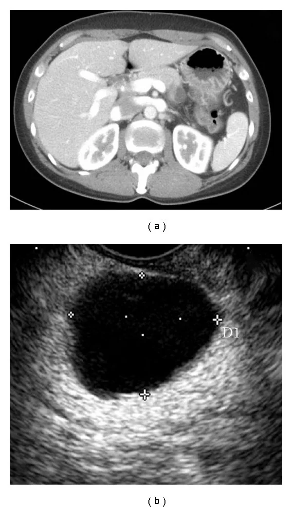
Pankreatik müsinöz kistik neoplazm, karakteristik olarak gövde-kuyrukta gelişen ve histolojik tanısı ovarian tip stroma gerektiren müsin üreten bir kistik neoplazmdır. MCN genellikle pankreas gövde veya kuyruğunda tek ya da az bölmeli kistik kitle olarak görülür; kontrastlı görüntülemede duvar ve septalar kontrastlanabilir. Ana pankreatik kanalla bağlantı genellikle yoktur; bu özellik yan dal IPMN'den ayrımda önem taşır. Mural nodülün gözden kaçması şüpheli bileşenin yanlışlıkla basit kist kabul edilmesine, kanal bağlantısının yanlış yorumlanması ise neoplazm alt tipinin karışmasına yol açabilir.
Faz ve pencere
- Portal tanısal
- MCN, pankreas gövde veya kuyruğunda tek ya da az bölmeli kistik kitle şeklinde görülebilir; BT’de duvar ve septal kontrastlanma ile varsa mural nodüller değerlendirilir.
- Arteriyel tanısal
- Kontrastlı BT'de kistik sıvı ile kontrastlanan duvar, septa veya mural nodül arasındaki atenüasyon farkı ayırt edilir; lezyonun komşu yapılarla ilişkisi değerlendirilir.
- Kontrastsız
- Kistik sıvı düşük atenüasyonludur; duvar veya septadaki kalsifikasyonları ve intrinsik hiperdens kan/protein içeriğini gösterebilir, ancak mural nodülün kontrastlanmasını tek başına belirleyemez.
Önerilen pencereler: Batın (G 400 / M 50), Yumuşak doku (G 400 / M 40).
BT bulguları
- Pankreas gövde veya kuyruğunda, bez parankiminden köken alan soliter makrokistik lezyon
- Tek boşluklu veya az sayıda büyük loküllü, düzgün ya da lobüle konturlu kistik kitle
- İnce olabilen kapsülün fokal kalınlaşması veya düzensiz duvar alanları
- Kontrast sonrası belirginleşen iç septalar veya duvar kalınlaşması
- Duvara oturan kontrastlanan mural nodül ya da kistik boşluğa uzanan solid komponent
- Ana pankreatik kanal ile görünür bağlantının olmaması ve kanalın lezyon tarafından belirgin biçimde genişletilmemesi
- Periferik, duvar veya septal kalsifikasyon; tek başına özgül değildir
- İleri olgularda komşu parankim, damar veya çevre organlara uzanım ve bölgesel nodlar
Normalde
Normal pankreas gövdesi çölyak arter düzeyinden splenik hilusa uzanan düzgün, kontrastlanan doku bandıdır; kuyruk dalak damarları boyunca devam eder ve içinde büyük sıvı boşluğu veya duvar nodülü bulunmaz. Aynı seviyede lezyonun pankreas dokusundan ayrı bir peripankreatik koleksiyon olup olmadığını, ana kanalın lezyondan bağımsız seyredip seyretmediğini ve mural dokunun sıvıya göre gerçekten kontrastlanıp kontrastlanmadığını kıyasla.
Normal BT ile kıyasla
Aynı bölgenin, kaynak veri setinde patoloji içermediği belirtilen bir BT incelemesini kesit kesit kaydırın; organ sınırlarını açarak anatomiyi hasta görüntüsüyle karşılaştırın. Küçük rastlantısal bulgular tümüyle dışlanmamıştır.
Kaynak: TotalSegmentator v2.01 veri seti (Wasserthal ve ark., Radiology: Artificial Intelligence 2023), CC BY 4.0, vaka s0541; organ sınırları veri setinin otomatik segmentasyonundan, birleştirilerek sadeleştirildi. Görüntüler 2,5 mm kesit aralığına yeniden örneklendi.
Ayırıcı tanı
- Yan dal IPMN
- çok odaklı üzüm salkımı biçimi ve pankreatik kanal dallarıyla iletişim IPMN lehinedir; MCN çoğunlukla tek kitle ve kanal bağlantısızdır.
- Seröz kistik neoplazm
- küçük mikrokistlerin bal peteği kümesi, ince septalar ve bazen santral skar SCN'yi destekler.
- Pankreatik psödokist
- geçirilmiş pankreatit veya travma, çevresel inflamasyon ve debris içeren koleksiyon lehinedir; MCN'de pankreatit öyküsü zorunlu değildir.
- Solid psödopapiller neoplazm
- solid komponent, hemoraji ve kistik dejenerasyon ve kapsüllü heterojen kitle görünümüyle ayrılır.
- Pankreatik duktal adenokarsinomda kanal obstrüksiyonuna, obstrüksiyonun gerisinde kanal genişlemesi ve parankim atrofisi eşlik edebilir.
- MCN orta yaşlı kadınlarda ve pankreas gövde-kuyruğunda daha sık görülür; bu özellikler tanıyı desteklese de görüntüleme bulguları diğer kistik neoplazmlarla örtüşebilir.
Tuzaklar
- Bir kesitte kanal bağlantısının görülmemesi kesin yokluk kanıtı değildir; ince kesit ve koronal/oblik rekonstrüksiyonlarda kanal-kist ilişkisini izle.
- Mural nodül ile kontrastlanmayan mukus/debrisi karıştırma; eşleşen kontrastsız ve kontrastlı görüntülerde fokal yumuşak dokunun atenüasyon değişimini kontrol et.
- MCN tanısı yalnızca görüntüyle histolojik olarak kesinleştirilemez; ovarian tip stroma patolojik tanımın parçasıdır.
- Ana pankreatik kanalla ilişki BT'de seçilemiyorsa, bu ilişkinin değerlendirilmesinde MRCP yardımcı olabilir.
Kendini dene
Orta yaşlı bir kadında pankreas kuyrukta tek makrokistik lezyon, ana pankreas kanalıyla bağlantı yok ve kontrastlanan mural nodül var. En olası neoplazm hangisidir?
Cevabı göster
Müsinöz kistik neoplazm. Kadın hastada gövde-kuyruk yerleşimli tek makrokistik lezyon ve ana kanalla bağlantı olmaması MCN’yi destekler; kontrastlanan mural nodül şüpheli solid bileşendir. Kistik PanNET genellikle arteriyel fazda canlı hipervasküler doku gösterir; görüntüleme ovarian tip stromayı kanıtlamaz ve histolojik alt tipi kesinleştirmez.
Bir kistik pankreas kitlesinde kontrastlanan fokal duvar çıkıntısının önemi nasıl değerlendirilmelidir?
Cevabı göster
Mural solid nodül olarak. Kontrastlanan ve duvara oturan yumuşak doku odağı mural nodül/solid komponent olarak tanımlanır ve önemle raporlanır. Kanal taşı lümen içinde yoğun odaktır; santral skar SCN'nin merkezinde, yağ lobülü ise kistik boşluk dışındadır.
İlgili konular
Kaynaklar
Bu sayfadaki 39 cümle ve 2 quiz sorusu aşağıdaki kaynaklardan alıntılarla otomatik olarak karşılaştırıldı; 6 cümle bu karşılaştırmada düzeltildi (2026-10-09).
- A resected case of pancreatic neuroendocrine neoplasm with completely cystic degeneration mimicking a mucinous cystic neoplasm: a case report and literature reviewpmc.ncbi.nlm.nih.gov
- Obstructive Jaundice Induced by Hilar Mucinous Cystic Neoplasm of the Liver: A Rare Case Report and Literature Reviewpmc.ncbi.nlm.nih.gov
- Radiomics advances in the evaluation of pancreatic cystic neoplasmspmc.ncbi.nlm.nih.gov
- The Incidental Pancreatic Cyst: When to Worry About Cancerpmc.ncbi.nlm.nih.gov
- Intraductal Papillary Mucinous Neoplasm of the Pancreas: A Challenging Diagnosispmc.ncbi.nlm.nih.gov
- Imaging of the Pancreas.pubmed.ncbi.nlm.nih.gov
- "Biliary Diseases with Pancreatic Counterparts": Cross-sectional Imaging Findings.pubmed.ncbi.nlm.nih.gov
- Mucinous Cystic Neoplasms of Pancreas.pubmed.ncbi.nlm.nih.gov
- What to do for the incidental pancreatic cystic lesion?pubmed.ncbi.nlm.nih.gov
- Pancreatic neoplasms in pregnancy: diagnosis, complications, and management.pubmed.ncbi.nlm.nih.gov
- Clinical approach to incidental pancreatic cysts.pubmed.ncbi.nlm.nih.gov
- Diagnostic approach to pancreatic cysts.pubmed.ncbi.nlm.nih.gov
- Management of mucinous cystic neoplasms of the pancreas.pubmed.ncbi.nlm.nih.gov
- Hepatobiliary mucinous cystic neoplasms and mimics.pubmed.ncbi.nlm.nih.gov
- [Diagnostic strategy and differential therapeutic approach for cystic lesions of the pancreas].pubmed.ncbi.nlm.nih.gov
- Primary pancreatic cystic neoplasms of the pancreas revisited. Part IV: rare cystic neoplasms.pubmed.ncbi.nlm.nih.gov
- [Differential diagnosis and radiological management of cystic pancreatic lesions].pubmed.ncbi.nlm.nih.gov
- Laparoscopic Resection of an Epithelial Cyst in an Intrapancreatic Accessory Spleen.pubmed.ncbi.nlm.nih.gov
- Uncommon liver tumors: Case report and literature review.pubmed.ncbi.nlm.nih.gov
- Anaplastic Carcinoma of the Pancreas Arising from Mucinous Cystic Neoplasm: A Case Report.pubmed.ncbi.nlm.nih.gov
- Beware of Radiological Diagnosis of Pancreatic Pseudocyst: Lesson Learned From an Interesting Case.pubmed.ncbi.nlm.nih.gov
- An Intraductal Papillary Mucinous Neoplasm-Like Lesion Manifesting as a Duodenal Polyp: A Rare Case Report.pubmed.ncbi.nlm.nih.gov
- Right-lobe hepatic mucinous cystic neoplasm with an adjacent large hemorrhagic-appearing cyst: Markedly elevated serum CEA and CA19-9 despite benign imaging/pathology.pubmed.ncbi.nlm.nih.gov
- Giant mucinous cystic neoplasm of the liver in a young asymptomatic woman: A case report.pubmed.ncbi.nlm.nih.gov
- Common cystic pancreatic neoplasms: A comprehensive magnetic resonance imaging-based review of typical and atypical imaging features.pubmed.ncbi.nlm.nih.gov
- A case of a pancreatic pseudocyst associated with alcoholic chronic pancreatitis that mimicked a mucinous cystic neoplasm.pubmed.ncbi.nlm.nih.gov
- Cystic liver lesions: a pictorial review.pubmed.ncbi.nlm.nih.gov
- New horizons in the endoscopic ultrasonography-based diagnosis of pancreatic cystic lesions.pubmed.ncbi.nlm.nih.gov
- Invasive biliary mucinous cystic neoplasm: a review.pubmed.ncbi.nlm.nih.gov
- A retrospective comparative study of CT and MRI findings of different types of pancreatic serous cystic neoplasms: Correlation with histopathology.pubmed.ncbi.nlm.nih.gov
- A case series of radiologically misdiagnosed visceral lymphatic malformations.pubmed.ncbi.nlm.nih.gov
- Case Report: The cystic masquerade in type 1 Von Hippel-Lindau disease: coexisting clear cell renal cell carcinoma and an imaging-suspected pancreatic neuroendocrine tumor.pubmed.ncbi.nlm.nih.gov
- Mucinous cystic neoplasms of the liver: current insights into epidemiology, diagnosis, and treatment.pubmed.ncbi.nlm.nih.gov
- Radiologic-Pathologic Correlation in Biliary Cystic Neoplasms: Insights From a Single-Centre Experience.pubmed.ncbi.nlm.nih.gov
- 불분명한 소아 복부 종괴: 영상 기반 진단 접근법.pubmed.ncbi.nlm.nih.gov
- ESUR female pelvis group approach to cystic female pelvic lesions.pubmed.ncbi.nlm.nih.gov
- Preliminary investigation of the clinical value of thoracic ultrasound in pediatric pleuropulmonary blastoma: a comparative analysis with computed tomography and pathology.pubmed.ncbi.nlm.nih.gov
- Case Report: A case of intrahepatic papillary neoplasm of the bile duct with invasive carcinoma in a young patient and review of the literature.pubmed.ncbi.nlm.nih.gov
- Endoscopic ultrasound-guided radiofrequency ablation for pancreatic cystic neoplasms: a comprehensive review.pubmed.ncbi.nlm.nih.gov
- Spectral attenuation curves of photon-counting detector CT scans for the differentiation between mucinous and non-mucinous cyst fluids of pancreatic lesions - a proof-of-concept study.pubmed.ncbi.nlm.nih.gov
- Rapid Interval Growth of a Presumed Simple Hepatic Cyst Revealing a Mucinous Cystic Neoplasm in a Rare Male Presentation.pubmed.ncbi.nlm.nih.gov
- Fertility-Sparing Surgery in Young Women with Ovarian Anaplastic Carcinoma: Two Case Reports and Literature Review.pubmed.ncbi.nlm.nih.gov
- Differentiating multilocular hepatic cysts from mucinous cystic neoplasms: characteristic imaging signs and a machine learning diagnostic framework.pubmed.ncbi.nlm.nih.gov
- Ovarian borderline mucinous tumor with carcinosarcomatous mural nodule: a case report of a rare entity and review of the literature.pubmed.ncbi.nlm.nih.gov
- Intraductal Papillary Mucinous Neoplasm (IPMN) of the Pancreas: History, Myths, and Realities Between Past and Future.pubmed.ncbi.nlm.nih.gov
- DIAGNOSIS AND MANAGEMENT OF INTRADUCTAL PAPILLARY MUCINOUS NEOPLASMS: A NARRATIVE REVIEW BASED ON INTERNATIONAL GUIDELINES.pubmed.ncbi.nlm.nih.gov
- Undifferentiated embryonal sarcoma of the liver presenting as a complex mixed solid-cystic hepatic lesion in a young adult: A case report.pubmed.ncbi.nlm.nih.gov
- Spontaneous rupture and hemorrhage of a giant simple hepatic cyst in a 94-year-old woman: a case report.pubmed.ncbi.nlm.nih.gov
- Schwannoma Likely Arising from the Pancreatic Head Nerve Plexus That Mimicked a Mucinous Cystic Neoplasm and Was Treated by Pancreaticoduodenectomy: A Case Report.pubmed.ncbi.nlm.nih.gov
- Multiparametric Abdominal Ultrasound as a Source of Imaging Biomarkers in Predictive and Personalized Medicine: A Narrative Review.pubmed.ncbi.nlm.nih.gov
- Imaging patterns and recommendations for diagnosis, staging, and management of lung cancer.pubmed.ncbi.nlm.nih.gov
- Multi-Disciplinary Care of Hilar Cholangiocarcinoma: Review of Guidelines and Recent Advancements.pubmed.ncbi.nlm.nih.gov
- A giant 31-cm pancreatic mucinous cystic neoplasm: a 4-year progression and surgical challenge.pubmed.ncbi.nlm.nih.gov
- Blunt abdominal trauma revealing a giant splenic artery aneurysm in a cirrhotic patient: a case report.pubmed.ncbi.nlm.nih.gov
- Splenic Torsion in a Patient with Polysplenia Syndrome and Dorsal Pancreatic Agenesis: A Case Report.pubmed.ncbi.nlm.nih.gov
- MASLD and Pancreatic Diseases: From Shared Mechanisms to Clinical Implications and Future Directions.pubmed.ncbi.nlm.nih.gov
- Clinical, Radiological, and Endoscopic Features of Pancreatic Pseudocyst and Walled-Off Necrosis: How to Diagnose and How to Drain Them.pubmed.ncbi.nlm.nih.gov
- Pancreatic Incidentaloma.pubmed.ncbi.nlm.nih.gov
- The Distribution and Predictive Factor of Extra-Pancreatic Malignancy Occurrence in Patients with Pancreatic Intraductal Papillary Mucinous Neoplasm-A Ten-Year Follow-Up Case-Control Study in Taiwan.pubmed.ncbi.nlm.nih.gov
- Special Issue: EACR 2026 Congress: Innovative Cancer Science, 8-11 June 2026.pubmed.ncbi.nlm.nih.gov
- EACR 2025 Congress: Innovative Cancer Science, 16-19 June 2025.pubmed.ncbi.nlm.nih.gov
- Imaging Findings of Biliary Adenofibroma of the Liver: A Case Report.pubmed.ncbi.nlm.nih.gov
- Beyond the Six-Week Rule: Contemporary Insights Into Pancreatic Pseudocyst.pubmed.ncbi.nlm.nih.gov
- Guidelines for endoscopic treatment of postinflammatory pancreatic and peripancreatic fluid collections. Recommendations of the Endoscopic Surgery Section of the Association of Polish Surgeons, the Endoscopy Section of the Polish Society of Gastroenterology, the Polish Pancreatic Club, the Laparoscopic and Robotic Surgery Section of the Association of Polish Surgeons, and the Pancreatic Surgery Section of the Association of Polish Surgeons; formulated by an expert panel led by Prof. Mateusz Jagielski, MD, PhD.pubmed.ncbi.nlm.nih.gov
- Pancreatic pseudocyst: The past, the present, and the future.pubmed.ncbi.nlm.nih.gov
- Large periampullary mass with vascular contact: Diagnostic and surgical decision-making in a solid pseudopapillary neoplasm.pubmed.ncbi.nlm.nih.gov
- Solid Pseudopapillary Neoplasm With Extensive Intratumoral Hemorrhage.pubmed.ncbi.nlm.nih.gov
- Frantz-Gruber tumor with atypical presentation as a pancreatic pseudocyst following abdominal trauma: a case report.pubmed.ncbi.nlm.nih.gov
- Basic Pancreatic Lesions: Radiologic-pathologic Correlation.pubmed.ncbi.nlm.nih.gov
- Total laparoscopic radical hysterectomy for cervical cancer in prolapsed uterus.pubmed.ncbi.nlm.nih.gov
- High-affinity alphavbeta3 integrin targeted optical probe as a new imaging biomarker for early atherosclerosis: initial studies in Watanabe rabbits.pubmed.ncbi.nlm.nih.gov
- Feasibility of deep learning-accelerated HASTE-FS for pancreatic cystic lesion surveillance: comparison with conventional HASTE and MRCP.pubmed.ncbi.nlm.nih.gov
- Evaluating the Quality of Optimal MRCP Image Using RT-2D-Compressed SENSE(CS)Turbo Spin Echo: Comparing Respiratory Triggering(RT)-2D-SENSE Turbo Spin Echo and Breath Hold-2D-Single-Shot Turbo Spin Echo.pubmed.ncbi.nlm.nih.gov
- 2.5-mm articulated endoluminal forceps using a compliant mechanism.pubmed.ncbi.nlm.nih.gov
- Primary Extrahepatic Biliary Mucinous Cystic Neoplasm Suspected Preoperatively and Treated by Robotic Pancreaticoduodenectomy: A Case Report.pubmed.ncbi.nlm.nih.gov
- The Great Mimicker: Variable Presentations of Xanthogranulomatous Salpingo-Oophoritis in a Case Series of Seven Patients From a Rural Tertiary Care Center.pubmed.ncbi.nlm.nih.gov
- Evaluation of cystic neoplasms using EUS, MRI, and CT scan and treatment strategy.pubmed.ncbi.nlm.nih.gov
- Pancreatic Endometriosis Coexisting with a Splenic Mesothelial Cyst: A Rare Case Report and Review of the Literature.pubmed.ncbi.nlm.nih.gov
- Imaging features of epidermoid cysts in intrapancreatic spleen: a single center study of 24 patients.pubmed.ncbi.nlm.nih.gov
- Spontaneous bacteremia and spontaneous bacterial peritonitis share similar prognosis in patients with cirrhosis: a cohort study.pubmed.ncbi.nlm.nih.gov
- Intraductal papillary mucinous neoplasm unveiling incidental multifocal pancreatic neuroendocrine tumors: a challenging case report.pubmed.ncbi.nlm.nih.gov
- Differentiating walled-off pancreatic necrosis with no documented pancreatitis from mucinous cystic neoplasms: a multicenter LASSO-based nomogram.pubmed.ncbi.nlm.nih.gov
- Pancreatic intraductal papillary mucinous neoplasm with invasive carcinoma and uterine metastasis: a case report.pubmed.ncbi.nlm.nih.gov
- Management of pancreatic cysts and guidelines: what the gastroenterologist needs to know.pubmed.ncbi.nlm.nih.gov
- Part 1: CT characterisation of pancreatic neoplasms: a pictorial essay.pubmed.ncbi.nlm.nih.gov
- Malignant potential of intraductal papillary mucinous neoplasm (IPMN) of the pancreas: imaging assessment of IPMN-derived cancer and concomitant pancreatic ductal adenocarcinoma.pubmed.ncbi.nlm.nih.gov
- Pancreatic serous cystic neoplasm mimicking intraductal papillary mucinous neoplasm: Two case reports and literature review.pubmed.ncbi.nlm.nih.gov
- The Role of Magnetic Resonance Imaging (MRI) in the Diagnosis of Pancreatic Cystic Lesions (PCLs).pubmed.ncbi.nlm.nih.gov
- Cystic Neoplasms of the Pancreas: Differential Diagnosis and Radiology Correlation.pubmed.ncbi.nlm.nih.gov
- Diffusion-Weighted Imaging in Oncology: An Update.pubmed.ncbi.nlm.nih.gov
- Case Report: A rare case of gastric cardia bronchogenic cyst: diagnostic challenges and EUS findings with elevated cyst-fluid tumor markers.pubmed.ncbi.nlm.nih.gov
- Primary mucinous sarcoma of the lung with EWSR1 translocation: a case report.pubmed.ncbi.nlm.nih.gov
- Endoscopic Ultrasound-Guided Pancreatic Tissue Sampling: Lesion Assessment, Needles, and Techniques.pubmed.ncbi.nlm.nih.gov
- Review of the Imaging Modalities in the Gynecological Neoplasms During Pregnancy.pubmed.ncbi.nlm.nih.gov
- Oncocytic intraductal papillary neoplasm of the pancreas: Multimodality imaging and cytopathologic correlation.pubmed.ncbi.nlm.nih.gov
- Umbilical hernia as the initial manifestation of pseudomyxoma peritonei: a case report.pubmed.ncbi.nlm.nih.gov
- Undifferentiated carcinoma of the pancreas arising from intraductal papillary mucinous neoplasm: a case report.pubmed.ncbi.nlm.nih.gov
- A Rare Pediatric Intraductal Papillary Mucinous Neoplasm Presenting as a Pancreatic Pseudocyst With Splenic Vein Thrombosis and Gastric Variceal Hemorrhage.pubmed.ncbi.nlm.nih.gov
- Pancreatic Cystic Lesions: From Basic Knowledge to Recent Guidelines.pubmed.ncbi.nlm.nih.gov
- Incidental cystic pancreatic neuroendocrine neoplasm and differential diagnosis.pubmed.ncbi.nlm.nih.gov
- Pancreatic Acinar Cell Carcinoma: A Rare Pancreatic Malignancy with Distinct Biology and Emerging Therapeutic Opportunities.pubmed.ncbi.nlm.nih.gov
- Vascularized jejunal pedicle graft for duodenal reconstruction in a cat: Case Report.pubmed.ncbi.nlm.nih.gov
- Role of interventional radiology in the management of hepatic hydatid disease.pubmed.ncbi.nlm.nih.gov
- Understanding Photon-Counting CT: Physics, Detector Technology, and Image Reconstructions.pubmed.ncbi.nlm.nih.gov
- How to implement a radiologist led whole-body MRI screening program.pubmed.ncbi.nlm.nih.gov
- Pancreatic Cyst Management: A Multimodal and Interdisciplinary Approach.pubmed.ncbi.nlm.nih.gov
- Magnetic Resonance Imaging Features of Hepatic Hydatid Disease: A Pictorial Review with Emphasis on Atypical Presentations and Differential Diagnosis.pubmed.ncbi.nlm.nih.gov
- A resected case of pancreatic neuroendocrine neoplasm with completely cystic degeneration mimicking a mucinous cystic neoplasm: a case report and literature review.pubmed.ncbi.nlm.nih.gov
- Monitoring of low-risk pancreatic cystic lesions with targeted abdominal ultrasound: diagnostic yield, clinical utility, and acceptance.pubmed.ncbi.nlm.nih.gov
- CT and MR evaluation of main pancreatic duct dilation: A radiologist's guide to differentiating benign from malignant causes.pubmed.ncbi.nlm.nih.gov
- Two Ducts, One Drug-Pembrolizumab-Induced Pancreatic Injury Manifesting as Double-Duct Sign.pubmed.ncbi.nlm.nih.gov
- Imaging-Based Risk Stratification of IPMN Using a Structured Imaging Score: A Retrospective Proof-of-Concept Study.pubmed.ncbi.nlm.nih.gov
- Pancreatic mucinous cystic neoplasms in a young female: A case report.pubmed.ncbi.nlm.nih.gov
Eğitim amaçlıdır; hasta başında tanı ve tedavi kararının yerine geçmez, tıbbi tavsiye değildir. Metinler yapay zekâ desteğiyle yazıldı ve otomatik bir adımda kaynak alıntılarıyla karşılaştırıldı; bu bir tıbbi doğrulama değildir ve içerik uzman radyolog incelemesinden geçmedi. Klinik kararlarda güncel kılavuzları, kurum protokollerini ve radyoloji raporunu esas alın.