Özet
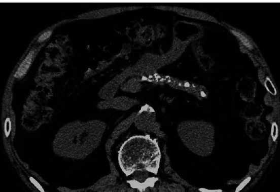
Ana pankreatik kanal taşı en sık kronik pankreatit zemininde gelişir ve kanal akımını bozarak tekrarlayan ağrı ya da pankreatit ataklarına eşlik edebilir. Kontrastsız BT, kalsifiye taşları ve bez içindeki kalsifikasyon dağılımını göstermede kullanışlıdır; kanalın genişleme ve darlıklarını ince kesitlerde izlemek gerekir. Taşın bulunduğu yerdeki obstrüksiyon, yukarı akım kanal genişlemesi ve bez atrofisi ile birlikte okunur. Kanaldaki ani geçişler yalnızca taşa bağlanmamalı; taş ile açıklanamayan fokal obstrüksiyonlarda altta yatan duktal tümör araştırılmalıdır.
Faz ve pencere
- Kontrastsız tanısal
- Kanal lümeninde yüksek atenüasyonlu, noktasal ya da kaba kalsifik odak ve bez içi parankimal kalsifikasyonlar seçilebilir; ince kesitlerde odağın ana kanal seyriyle aynı hatta olup olmadığını doğrula. Bu faz taşın kalsifik yapısını ve dağılımını gösterir, eşlik eden yumuşak doku kitlesini dışlamaz.
- Portal
- Kontrastlı portal venöz görüntülerde obstrüksiyonun proksimalindeki kanal genişlemesi, bez hacim kaybı ve kalsifikasyonun çevresindeki hipoenhans kitle araştırılır; kronik pankreatitte fibrotik parankim heterojen olabilir. Kitle şüphesi varsa pankreas protokolü ince kesitleriyle değerlendirme gerekir.
Önerilen pencereler: Batın (G 400 / M 50), Yumuşak doku (G 400 / M 40).
BT bulguları
- Ana kanal hattında lümen içi kalsifik odak, komşu damar kalsifikasyonundan ayrı olarak kanalın uzunlamasına seyriyle eşleşir.
- Taşın proksimalindeki ana kanal segmenti genişleyebilir; darlık ve genişleme alanları ardışık kesitlerde değişken kalibre oluşturabilir.
- Kronik obstrüksiyonda pankreas gövde-kuyruk parankimi incelmiş, konturu düzensiz ve lobülasyonu azalmış olabilir.
- Kanal çevresinde ve bez içinde dağınık kaba kalsifikasyonlar kronik pankreatit örüntüsünü destekler.
- Tek, keskin kanal geçişi çevresinde fokal yumuşak doku kalınlaşması veya kontur kabarıklığı varsa taş dışında duktal kitle aranır.
- Pankreas başındaki obstrüksiyonla birlikte koledok genişlemesi görülebilir; iki kanalın ampulla çevresindeki devamı izlenir.
Normalde
Normal incelemede ana pankreatik kanal baş, gövde ve kuyruk boyunca ince, düzenli kalibreli bir çizgi olarak izlenir; bez parankiminde kaba kalsifik odak bulunmaz. Taş şüphesinde aynı düzeyde lümen içi kalsifik odağın kanal ile aynı hatta olup olmadığına, ardından kanalın yukarı akım çapına ve o bölgedeki parankim kalınlığına bakılır.
Normal BT ile kıyasla
Aynı bölgenin, kaynak veri setinde patoloji içermediği belirtilen bir BT incelemesini kesit kesit kaydırın; organ sınırlarını açarak anatomiyi hasta görüntüsüyle karşılaştırın. Küçük rastlantısal bulgular tümüyle dışlanmamıştır.
Kaynak: TotalSegmentator v2.01 veri seti (Wasserthal ve ark., Radiology: Artificial Intelligence 2023), CC BY 4.0, vaka s0541; organ sınırları veri setinin otomatik segmentasyonundan, birleştirilerek sadeleştirildi. Görüntüler 2,5 mm kesit aralığına yeniden örneklendi.
Ayırıcı tanı
- Pankreatik parankimal kalsifikasyon
- odak kanal lümeninde değil, bez dokusu içinde veya kanal dışındadır.
- PDAC
- kanal geçişinde hipoenhans yumuşak doku, kontur bozulması veya çift kanal genişlemesi eşlik edebilir.
- Kronik pankreatite bağlı duktal protein tıkacı
- kanal genişlemesi yapabilir ancak BT'de kalsifiye taş gibi belirgin yoğunluk göstermeyebilir.
Tuzaklar
- BT’de kronik pankreatit değerlendirilirken pankreas kalsifikasyonları, kanal genişlemesi ve atrofi birlikte dikkate alınmalıdır.
- Kronik pankreatit kalsifikasyonları kanal taşına eşlik edebilir; tüm bezdeki dağılımı incelemeden tek odağı obstrüksiyonun tek nedeni sayma.
- Kalsifiye taş, çevresindeki küçük izodens PDAC odağını görünmez kılabilir; kanalın ani kesildiği kesitlerde parankim kontrastlanmasını ayrıca tara.
Kendini dene
Kontrastsız BT'de kanal ekseninde kalsifik odak ve kuyruk yönünde genişleme var. En olası açıklama nedir?
Cevabı göster
Pankreatik kanal taşı. Kanal seyriyle aynı hatta bulunan kalsifik odak ve proksimal kanal genişlemesi obstrüktif duktal taşı destekler. Anevrizma damar lümeniyle devam eder; kistadenom kistik kitle, safra taşı ise safra yolları veya kesede yerleşir.
Kanalın aniden kesildiği düzeyde fokal hipoenhans doku da görülüyor. Hangi eşlikçi tanı dışlanmalıdır?
Cevabı göster
Duktal adenokarsinom. Ani kanal geçişiyle aynı noktadaki hipoenhans doku obstrüksiyon yapan pankreatik kitleyi düşündürür. Psödokist sıvı dansitesinde kapsüllü koleksiyondur; dalak enfarktı dalak parankiminde, divertikül ise duodenum lümeniyle ilişkili bulunur.
Kaynaklar
Bu sayfadaki 35 cümle ve 2 quiz sorusu aşağıdaki kaynaklardan alıntılarla tek tek karşılaştırıldı; 4 cümle bu karşılaştırmada düzeltildi (2026-10-09).
- Staged management of a giant adult Todani type IVa choledochal cyst mimicking gallbladder hydropspmc.ncbi.nlm.nih.gov
- A Pancreas Out of Place: Acute Interstitial Pancreatitis Caused by Pancreatic Herniation in a Type IV Paraesophageal Herniapmc.ncbi.nlm.nih.gov
- Clinical Characteristics, ERCP Management, and Early Adverse Events Among Patients with Choledocholithiasis at a Romanian Tertiary Referral Center: A Retrospective Cohortpmc.ncbi.nlm.nih.gov
- Acute Emergent Biliary Pathologies Identified Using Point-of-Care Ultrasoundpmc.ncbi.nlm.nih.gov
- BILIARY SLUDGE-ASSOCIATED ACUTE PANCREATITIS: CLINICAL SPECTRUM, DIAGNOSIS, AND MANAGEMENTpmc.ncbi.nlm.nih.gov
- Diagnosis and Management of Chronic Pancreatitis: A Review.pubmed.ncbi.nlm.nih.gov
- Subcapsular Hepatic Hematoma After Endoscopic Retrograde Cholangiopancreatography: A Case Report and Review of Literature.pubmed.ncbi.nlm.nih.gov
- Current advances in the management of chronic pancreatitis.pubmed.ncbi.nlm.nih.gov
- IgG4-related disease coexisting with autoimmune haemolytic anaemia.pubmed.ncbi.nlm.nih.gov
- Obstructive jaundice due to a blood clot after ERCP: a case report and review of the literature.pubmed.ncbi.nlm.nih.gov
- Asian chronic pancreatitis: the common and the unique.pubmed.ncbi.nlm.nih.gov
- Pre-ERCP imaging of the bile duct and gallbladder.pubmed.ncbi.nlm.nih.gov
- Laparoscopic Puestow: lateral pancreaticojejunostomy.pubmed.ncbi.nlm.nih.gov
- Mirizzi syndrome with endoscopic ultrasound image.pubmed.ncbi.nlm.nih.gov
- Pancreatic stones treated via an EUS-guided pancreaticogastrostomy with a fully-covered self-expandable metal stent.pubmed.ncbi.nlm.nih.gov
- Obstructive jaundice due to a rare periampullary tumor.pubmed.ncbi.nlm.nih.gov
- Computed tomography based scoring system in a prospectively ascertained cohort of patients with chronic pancreatitis.pubmed.ncbi.nlm.nih.gov
- Portal hypertensive biliopathy: A single center experience and literature review.pubmed.ncbi.nlm.nih.gov
- A unusual case of multifocal pyogenic abscess formation following ERCP procedure.pubmed.ncbi.nlm.nih.gov
- Risk Analysis of Pancreatitis With Pancreatolithiasis in Relation to Dyslipidemia as a Cofactor.pubmed.ncbi.nlm.nih.gov
- Pancreatopleural Fistula Presenting as Recurrent Right-Sided Pleural Effusion: A Case Report.pubmed.ncbi.nlm.nih.gov
- Artificial Intelligence-Based Prediction of Pancreatic Stone Clearance in Pancreatolithiasis Using Pretreatment CT Images and Clinical Features.pubmed.ncbi.nlm.nih.gov
- Spontaneous bacteremia and spontaneous bacterial peritonitis share similar prognosis in patients with cirrhosis: a cohort study.pubmed.ncbi.nlm.nih.gov
- Pancreatic Calculi: Two Case Reports and Literature Review.pubmed.ncbi.nlm.nih.gov
- Intraductal Papillary Mucinous Neoplasm (IPMN) of the Pancreas: History, Myths, and Realities Between Past and Future.pubmed.ncbi.nlm.nih.gov
- Focal Pancreatic Parenchymal Atrophy in Main Duct Intraductal Papillary Mucinous Neoplasms of the Pancreas.pubmed.ncbi.nlm.nih.gov
- A Case of Focal Liver Necrosis Following Whipple's Procedure: Presentation, Workup, and Management.pubmed.ncbi.nlm.nih.gov
- A scoring model for preoperative differentiation of high-enhancement pancreatic ductal adenocarcinoma from mass-forming chronic pancreatitis.pubmed.ncbi.nlm.nih.gov
- The Differential Diagnosis and Management of Pancreatic Cystic Neoplasms.pubmed.ncbi.nlm.nih.gov
- Imaging of Acute Abdominopelvic Pain in Pregnancy and Puerperium-Part II: Non-Obstetric Complications.pubmed.ncbi.nlm.nih.gov
- Value and Accuracy of Multidetector Computed Tomography in Obstructive Jaundice.pubmed.ncbi.nlm.nih.gov
- Perioperative characteristics, histologic diagnosis, complications, and outcomes of dogs undergoing percutaneous drainage, sclerotherapy or surgical management of intrarenal cystic lesions: 18 dogs (2004-2021).pubmed.ncbi.nlm.nih.gov
- Improvement in the Image Quality of Bile and Pancreatic Ducts with Dual- Layer Spectral CT.pubmed.ncbi.nlm.nih.gov
- Surgical management strategies and postoperative outcomes of choledochal cysts in children: the role of preoperative radiological assessment in a retrospective observational study.pubmed.ncbi.nlm.nih.gov
- Giant Asymptomatic Submandibular Sialolith: A Case Report Accompanied by Systematic Review.pubmed.ncbi.nlm.nih.gov
- Adult Hepatic Infarction for Internists: A Practical Diagnostic and Management Pathway to Avoid Misdiagnosis, Unnecessary Drainage, and Delayed Vascular Recognition.pubmed.ncbi.nlm.nih.gov
- An update on pancreatic cancer imaging, staging, and use of the PACT-UK radiology template pre- and post-neoadjuvant treatment.pubmed.ncbi.nlm.nih.gov
- Bridging Imaging and Pathohistology in Pancreatic Hamartoma: A Systematic Review of the Literature with an Integrated Case Report.pubmed.ncbi.nlm.nih.gov
- Congenital pulmonary airway malformation mimicking lung cancer in adults: a pitfall in oncologic diagnosis.pubmed.ncbi.nlm.nih.gov
- Beyond the Expected: A Case-Based Exploration and Literature Review on the Rare Evolution of Pseudomyxoma Peritonei From a Side-Branch Intraductal Papillary Mucinous Neoplasm.pubmed.ncbi.nlm.nih.gov
- Clinical features and outcomes of adult primary retroperitoneal lymphangioma: insights from a high-volume sarcoma center.pubmed.ncbi.nlm.nih.gov
- Medical hyperspectral imaging: an updated review of technology advancements and biomedical applications.pubmed.ncbi.nlm.nih.gov
- The SLC-ome of membrane transport: From molecular discovery to physiology and clinical applications.pubmed.ncbi.nlm.nih.gov
- Mathematical models of cystic fibrosis as a systemic disease.pubmed.ncbi.nlm.nih.gov
- Common Bile Duct Access After Bariatric Surgery: A Narrative Review of Endoscopic Considerations for Treatment of Choledocholithiasis.pubmed.ncbi.nlm.nih.gov
- High-Frequency Miniprobe Endoscopic Ultrasonography Across the Gastrointestinal Tract.pubmed.ncbi.nlm.nih.gov
- Prevention and Management of Endoscopic Retrograde Cholangiopancreatography-Related Perforation: A Guideline-Based Narrative Review.pubmed.ncbi.nlm.nih.gov
- Pancreatic head resection alongside side-to-side pancreatic duct-jejunostomy for pancreatic stones: A case report and review of literature.pubmed.ncbi.nlm.nih.gov
- Common cystic pancreatic neoplasms: A comprehensive magnetic resonance imaging-based review of typical and atypical imaging features.pubmed.ncbi.nlm.nih.gov
- When Cancer Clots: An Extensive Radiologic Analysis of Cancer-Associated Thromboembolism.pubmed.ncbi.nlm.nih.gov
- Case Report: A rare case of IgG4-related disease initially manifesting with diarrhea and jaundice.pubmed.ncbi.nlm.nih.gov
- Case report and literature review of IgG4-related autoimmune pancreatitis secondary to pancreatic involvement of IgG4-related sclerosing disease.pubmed.ncbi.nlm.nih.gov
- Multiple Incidental Pancreatic Neuroendocrine Tumors in a Patient With Suspected Duodenal Papillary Adenocarcinoma: A Case Report.pubmed.ncbi.nlm.nih.gov
- Synchronous medullary carcinoma of the pancreas and non-ampullary duodenal adenocarcinoma.pubmed.ncbi.nlm.nih.gov
- Not All Painless Jaundice in the Elderly Is Primary Pancreatic Cancer.pubmed.ncbi.nlm.nih.gov
- Prolonged Retention of a Spontaneously Dislodging Pancreatic Duct Stent in a Patient with Surgically Altered Anatomy: A Case Report.pubmed.ncbi.nlm.nih.gov
- Beyond the Six-Week Rule: Contemporary Insights Into Pancreatic Pseudocyst.pubmed.ncbi.nlm.nih.gov
- Special Issue: EACR 2026 Congress: Innovative Cancer Science, 8-11 June 2026.pubmed.ncbi.nlm.nih.gov
- Long-term safety and effectiveness of the "OptEase" vena cava filter.pubmed.ncbi.nlm.nih.gov
- Commentary: an eye on PET quantification.pubmed.ncbi.nlm.nih.gov
- Virtual non-contrast images from dual-layer spectral CT: comparison with true non-contrast across abdominal structures.pubmed.ncbi.nlm.nih.gov
- Histopathological effect of hypervitaminosis D<sub>3</sub> on the submandibular salivary gland of albino rats and the possible protective role of vitamin K "histological & ultrastructural study".pubmed.ncbi.nlm.nih.gov
- Big data analytics for CLEC5A dynamics based on single cell genomics and proteomics reveal its diverse functions in human diseases.pubmed.ncbi.nlm.nih.gov
- Imaging of pancreatic hepatoid adenocarcinoma: a scoping review.pubmed.ncbi.nlm.nih.gov
- Case Report: Pancreatic sclerosing epithelioid mesenchymal neoplasm (SEMN) with novel pancreatic duct obstruction in a middle-aged female.pubmed.ncbi.nlm.nih.gov
- Dynamic volume perfusion CT for preoperative multidimensional resectability assessment of pancreatic ductal adenocarcinoma.pubmed.ncbi.nlm.nih.gov
- Mixed Neuroendocrine Non-Neuroendocrine Neoplasm of the Ampulla of Vater: Report of a Rare Location.pubmed.ncbi.nlm.nih.gov
- Diagnosis and Surgical Management for Advanced Pancreatic Cancer Requiring Vascular Resection.pubmed.ncbi.nlm.nih.gov
- Pancreatic tail cancer in the setting of pancreatitis with a review of the literature: A case report.pubmed.ncbi.nlm.nih.gov
- Outcomes of surgical treatment in chronic pancreatitis - a single centre study.pubmed.ncbi.nlm.nih.gov
- Endovascular management of hemosuccus pancreaticus secondary to splenic artery pseudoaneurysm: a case report.pubmed.ncbi.nlm.nih.gov
- Patterns of injury and violence in Yaoundé Cameroon: an analysis of hospital data.pubmed.ncbi.nlm.nih.gov
- Acute Pancreatitis as an Initial Presentation of Hereditary Chronic Calcific Pancreatitis with Pancreatic Duct Stones: A Case Report.pubmed.ncbi.nlm.nih.gov
- Chronic Pancreatitis: Role of Endoscopic Procedures.pubmed.ncbi.nlm.nih.gov
- 2025 Heart Disease and Stroke Statistics: A Report of US and Global Data From the American Heart Association.pubmed.ncbi.nlm.nih.gov
- 2024 Heart Disease and Stroke Statistics: A Report of US and Global Data From the American Heart Association.pubmed.ncbi.nlm.nih.gov
- Heart Disease and Stroke Statistics-2023 Update: A Report From the American Heart Association.pubmed.ncbi.nlm.nih.gov
- Age-Associated Differences in Ductal Involvement in Biliary Lithiasis: A Consecutive MRCP Cohort Study.pubmed.ncbi.nlm.nih.gov
- Cone Beam CT Sialography for the Assessment of Submandibular Gland Structure in Diabetes Mellitus.pubmed.ncbi.nlm.nih.gov
- Technical success, clinical efficacy, and safety of endoscopic minor papilla interventions for symptomatic pancreatic diseases.pubmed.ncbi.nlm.nih.gov
- The pseudocirrhotic liver: Hepar lobatum carcinomatosum in a patient with triple-negative breast cancer.pubmed.ncbi.nlm.nih.gov
- Multimodal imaging features to diagnose abdominal complications of sickle cell disease.pubmed.ncbi.nlm.nih.gov
- Testicular Disorders in Equids: A Case Series.pubmed.ncbi.nlm.nih.gov
- Diagnostic Uncertainty in Pancreatic Masses: The Critical Role of excluding Malignancy in Suspected Autoimmune Pancreatitis.pubmed.ncbi.nlm.nih.gov
- A case of chronic cholangitis caused by calculi with foreign matter mimicking perihilar cholangiocarcinoma.pubmed.ncbi.nlm.nih.gov
- Extraordinary enlargement of the main pancreatic duct dilatation in pancreatic ductal adenocarcinoma - a case report.pubmed.ncbi.nlm.nih.gov
- Chronic pancreatitis with pancreatic duct stones complicated by acute obstructive suppurative pancreatic ductitis: Report of a case and review of the literature.pubmed.ncbi.nlm.nih.gov
- Advances in MRI in the Evaluation of Chronic Pancreatitis and the Less Common Forms of Pancreatitis.pubmed.ncbi.nlm.nih.gov
- Pancreatic masses clinically diagnosed as tuberculosis: Case reports.pubmed.ncbi.nlm.nih.gov
- EUS-Guided Shear-Wave-Based Elastography: Current Evidence and the Emerging Role of Two-Dimensional Shear-Wave Elastography.pubmed.ncbi.nlm.nih.gov
- Mucinous cystic neoplasms of the liver: current insights into epidemiology, diagnosis, and treatment.pubmed.ncbi.nlm.nih.gov
- Staged management of a giant adult Todani type IVa choledochal cyst mimicking gallbladder hydrops.pubmed.ncbi.nlm.nih.gov
- Clinical Characteristics, ERCP Management, and Early Adverse Events Among Patients with Choledocholithiasis at a Romanian Tertiary Referral Center: A Retrospective Cohort.pubmed.ncbi.nlm.nih.gov
- A Pancreas Out of Place: Acute Interstitial Pancreatitis Caused by Pancreatic Herniation in a Type IV Paraesophageal Hernia.pubmed.ncbi.nlm.nih.gov
- Changes Connected to Early Chronic Pancreatitis and Early Pancreatic Cancer in Endoscopic Ultrasonography (EUS): Clinical Implications.pubmed.ncbi.nlm.nih.gov
- Hepato-pancreato-biliary tuberculosis: A review.pubmed.ncbi.nlm.nih.gov
- The Role of Endoscopic Ultrasound and Ancillary Techniques in the Diagnosis of Autoimmune Pancreatitis: A Comprehensive Review.pubmed.ncbi.nlm.nih.gov
- Imaging of inflammatory disease of the pancreas.pubmed.ncbi.nlm.nih.gov
- Mass-forming pancreatitis versus pancreatic ductal adenocarcinoma: CT and MR imaging for differentiation.pubmed.ncbi.nlm.nih.gov
- Duodenal imaging on the spotlight: from A to Z.pubmed.ncbi.nlm.nih.gov
- Unusual Configuration of a Giant Trans-Spatial Pancreatic Pseudocyst with Spontaneous Shrinkage: A Rare Case Report.pubmed.ncbi.nlm.nih.gov
- Pancreatic duct stones treated by Whipple as a last resort: A case report.pubmed.ncbi.nlm.nih.gov
- The Role of Imaging in Portal Vein Thrombosis: From the Diagnosis to the Interventional Radiological Management.pubmed.ncbi.nlm.nih.gov
- Extravertebral low back pain: a scoping review.pubmed.ncbi.nlm.nih.gov
- Adoption of multi-slice spiral computed tomography and multiparametric magnetic resonance imaging in the differential diagnosis of pancreatic cancer and chronic pancreatitis.pubmed.ncbi.nlm.nih.gov
- Portal Vein Thrombosis: State-of-the-Art Review.pubmed.ncbi.nlm.nih.gov
- Risk assessment and prognostic analysis of patients with splenic infarction in emergency department: a multicenter retrospective study.pubmed.ncbi.nlm.nih.gov
- Research on the classification of pancreatic pseudocysts based on individualized treatment protocols.pubmed.ncbi.nlm.nih.gov
- Acute Necrotizing Pancreatitis Leading to Hemosuccus Pancreaticus and Hemorrhagic Shock in the Setting of Decompensated Cirrhosis.pubmed.ncbi.nlm.nih.gov
- Cystic pancreatic lesions: MR imaging findings and management.pubmed.ncbi.nlm.nih.gov
- Ultrasound of Bile Ducts-An Update on Measurements, Reference Values, and Their Influencing Factors.pubmed.ncbi.nlm.nih.gov
- Machine Learning Models for Pancreatic Cancer Risk Prediction Using Electronic Health Record Data-A Systematic Review and Assessment.pubmed.ncbi.nlm.nih.gov
- Prediction of Pancreatic Cancer Based on Imaging Features in Patients With Duct Abnormalities.pubmed.ncbi.nlm.nih.gov
- Turkish Society of Gastroenterology: Pancreas Working Group, Acute Pancreatitis Committee Consensus Report.pubmed.ncbi.nlm.nih.gov
- Diseases of bile duct in children.pubmed.ncbi.nlm.nih.gov
Eğitim amaçlıdır; hasta başında tanı ve tedavi kararının yerine geçmez. İçerik yapay zekâ destekli bir süreçle güncel literatüre göre cümle cümle kaynaklarla karşılaştırıldı, ancak uzman radyolog incelemesinden geçmedi. Klinik kararlarda güncel kılavuzları, kurum protokollerini ve radyoloji raporunu esas alın.