Özet
Pulmoner amiloidoz, akciğerde anormal protein birikiminin nodül, hava yolu duvarı veya alveoler septa ağırlıklı görünümler oluşturduğu nadir bir hastalıktır; tutulum lokal kalabilir ya da sistemik amiloidozun parçası olabilir. BT nodüler, diffüz alveoler-septal ve trakeobronşiyal örüntüleri ve hastalığın yayılımını gösterebilir; trakeobronşiyal tutulum hava yolu darlığına yol açabilir, amiloid tipinin belirlenmesi için doku örneği ve immünohistokimyasal inceleme gerekir. Özellikle kalsifiye nodüller maligniteyi dışlamaz, bronş duvarındaki kalınlaşma ise distal akciğer değişikliklerinin sebebini açıklayabilir. Hava yolu daralmasının fark edilmemesi tekrarlayan enfeksiyon veya atelektazinin altında yatan nedeni gizleyebilir.
Faz ve pencere
- HRCT tanısal
- BT’de periferik nodüller, trakea duvarında kalınlaşma ve diffüz tutulumda buzlu cam opasiteleri ile interlobüler septal kalınlaşma araştırılır.
Önerilen pencereler: Akciğer (G 1500 / M -600), Mediasten (G 350 / M 50).
BT bulguları
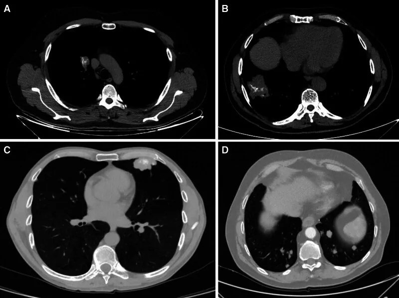
- Periferik nodüler birikim — iyi sınırlı tek veya çoklu parankim nodülleri plevraya yakın yerleşebilir; bazı nodüllerde noktasal ya da kaba kalsiyum bulunur.
- Trakeobronşiyal plaklar — submukozal birikim trakea ve büyük bronş duvarında yamalı, nodüler veya düzgün çevresel kalınlık oluşturabilir.
- Hava yolu kalsifikasyonu — duvar içi kalsifik odaklar mediasten penceresinde seçilir ve kıkırdak halkalardan bağımsız dağılım gösterebilir.
- Bronş lümeni daralması — duvar birikimi lümeni düzensizleştirip daraltabilir; tıkanıklık distalinde hava hapsi veya atelektazi gelişebilir.
- Alveoler-septal örüntü — interlobüler septalarda ve interstisyumda kalınlaşma, ince retikülasyon ve buzlu cam alanları birlikte görülebilir.
- Komşu subplevral mikronodüller — diffüz septal süreçte küçük periferal nodüller retiküler alanlara eşlik edebilir.
- Kistik eşlik — özellikle lenfositik interstisyel pnömoniyle birlikte ince duvarlı kistler nodüler amiloid birikimine eşlik edebilir.
- Çok kompartımanlı hastalık — bronş duvarı, parankimal nodül ve septal tutulum tek hastada birlikte bulunabilir.
Ölçütler ve sınıflamalar
- Nodüler parankimal tutulum
- Bir veya çok sayıda periferik, iyi sınırlı nodül baskındır; kalsifikasyon bulunabilir.
- Trakeobronşiyal tutulum
- Trakea ve bronş duvarındaki birikim plak, nodül veya çevresel kalınlaşma biçimindedir; lümen daralması eşlik edebilir.
- Diffüz alveoler-septal tutulum
- İnterstisyum ve küçük damar çevresindeki birikim retiküler opasiteler, septal kalınlaşma ve buzlu cam oluşturabilir.
Normalde
Normal trakea ve ana bronşların duvarı ince, konturu düzgün ve hava sütunu açıktır; akciğer parankiminde belirgin septal çizgiler, yaygın nodül veya buzlu cam bulunmaz. Mediasten penceresinde duvar kalınlaşması ile lümen içi lezyon ayrımı değerlendirilebilir ve hastalarda distal parankim değişiklikleri saptanabilir.
Normal BT ile kıyasla
Aynı bölgenin, kaynak veri setinde patoloji içermediği belirtilen bir BT incelemesini kesit kesit kaydırın; organ sınırlarını açarak anatomiyi hasta görüntüsüyle karşılaştırın. Küçük rastlantısal bulgular tümüyle dışlanmamıştır.
Kaynak: TotalSegmentator v2.01 veri seti (Wasserthal ve ark., Radiology: Artificial Intelligence 2023), CC BY 4.0, vaka s1278; organ sınırları veri setinin otomatik segmentasyonundan, birleştirilerek sadeleştirildi. Görüntüler 2,5 mm kesit aralığına yeniden örneklendi.
Ayırıcı tanı
- Kalsifiye metastatik nodüller
- bazı müsinöz veya kemik oluşturan primer tümörlerden yayılımda nodül kalsifiye olabilir; bilinen malignite ve dağılım ipuçları önemlidir.
- Granülomatöz enfeksiyon sekeli
- eski enfeksiyon da kalsifiye parankimal odaklar bırakabilir; aktif enfeksiyon lehine kavite, tomurcuklu dal veya nekrotik düğüm aranır.
- Trakeobronkopati osteokondroplastika
- kemik-kıkırdak nodülleri çoğunlukla ön ve yan trakea duvarındadır, arka membranöz duvarı koruma eğilimindedir.
- Relapsing polikondrit
- kıkırdak halkalarda kalınlaşma ve kalsifikasyon yapabilir; posterior membranöz yüz genellikle korunur ve klinik kıkırdak inflamasyonu eşlik edebilir.
- Lenfositik interstisyel pnömoni
- buzlu cam, retikülasyon ve kistler oluşturabilir; Sjögren bağlamı ve eşlik eden amiloid nodülleri ayrımı destekler.
Tuzaklar
- Kalsifiye nodül tek başına amiloidoz kanıtı değildir; eski enfeksiyon ve bazı metastazlar da benzer yoğunluk verir.
- Trakeobronşiyal amiloidozda arka membranöz duvar korunumu görülebilir; bu özellik tek başına amiloidoz tanısını doğrulamaz.
- Küçük lümenli bronşlardaki tıkanma tek kesitte fark edilmeyebilir; hava yolu boyunca takip edip distal lobüler havalanmayı kontrol edin.
- Diffüz septal kalınlaşma özgül değildir; BT amiloid örüntüsünü düşündürür, kesin sınıflama histopatoloji ve klinik korelasyon gerektirir.
Kendini dene
BT'de ana hava yollarında kalsifiye duvar birikimi lümeni daraltıyor ve distal akciğerde tekrarlayan kollaps alanları var. Hangi hastalık en olasıdır?
Cevabı göster
Pulmoner AL amiloidoz. Duvar boyunca yaygın kalsifiye submukozal birikim, hava yolu darlığıyla birlikte amiloid tutulumunu düşündürür. Diğer endobronşiyal süreçler genellikle fokal lezyonlar oluştururken; yaygın duvar kalsifikasyonu ve daralma amiloidozun karakteristik özelliğidir.
Akciğer kenarlarında kalsifiye olabilen nodüllerin yanı sıra septal retikülasyon ve buzlu cam alanları bulunuyor. Hangi depo hastalığı bu çoklu örüntüyü açıklayabilir?
Cevabı göster
Pulmoner amiloidoz. Amiloidoz nodüler parankimal ve diffüz alveoler-septal görünümleri aynı hastada oluşturabilir. Amiloidoz nodüler parankimal ve diffüz alveoler-septal görünümleri aynı hastada oluşturabilir; bu kombinasyon diğer deposyon hastalıklarıyla ayırt edilmelidir.
İlgili konular
Kaynaklar
Bu sayfadaki 43 cümle ve 2 quiz sorusu aşağıdaki kaynaklardan alıntılarla otomatik olarak karşılaştırıldı; 8 cümle bu karşılaştırmada düzeltildi (2026-10-09).
- Pulmonary amyloidosis, gastric MALT lymphoma, and multiple sclerosis in a patient with Sjögren’s disease: a case report and review of the literaturepmc.ncbi.nlm.nih.gov
- Association Between Common Variable Immunodeficiency and Pulmonary Amyloidosis: Reviewpmc.ncbi.nlm.nih.gov
- Review of research on pulmonary cysts in sjögren’s syndromepmc.ncbi.nlm.nih.gov
- Pulmonary AL amyloidosis: A review and update on treatment optionspmc.ncbi.nlm.nih.gov
- Diffuse parenchymal pulmonary amyloidosis associated with multiple myeloma: a case report and systematic review of the literaturepmc.ncbi.nlm.nih.gov
- Pulmonary Calcification and Ossification: Pathogenesis, CT Appearance, and Specific Disorders.pubmed.ncbi.nlm.nih.gov
- The lung in amyloidosis.pubmed.ncbi.nlm.nih.gov
- Radiographic and Pathologic Manifestations of Uncommon and Rare Pulmonary Lesions.pubmed.ncbi.nlm.nih.gov
- Review of research on pulmonary cysts in sjögren's syndrome.pubmed.ncbi.nlm.nih.gov
- The spectrum of pulmonary amyloidosis.pubmed.ncbi.nlm.nih.gov
- Pulmonary AL amyloidosis: A review and update on treatment options.pubmed.ncbi.nlm.nih.gov
- 18F-FDG PET/CT in patients with amyloid light-chain amyloidosis: case-series and literature review.pubmed.ncbi.nlm.nih.gov
- Clinical, radiologic, and pathologic features and outcomes of pulmonary transthyretin amyloidosis.pubmed.ncbi.nlm.nih.gov
- Nodular Pulmonary Amyloidosis Mimicking Metastatic Pulmonary Nodules: A case report and review of the literature.pubmed.ncbi.nlm.nih.gov
- Diagnosis of cystic lung diseases: a position statement from the UK Cystic Lung Disease Rare Disease Collaborative Network.pubmed.ncbi.nlm.nih.gov
- Pathological entities that may affect the lungs and the myocardium. Evaluation with chest CT and cardiac MR.pubmed.ncbi.nlm.nih.gov
- Pulmonary amyloidosis and cystic lung disease in primary Sjögren's syndrome: a case report and literature review.pubmed.ncbi.nlm.nih.gov
- [Pulmonary amyloidosis in Sjögren's syndrome: a case report].pubmed.ncbi.nlm.nih.gov
- Pulmonary amyloidosis: A case series.pubmed.ncbi.nlm.nih.gov
- Solitary nodular pulmonary amyloidosis mimicking primary lung cancer: A case report.pubmed.ncbi.nlm.nih.gov
- Diffuse cystic lung diseases-a primer for radiologists.pubmed.ncbi.nlm.nih.gov
- Radiologic patterns of interstitial lung disease: a pictorial review based on the 2025 European Respiratory Society/American Thoracic Society Classification.pubmed.ncbi.nlm.nih.gov
- Pulmonary AL Amyloidosis as a Cause of Interstitial Lung Disease Diagnosed With Transbronchial Lung Biopsy.pubmed.ncbi.nlm.nih.gov
- Diagnostic Sensitivity of [18F]-Florbetaben-PET/CT in Detecting Cardiac Involvement in AL Amyloidosis: The Prospective MoRBiDA Study.pubmed.ncbi.nlm.nih.gov
- An Uncommon Cause of Cystic Lung Disease.pubmed.ncbi.nlm.nih.gov
- Symptomatic diffuse pulmonary meningotheliomatosis with persistent diffusing-capacity impairment.pubmed.ncbi.nlm.nih.gov
- Primary nodular pulmonary amyloidosis: A case report.pubmed.ncbi.nlm.nih.gov
- Probable Pleural-Based Pulmonary Tuberculoma Mimicking Lung Malignancy: A Case Report.pubmed.ncbi.nlm.nih.gov
- Multimodal interventional bronchoscopy for bronchial myelolipoma complicated with airway obstruction: a case report and literature review.pubmed.ncbi.nlm.nih.gov
- Relapsing polychondritis: tracheobronchial involvement and differential diagnoses.pubmed.ncbi.nlm.nih.gov
- Tracheobronchial ossification in children: a case report and review of the literature.pubmed.ncbi.nlm.nih.gov
- A scoping review of photon-counting detector computed tomography in cardiovascular imaging.pubmed.ncbi.nlm.nih.gov
- High-Resolution CT Findings in Interstitial Lung Disease Associated with Connective Tissue Diseases: Differentiating Patterns for Clinical Practice-A Systematic Review with Meta-Analysis.pubmed.ncbi.nlm.nih.gov
- Diagnosis and Treatment of Adult Patients with Cough.pubmed.ncbi.nlm.nih.gov
- Morphological Diversity and Diagnostic Evaluation of Thoracic Amyloidosis: Insights From a Tertiary Referral Center in Vietnam.pubmed.ncbi.nlm.nih.gov
- Localized primary tracheobronchial AL amyloidosis with posterior wall sparing: A case report.pubmed.ncbi.nlm.nih.gov
- Classic Bronchoscopic Nodularity Sparing the Posterior Wall: A Case of Tracheobronchopathia Osteochondroplastica.pubmed.ncbi.nlm.nih.gov
- 60-Year-Old Man With Recurrent Hemoptysis, Pulmonary Nodules, and Mediastinal Lymphadenopathy.pubmed.ncbi.nlm.nih.gov
- Pulmonary plasma cell disorders: histopathology, diagnosis, and clinical perspectives.pubmed.ncbi.nlm.nih.gov
- Uncommon Causes of Interlobular Septal Thickening on CT Images and Their Distinguishing Features.pubmed.ncbi.nlm.nih.gov
- Systemic sclerosis associated with silicosis in an agricultural worker: A case of Erasmus syndrome.pubmed.ncbi.nlm.nih.gov
- Spontaneous Pneumothorax Revealing Pulmonary Alveolar Microlithiasis: A Case Report.pubmed.ncbi.nlm.nih.gov
- Lymphoid interstitial pneumonia in Sjögren disease: clinical course and comparison with other ILD patterns.pubmed.ncbi.nlm.nih.gov
- Histologic patterns of chronic interstitial lung diseases in dogs, cats, and humans: a review.pubmed.ncbi.nlm.nih.gov
- Pulmonary amyloidosis: a case report.pubmed.ncbi.nlm.nih.gov
- Prevalence of Central Airway Anatomical Variations and Structural Abnormalities Using Virtual Bronchoscopy: A Retrospective Study From a Tertiary Care Center.pubmed.ncbi.nlm.nih.gov
- CT Anatomy of the Lower Respiratory Tracts of the Testudo graeca.pubmed.ncbi.nlm.nih.gov
- Retrospective Review Is the Best View: A Primer for Residents and Radiologists on Missed Findings in Chest Radiography.pubmed.ncbi.nlm.nih.gov
- Pediatric Congenital Tracheobronchial Variants and Their Clinical Significance for Endotracheal Intubation and Airway Management: A Scoping Review of Fetal, Neonatal, and Pediatric Cases.pubmed.ncbi.nlm.nih.gov
- Anatomical Variations in Critical Structures in Esophageal Surgery: Implications for Personalized Surgery.pubmed.ncbi.nlm.nih.gov
- Anatomic reference measures for central airway anatomy in Indian adults: implications for precision airway management in surgical patient safety.pubmed.ncbi.nlm.nih.gov
- Gradually Enlarging Nodular Pulmonary Amyloidosis Associated With Sjögren's Syndrome.pubmed.ncbi.nlm.nih.gov
- Pulmonary amyloidosis, gastric MALT lymphoma, and multiple sclerosis in a patient with Sjögren's disease: a case report and review of the literature.pubmed.ncbi.nlm.nih.gov
- Localized Pulmonary Amyloidosis Associated With Sjögren's Syndrome, Coexisting Lymphoid Interstitial Pneumonia, and a Severe Double Aortic Lesion: A Case Report and Literature Review.pubmed.ncbi.nlm.nih.gov
- Relapsing Polychondritis Mimicking ANCA-Negative Granulomatosis with Polyangiitis: Diagnostic Value of <sup>18</sup>F-FDG PET/CT.pubmed.ncbi.nlm.nih.gov
- Tracheobronchial amyloidosis in a 64-year-old woman with type 2 diabetes mellitus and Sjögren's syndrome: a case report.pubmed.ncbi.nlm.nih.gov
- Epstein-Barr virus-associated inflammatory polyp of the proximal cervical trachea causing central airway obstruction: a case report.pubmed.ncbi.nlm.nih.gov
- Pulmonary Amyloidosis Diagnosed by Transbronchial Lung Biopsy: A Three-Case Series With Diverse Radiological Manifestations.pubmed.ncbi.nlm.nih.gov
- Incidental Lung Lesions on the CT Component of Oncologic FDG PET/CT: A Pictorial Review of Interpretive Pitfalls and Diagnostic Clues.pubmed.ncbi.nlm.nih.gov
- Pulmonary Tuberculosis Mimicking Metastatic Disease in a Patient With Two Previous Primary Malignancies: A Thoracoscopic Diagnostic Challenge.pubmed.ncbi.nlm.nih.gov
- Case Report: Synchronous primary lung and thyroid cancers - management informed by multimodal imaging and pathologic correlation.pubmed.ncbi.nlm.nih.gov
- Silent Renal Autodestruction: Incidental Discovery of End-Stage Genitourinary Tuberculosis (Putty Kidney) 14 Years After Treatment for Pulmonary Tuberculosis With Multisystem Sequelae.pubmed.ncbi.nlm.nih.gov
- An Asymptomatic 42-Year-Old Woman With Randomly Distributed Semicalcified Pulmonary Nodules.pubmed.ncbi.nlm.nih.gov
- Focus on Rare Tracheal Pathologies You Should Not Miss.pubmed.ncbi.nlm.nih.gov
- Unexpected difficult intubation due to tracheobronchopathia osteochondroplastica.pubmed.ncbi.nlm.nih.gov
- A rare case of tracheobronchopathia osteochondroplastica.pubmed.ncbi.nlm.nih.gov
- Diagnosis and treatment strategies for airway involvement in relapsing polychondritis: a comprehensive review.pubmed.ncbi.nlm.nih.gov
- Relapsing polychondritis with isolated tracheal involvement: Imaging findings on CT, MRI, and PET-CT: A case report and literature review.pubmed.ncbi.nlm.nih.gov
- Lymphocytic interstitial pneumonia and pseudolymphomatous lymphadenopathy as the first manifestation of primary Sjögren syndrome.pubmed.ncbi.nlm.nih.gov
- Pulmonary coexistence of immunoglobulin light chain amyloidosis and light chain deposition disease: A case report.pubmed.ncbi.nlm.nih.gov
- Clinical Relevance of Calcifications in Osteosarcoma Lung Metastases: Correlations Among Radiological Patterns, Histological Subtypes, and Chemotherapy Responses.pubmed.ncbi.nlm.nih.gov
- CT Features of an Extraskeletal Osteosarcoma Associated With a Retained Surgical Sponge in a Dog.pubmed.ncbi.nlm.nih.gov
- Presence of Antinuclear Antibodies in the Cerebrospinal Fluid and in the Serum in a Patient With Sjögren's Syndrome and Systemic Lupus: A Case Report.pubmed.ncbi.nlm.nih.gov
- Case Report: Severe bronchiectasis in three kittens with detection of <i>Mycoplasma felis</i>: radiographic and computed tomography findings.pubmed.ncbi.nlm.nih.gov
- Morphological Analysis of the Flow-Volume Curve for Identifying Fixed Airflow Obstruction: A Scoping Review.pubmed.ncbi.nlm.nih.gov
- When Tuberculosis Left the Heart in a Cage: Multimodality Imaging Revealing Constrictive Pericarditis in a Young Adult.pubmed.ncbi.nlm.nih.gov
- Imaging Cardiac Amyloidosis: From Early Diagnosis to Risk Stratification and Evaluation of Treatment Efficacy.pubmed.ncbi.nlm.nih.gov
- Isolated nasal septal involvement in granulomatosis with polyangiitis: Radiologic features and literature review.pubmed.ncbi.nlm.nih.gov
- Quantitative Imaging Metrics for the Assessment of Pulmonary Pathophysiology: An Official American Thoracic Society and Fleischner Society Joint Workshop Report.pubmed.ncbi.nlm.nih.gov
- The lung physiome: merging imaging-based measures with predictive computational models.pubmed.ncbi.nlm.nih.gov
- Particle transport and deposition: basic physics of particle kinetics.pubmed.ncbi.nlm.nih.gov
- Stepwise diagnostic algorithm for high-attenuation pulmonary abnormalities on CT.pubmed.ncbi.nlm.nih.gov
- Tracheal mucosal keratosis: Case discussion and literature review.pubmed.ncbi.nlm.nih.gov
- Three Cases of Primary Central Airway Tumors of Neurological and Neuroendocrine Origin in Children.pubmed.ncbi.nlm.nih.gov
- A 29-Year Old Man With an Enlarging Lung Lesion With Calcifications Giving Finger-in-Glove Appearance.pubmed.ncbi.nlm.nih.gov
- The first CT-based classification system for pulmonary actinomycosis: correlating imaging patterns with therapeutic strategies and prognosis in a highly misdiagnosed disease.pubmed.ncbi.nlm.nih.gov
- Pulmonary Manifestations in Patients With Hematologic Malignancies: In Pursuit of an Accurate Diagnosis.pubmed.ncbi.nlm.nih.gov
- Tips and Tricks in Thoracic Radiology for Beginners: A Findings-Based Approach.pubmed.ncbi.nlm.nih.gov
- A descriptive analysis of 21 patients with pulmonary amyloidosis: An observational study.pubmed.ncbi.nlm.nih.gov
- Comprehensive Interventional treatment for severe central airway collapse caused by Relapsing Polychondritis: A case report.pubmed.ncbi.nlm.nih.gov
- Preclinical and human tracheal replacement with aortic grafts: a narrative review.pubmed.ncbi.nlm.nih.gov
- New insights into imaging of pulmonary metastases from extra-thoracic neoplasms.pubmed.ncbi.nlm.nih.gov
- ERDHEIM-Chester Disease: Two Fatal Cases With Unusual Presentations.pubmed.ncbi.nlm.nih.gov
- Association Between Common Variable Immunodeficiency and Pulmonary Amyloidosis: Review.pubmed.ncbi.nlm.nih.gov
- AWMF S2k Guideline: Diagnosis and Assessment of Lung Disease Caused by Occupational Silica Dust Exposure (Silicosis) - Disease No. 4101 as per Occupational Diseases Ordinance (BKV) Annex 1.pubmed.ncbi.nlm.nih.gov
- Artificial Intelligence Enabled Lung Sound Auscultation in the Early Diagnosis and Subtyping of Interstitial Lung Disease.pubmed.ncbi.nlm.nih.gov
- Korean Guidelines for Diagnosis and Management of Interstitial Lung Diseases.pubmed.ncbi.nlm.nih.gov
- First Whole-Genome Sequencing Analysis of Tracheobronchopathia Osteochondroplastica with Critical Vocal Cord Involvement: Proposing a Novel Pathophysiological Model.pubmed.ncbi.nlm.nih.gov
- Animal models of benign airway stenosis: Advances in construction techniques, evaluation systems, and perspectives.pubmed.ncbi.nlm.nih.gov
- Identification of imaging-based pulmonary and extrapulmonary treatable traits in COPD: a review.pubmed.ncbi.nlm.nih.gov
- CT-Occult Primary Benign Tracheobronchial Neoplasms: A Single-Center 40-Case Clinicopathologic Series.pubmed.ncbi.nlm.nih.gov
- Tracheobronchopathia osteochondroplastica: A rare case report.pubmed.ncbi.nlm.nih.gov
- Submucosal laryngeal lesions: A puzzling diagnostic conundrum.pubmed.ncbi.nlm.nih.gov
- Case Report: Middle lobe syndrome: a rare presentation in eosinophilic granulomatosis with polyangiitis.pubmed.ncbi.nlm.nih.gov
- CT Findings of Central Airway Lesions Causing Airway Stenosis-Visualization and Quantification: A Pictorial Essay.pubmed.ncbi.nlm.nih.gov
- Tracheobronchial amyloidosis: an uncommon disease with a common presentation.pubmed.ncbi.nlm.nih.gov
- CT Findings of Asthma and Asthma-Mimicking Conditions.pubmed.ncbi.nlm.nih.gov
- Correlation analysis of chest HRCT quantitative parameters, exhaled nitric oxide, and pulmonary function in patients with chronic obstructive pulmonary disease.pubmed.ncbi.nlm.nih.gov
- Emergency repair of complete right main bronchial transection after blunt chest trauma: a case report.pubmed.ncbi.nlm.nih.gov
- Precision treatment of COPD based on novel imaging phenotypes: a treatable traits approach.pubmed.ncbi.nlm.nih.gov
- Pulmonary amyloidosis: clinicopathologic spectrum and diagnostic pitfalls in a 12-patient case series.pubmed.ncbi.nlm.nih.gov
- Vasculitis syndromes: large vessel vasculitides and ANCA-associated vasculitides from a neuroradiologist's perspective.pubmed.ncbi.nlm.nih.gov
- Comments and Illustrations of the European Federation of Societies for Ultrasound in Medicine Guidelines: Benign Pleura Lesions (Benign Pleura Thickening, Lesions and Masses)-What Can Be Seen on Transthoracic Ultrasound?pubmed.ncbi.nlm.nih.gov
- Recurrent Cryptogenic Ulcerative Tracheobronchitis 6 Years After Lung Transplantation: Case Report.pubmed.ncbi.nlm.nih.gov
- Central airway pathology: clinic features, CT findings with pathologic and virtual endoscopy correlation.pubmed.ncbi.nlm.nih.gov
Eğitim amaçlıdır; hasta başında tanı ve tedavi kararının yerine geçmez, tıbbi tavsiye değildir. Metinler yapay zekâ desteğiyle yazıldı ve otomatik bir adımda kaynak alıntılarıyla karşılaştırıldı; bu bir tıbbi doğrulama değildir ve içerik uzman radyolog incelemesinden geçmedi. Klinik kararlarda güncel kılavuzları, kurum protokollerini ve radyoloji raporunu esas alın.