Özet
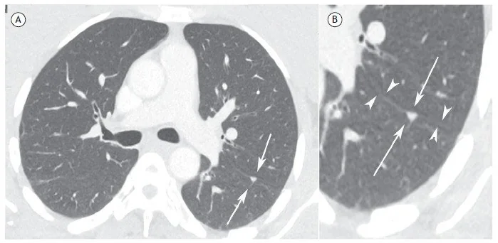
Perifissüral nodül, çoğunlukla akciğer içi lenf noduna karşılık gelen, fissür veya plevra yakınında görülen solid pulmoner odaktır. Aksiyel kesitteki üçgen ya da oval görünüm tek başına yeterli değildir; koronal ve sagittal düzlemlerde yassı/lentiform biçimin ve fissürle ilişkisinin doğrulanması yanlış metastaz etiketini önler. Tipik morfolojinin tanınması, özellikle kanser evrelemesinde gereksiz ileri incelemeleri ve yanlış evre yükseltmesini azaltır. Üst lob, düzensiz veya damara yapışık nodüller tipik intrapulmoner lenf nodu kabul edilmemelidir.
Faz ve pencere
- Kontrastsız tanısal
- Kontrastsız ince kesit BT'de perifissüral lenf nodu, keskin sınırlı solid ve genellikle kalsifiye olmayan odak olarak görülür; fissüre ince yumuşak doku bağlantısı olabilir ve multiplanar rekonstrüksiyon şekil/konum ilişkisini gösterir.
Önerilen pencereler: Akciğer (G 1500 / M -600), Mediasten (G 350 / M 50).
BT bulguları
- Düzgün ve keskin sınırlı solid nodül; tipik intrapulmoner lenf nodu genellikle kalsifiye değildir.
- Aksiyel görüntüde oval, yuvarlak, üçgen veya poligonal şekil; şekil bir ortogonal düzlemde tekrar doğrulanır.
- Koronal veya sagittal düzlemde yassı, lentiform ya da üçgen morfoloji.
- Majör/minör fissüre bitişiklik veya nodülden fissüre uzanan ince doğrusal bağlantı.
- Orta ya da alt lob yerleşimi ve plevraya yakınlık; üst lob yerleşimi tipik morfolojiye rağmen daha temkinli yorum gerektirir.
Ölçütler ve sınıflamalar
- İntrapulmoner lenf nodlarının çoğunda plevraya bildirilen uzaklık
- Tipik intrapulmoner lenf nodları pleural yüzeyden genellikle 15 mm mesafede veya daha yakında bulunur; bu ölçüt kesin dışlama kriteri değildir.
- Perifissüral nodül olarak yanlış sınıflamayı azaltmaya yönelik önerilen üst çap
- Perifissüral nodüller için yanlış pozitifleri azaltmaya yönelik önerilen üst çap sınırı 10 mm'dir.
- Tipik intrapulmoner lenf nodu morfolojisi varsa Fleischner takip istisnası
- Ortalama çap >6 mm olsa bile rutin takip önerilmez
Normalde
Normal aynı düzeyde fissür, visseral plevranın iki lob arasına uzanan ince çizgisel yaprağı olarak izlenir; çevresindeki lob parankimi havalıdır ve fissür içinde nodüler yumuşak doku bulunmaz. Nodülün şeklinin yalnız aksiyel kesitte değil koronal/sagittal düzlemde de lenf nodu morfolojisi göstermesine ve fissürle gerçek temas/ince bağlantı ilişkisine bakılır.
Normal BT ile kıyasla
Aynı bölgenin, kaynak veri setinde patoloji içermediği belirtilen bir BT incelemesini kesit kesit kaydırın; organ sınırlarını açarak anatomiyi hasta görüntüsüyle karşılaştırın. Küçük rastlantısal bulgular tümüyle dışlanmamıştır.
Kaynak: TotalSegmentator v2.01 veri seti (Wasserthal ve ark., Radiology: Artificial Intelligence 2023), CC BY 4.0, vaka s1278; organ sınırları veri setinin otomatik segmentasyonundan, birleştirilerek sadeleştirildi. Görüntüler 2,5 mm kesit aralığına yeniden örneklendi.
Ayırıcı tanı
- Subplevral metastaz
- yuvarlak veya lobüle yumuşak doku nodülü olabilir; tipik lenf nodunun üç boyutlu yassı/üçgen geometrisini ve fissür bağlantısını göstermeyebilir.
- Kalsifiye granülom
- kalsiyum odağı içerir ve çoğunlukla fissüre özgü yassı morfoloji taşımaz.
- Akciğer adenokarsinomu
- üst lob yerleşimi, spikülasyon, fissür çekintisi veya subsolid bileşen tipik lenf nodu görünümüne karşıdır.
- Fissüral plak veya nodüler plevral kalınlaşma
- fissüral plevrada kalınlaşma şeklinde görülebilir; intrapulmoner lenf nodları ise keskin sınırlı oval, yuvarlak, üçgen veya trapezoid nodüllerdir.
Tuzaklar
- Aksiyel kesitteki yuvarlak görünüm, üç boyutlu yassı lenf nodunun enine kesiti olabilir; nodül şeklini koronal ve sagittal rekonstrüksiyonda kontrol et.
- Bir perifissüral nodül büyüyebilir ve yine de benign kalabilir; büyüme tek başına kanser tanısı koydurmaz, ancak morfoloji artık tipik değilse yeniden sınıflandır.
- Kanserli hastada her fissür komşuluğu odağını otomatik olarak lenf nodu saymak yanlış evrelemeye yol açar; arterle bağlantı, lob yerleşimi, biçim ve plevral yüzey ilişkisini birlikte incele.
- Üst lobdaki düzgün odak veya pulmoner artere yapışık nodül, bazı tanımlardaki tipik intrapulmoner lenf nodu aday sınırlarının dışındadır.
Kendini dene
Tarama BT'sinde alt lobda, fissüre temas eden, aksiyelde oval ve koronal planda lentiform, keskin sınırlı solid nodül izleniyor. En olası yapı nedir?
Cevabı göster
İntrapulmoner lenf nodu. Alt lob yerleşimi, fissür teması ve ortogonal planda lentiform şekil intrapulmoner lenf nodunu destekler. Metastaz genellikle bu üç boyutlu biçim-fissür uyumunu göstermez; arteriol damar boyunca devam eder, granülomda kalsiyum görülebilir.
Akciğer kanserli hastada üst lobda spiküle, fissürü çeken ve damara yapışık solid nodül görülüyor. Hangi yorum daha uygundur?
Cevabı göster
Şüpheli nodül; tipik intrapulmoner lenf nodu sayılmaz. Spikülasyon ve fissür çekintisi tipik lenf nodu morfolojisine uymaz; bu nodül şüpheli kabul edilir. BT tek başına bunun yeni primer mi yoksa metastaz mı olduğunu belirlemez. Granülomda kalsifikasyon, hamartomda yağ veya kondroid kalsifikasyon aranır.
İlgili konular
Kaynaklar
Bu sayfadaki 37 cümle ve 2 quiz sorusu aşağıdaki kaynaklardan alıntılarla tek tek karşılaştırıldı; 3 cümle bu karşılaştırmada düzeltildi (2026-10-09).
- Intrapulmonary lymph node: a common and underrecognized tomography findingpmc.ncbi.nlm.nih.gov
- Initial LDCT appearance of incident lung cancers in the ITALUNG trial.pubmed.ncbi.nlm.nih.gov
- Clinical significance of perifissural nodules in the oncologic population.pubmed.ncbi.nlm.nih.gov
- Strategies for Reducing False-Positive Screening Results for Intermediate-Size Nodules Evaluated Using Lung-RADS: A Secondary Analysis of National Lung Screening Trial Data.pubmed.ncbi.nlm.nih.gov
- Typical CT Features of Intrapulmonary Lymph Nodes: A Review.pubmed.ncbi.nlm.nih.gov
- The impact of reconstruction techniques on observer performance for the detection and characterization of small pulmonary nodules in chest CT of children under 13 years.pubmed.ncbi.nlm.nih.gov
- CT Features of Benign Intrapulmonary Lymph Nodes in Pediatric Patients With Known Extrapulmonary Solid Malignancy.pubmed.ncbi.nlm.nih.gov
- Association between the number and size of intrapulmonary lymph nodes and chronic obstructive pulmonary disease severity.pubmed.ncbi.nlm.nih.gov
- A 10-Year Comprehensive, Single-Center, Retrospective Analysis on Juxtapleural Nodules: Insights into Classification and Risk.pubmed.ncbi.nlm.nih.gov
- Ultrasmall intrapulmonary lymph node: usual high-resolution computed tomographic findings with histopathologic correlation.pubmed.ncbi.nlm.nih.gov
- Morphologic and molecular analysis of 39 spontaneous feline pulmonary carcinomas.pubmed.ncbi.nlm.nih.gov
- Patterns of allelic loss of synchronous adenocarcinomas of the lung.pubmed.ncbi.nlm.nih.gov
- The Lung Reporting and Data System (LU-RADS): a proposal for computed tomography screening.pubmed.ncbi.nlm.nih.gov
- Classification of CT Pulmonary Opacities as Perifissural Nodules: Reader Variability.pubmed.ncbi.nlm.nih.gov
- Canine intrahepatic portosystemic shunts: Interlobar and intralobar classifications.pubmed.ncbi.nlm.nih.gov
- Conditions and mechanism for the appearance of interlobar fissures as 5-line signs in axial maximum intensity projection images.pubmed.ncbi.nlm.nih.gov
- Short linear shadows connecting pulmonary segmental arteries to oblique fissures in volumetric thin-section CT images: comparing CT, micro-CT and histopathology.pubmed.ncbi.nlm.nih.gov
- A Curious Case of Clear Cell Morphology in a Patient with Lung Cancer: Diagnostic Challenges.pubmed.ncbi.nlm.nih.gov
- Analysis of risk factors for stage I lung adenocarcinoma using low-dose high-resolution computed tomography.pubmed.ncbi.nlm.nih.gov
- Rare coexistence of sarcoidosis and lung adenocarcinoma.pubmed.ncbi.nlm.nih.gov
- Multidetector CT Findings and Differential Diagnoses of Malignant Pleural Mesothelioma and Metastatic Pleural Diseases in Korea.pubmed.ncbi.nlm.nih.gov
- Pulmonary perifissural nodules on CT scans: rapid growth is not a predictor of malignancy.pubmed.ncbi.nlm.nih.gov
- Solitary fibrous tumor of the pleura manifesting as an air-containing cystic mass: radiologic and histopathologic correlation.pubmed.ncbi.nlm.nih.gov
- Evaluating the Patient With a Pulmonary Nodule: A Review.pubmed.ncbi.nlm.nih.gov
- Identification of Nonaggressive Pulmonary Nodules Using an Optimized Scoring System.pubmed.ncbi.nlm.nih.gov
- CT imaging features regarding ground-glass nodules and solid lesions reflect prognostication of synchronous multiple lung adenocarcinoma.pubmed.ncbi.nlm.nih.gov
- Multiple primary lung cancer versus intrapulmonary metastatic cancer: A case of multiple pulmonary nodules.pubmed.ncbi.nlm.nih.gov
- Indication and Planning for Lobar Versus Sublobar Resection.pubmed.ncbi.nlm.nih.gov
- ENDOCRINE TUMOURS: Calcitonin in thyroid and extra-thyroid neuroendocrine neoplasms: the two-faced Janus.pubmed.ncbi.nlm.nih.gov
- Experience With a CT Screening Program for Individuals at High Risk for Developing Lung Cancer.pubmed.ncbi.nlm.nih.gov
- Experience with a CT screening program for individuals at high risk for developing lung cancer.pubmed.ncbi.nlm.nih.gov
- (18)F-FDG-PET/CT findings in pancreatic metastasis.pubmed.ncbi.nlm.nih.gov
- A Case Report of Hodgkin's Lymphoma in a Patient with Carcinosarcoma of the Bladder.pubmed.ncbi.nlm.nih.gov
- Lung cancer screening with low-dose CT: definition of positive, indeterminate, and negative screen results. A nodule management recommendation from the European Society of Thoracic Imaging.pubmed.ncbi.nlm.nih.gov
- ESR Essentials: lung cancer screening with low-dose CT-practice recommendations by the European Society of Thoracic Imaging.pubmed.ncbi.nlm.nih.gov
- Pediatric pulmonary nodules: current state of knowledge, AI applications, and future directions.pubmed.ncbi.nlm.nih.gov
- Management of incidental lung nodules <8 mm in diameter.pubmed.ncbi.nlm.nih.gov
- The TSANZ Practical Guide for Clinicians in the Management of Screen- and Incidentally-Detected Nodules.pubmed.ncbi.nlm.nih.gov
- RADS ALPHABET: news and tips for young and general radiologists.pubmed.ncbi.nlm.nih.gov
- Nonproliferative and Proliferative Lesions of the Gastrointestinal Tract, Pancreas and Salivary Glands of the Rat and Mouse.pubmed.ncbi.nlm.nih.gov
- Case Report: Primary gastric extraskeletal osteosarcoma and literature review.pubmed.ncbi.nlm.nih.gov
- Early stage nonsmall cell lung cancer: Toward a risk-adaptive paradigm in the era of biologic precision.pubmed.ncbi.nlm.nih.gov
- Prediction of Metastasis in Small Pulmonary Oligonodules Detected in Breast Cancer Patients at Baseline CT.pubmed.ncbi.nlm.nih.gov
- Evaluation of the solitary pulmonary nodule: size matters, but do not ignore the power of morphology.pubmed.ncbi.nlm.nih.gov
- New insights into imaging of pulmonary metastases from extra-thoracic neoplasms.pubmed.ncbi.nlm.nih.gov
- Transcatheter arterial embolization of coronary feeder vessels to primary cardiac paraganglioma: a case report.pubmed.ncbi.nlm.nih.gov
- Calcified Chondroid Mesenchymal Neoplasm of the Temporomandibular Joint: A Rare and Recently Described Benign Tumor.pubmed.ncbi.nlm.nih.gov
- Diagnosis of Mucormycosis: Current Situation, Challenges and Future Prospects.pubmed.ncbi.nlm.nih.gov
- Tool in lesion verification of shape-sensing robotic-assisted bronchoscopy with cone beam CT in sampling peripheral pulmonary nodules.pubmed.ncbi.nlm.nih.gov
- Incidental Lung Lesions on the CT Component of Oncologic FDG PET/CT: A Pictorial Review of Interpretive Pitfalls and Diagnostic Clues.pubmed.ncbi.nlm.nih.gov
- Risk of Lung Cancer in Peripheral Pulmonary Nodules.pubmed.ncbi.nlm.nih.gov
- Long-term safety and effectiveness of the "OptEase" vena cava filter.pubmed.ncbi.nlm.nih.gov
- Commentary: an eye on PET quantification.pubmed.ncbi.nlm.nih.gov
- Lung cancer screening: nodule identification and characterization.pubmed.ncbi.nlm.nih.gov
- Consensus guidelines for the use and interpretation of angiogenesis assays.pubmed.ncbi.nlm.nih.gov
- Tips and Tricks in Thoracic Radiology for Beginners: A Findings-Based Approach.pubmed.ncbi.nlm.nih.gov
- Combined two-stage total pancreatoduodenectomy and esophagectomy for synchronous malignancy of the pancreatic corpus and the esophagus: A surgical case report.pubmed.ncbi.nlm.nih.gov
- Recent advances in artificial intelligence across interventional pulmonology: a narrative review.pubmed.ncbi.nlm.nih.gov
- Beyond the usual - Atypical imaging presentation in lung cancer and implications for TNM-staging.pubmed.ncbi.nlm.nih.gov
- Bronchogenic cyst: a rare case of malignant transformation.pubmed.ncbi.nlm.nih.gov
- High-affinity alphavbeta3 integrin targeted optical probe as a new imaging biomarker for early atherosclerosis: initial studies in Watanabe rabbits.pubmed.ncbi.nlm.nih.gov
- In vitro expanded human CD4+CD25+ regulatory T cells suppress effector T cell proliferation.pubmed.ncbi.nlm.nih.gov
- IILS: Intelligent imaging layout system for automatic imaging report standardization and intra-interdisciplinary clinical workflow optimization.pubmed.ncbi.nlm.nih.gov
- Orbital tumours and tumour-like lesions: exploring the armamentarium of multiparametric imaging.pubmed.ncbi.nlm.nih.gov
- MDCT Imaging of Non-Traumatic Thoracic Aortic Emergencies and Its Impact on Diagnosis and Management-A Reappraisal.pubmed.ncbi.nlm.nih.gov
- Body Biofluids for Minimally-Invasive Diagnostics: Insights, Challenges, Emerging Technologies, and Clinical Potential.pubmed.ncbi.nlm.nih.gov
- Ultrathin Bronchoscopy Without Virtual Navigation for Diagnosis of Peripheral Lung Lesions.pubmed.ncbi.nlm.nih.gov
- International Society of Paediatric Surgical Oncology (IPSO) Minimally Invasive Surgery (MIS) Guidelines.pubmed.ncbi.nlm.nih.gov
- Diagnosis and management of peripheral lung nodule.pubmed.ncbi.nlm.nih.gov
- Intravascular Pulmonary Artery Tumour Mimicking Nodal Recurrence and Inadvertently Sampled by EBUS-TBNA: A Case Report.pubmed.ncbi.nlm.nih.gov
- Two Cases of Lung Cancer-Associated Horner Syndrome With Divergent Outcomes Following Radiotherapy.pubmed.ncbi.nlm.nih.gov
- Lymphadenectomy in pulmonary metastasectomy.pubmed.ncbi.nlm.nih.gov
- Features of low-dose CT-detected lung nodules: individuals who never smoked vs. who smoke(d) in a Chinese general population.pubmed.ncbi.nlm.nih.gov
- New developments in the imaging of lung cancer.pubmed.ncbi.nlm.nih.gov
- Inter-Slice Representation Outweighs Bounding-Box Supervision Extent in Lightweight 2.5D Pulmonary Nodule Detection: A Whole-Volume Benchmark on LUNA16.pubmed.ncbi.nlm.nih.gov
- Impact of CT slice thickness reduction algorithm on AI-based lung nodule detection in chest CTs of colorectal cancer patients.pubmed.ncbi.nlm.nih.gov
- Application of an improved YOLOv5s-based deep learning model for automated detection of pulmonary adenocarcinoma <i>in situ</i> and minimally invasive adenocarcinoma.pubmed.ncbi.nlm.nih.gov
- Rare Unicentric Intrapulmonary Castleman Disease: A Systematic Review and Report of a Case.pubmed.ncbi.nlm.nih.gov
- Discriminating bronchiolar adenoma from peripheral lung cancer by thin-section computed tomography (CT): a 2-center study.pubmed.ncbi.nlm.nih.gov
- Bronchogenic cyst: a rare cause of palpitations and atrial fibrillation.pubmed.ncbi.nlm.nih.gov
- Synchrotron-Based Phase-Contrast Micro-CT Combined With Histology to Decipher Differences Between Hereditary and Sporadic Pediatric Pulmonary Veno-Occlusive Disease.pubmed.ncbi.nlm.nih.gov
- EACR 2025 Congress: Innovative Cancer Science, 16-19 June 2025.pubmed.ncbi.nlm.nih.gov
- International Harmonization of Nomenclature and Diagnostic Criteria (INHAND): Nonproliferative and Proliferative Lesions of the Rabbit.pubmed.ncbi.nlm.nih.gov
- Surgery for thyroid cancer.pubmed.ncbi.nlm.nih.gov
- Lung nodules: A comprehensive review on current approach and management.pubmed.ncbi.nlm.nih.gov
Eğitim amaçlıdır; hasta başında tanı ve tedavi kararının yerine geçmez. İçerik yapay zekâ destekli bir süreçle güncel literatüre göre cümle cümle kaynaklarla karşılaştırıldı, ancak uzman radyolog incelemesinden geçmedi. Klinik kararlarda güncel kılavuzları, kurum protokollerini ve radyoloji raporunu esas alın.