
Özet
Over neoplazilerinin görüntüleme bulguları alt tipler arasında örtüşebilir; bu durum doğru ayırıcı tanıda zorluk yaratabilir ve kesin sınıflama için histopatoloji gereklidir. Seröz tümörlerde papiller veya solid kontrastlanan bileşenler ve peritoneal yayılım görülebilirken, müsinöz neoplaziler sıklıkla büyük multiloküle kistik kitle oluşturur. Bilateral solid-kistik kitleler ve eşlik eden gastrointestinal odak, over metastazı olasılığını gündeme getirir. Örüntüleri birbirine karıştırmak primer tümör ile metastaz ayrımını ve doğru evreleme incelemesini geciktirebilir; kesin sınıflama histopatoloji gerektirir.
Faz ve pencere
- Portal tanısal
- Kontrastlı portal venöz seride kist duvarı ve septaların düzensiz kalınlaşması, kontrastlanan papiller/solid bileşenler, nekroz, bilateral kitleler ve peritoneal yayılım aranır; BT, MRG dinamik kontrast karakterizasyonunun yerini tutmaz.
Önerilen pencereler: Batın (G 400 / M 50), Yumuşak doku (G 400 / M 40).
BT bulguları
- Seröz örüntü: papiller çıkıntılı veya solid-kistik adneksiyel kitle; eşlik eden asit ve peritoneal implantlar olabilir
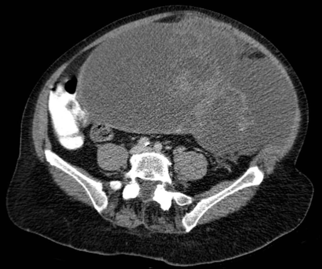
- Müsinöz örüntü: çoğunlukla tek taraflı, büyük ve multiloküle kistik kitle; loküller farklı içerik yoğunlukları gösterebilir
- Kalın, düzensiz septa veya duvar ve kontrastlanan mural nodül; sınırda ya da malign epitelyal neoplazi kuşkusunu artırır, fakat özgül değildir
- Belirgin solid bileşen, nekroz veya çevre organa uzanım; invaziv tümör morfolojisi lehine
- Bilateral, lobüle ve solid-kistik adneksiyel kitleler; özellikle eşlik eden mide, kolon veya apendiks tümörlerinde sekonder metastaz olasılığını düşündürür.
- Omental kek, yüzey implantları, asit ve mezenterik nodüller; yayılım paternini ve primer-metastatik ayrımını araştırmayı gerektirir
- Müsinöz kitle yanında lokalize müsinöz asit ve peritoneal yayılım; psödomiksoma peritonei ve apendiks kaynağı olasılığını düşündürür.
Ölçütler ve sınıflamalar
- Morfolojik örüntü (histolojik tanı değildir)
- Seröz: papiller/solid-kistik kitle ve peritoneal yayılım örüntüsü. Müsinöz: genellikle büyük, tek taraflı, multiloküle kistik kitle. Sekonder/metastatik: sıklıkla bilateral, lobüle solid-kistik kitle ve olası gastrointestinal primer. Bu görünümler örtüşür; alt tip patolojiyle belirlenir.
Normalde
Normal overler uterus yanında küçük, düzgün konturlu yapılar olarak izlenir; multiloküle kistik kitle, kalın septa veya kontrastlanan papiller doku bulunmaz. Karşı adnekle boyut ve taraflığı karşılaştırınız; ayrıca omentum, periton ve bağırsak kökenli olası primer odakları da değerlendirmelisiniz.
Normal BT ile kıyasla
Aynı bölgenin, kaynak veri setinde patoloji içermediği belirtilen bir BT incelemesini kesit kesit kaydırın; organ sınırlarını açarak anatomiyi hasta görüntüsüyle karşılaştırın. Küçük rastlantısal bulgular tümüyle dışlanmamıştır.
Kaynak: TotalSegmentator v2.01 veri seti (Wasserthal ve ark., Radiology: Artificial Intelligence 2023), CC BY 4.0, vaka s0119; organ sınırları veri setinin otomatik segmentasyonundan, birleştirilerek sadeleştirildi. Görüntüler 2,5 mm kesit aralığına yeniden örneklendi.
Ayırıcı tanı
- Matür kistik teratom
- makroskopik yağ, yağ-sıvı seviyesi ve diş benzeri kalsifikasyon malign epitelyal kitleden ayrılır.
- Endometrioma ayırıcı tanıda yer alır; BT'de heterojen kontrastlanma gösterebilir.
- Tubo-ovaryan apse, adneksiyel kitle ayırıcı tanısında yer alır; BT bu apseyi saptamada kullanılabilir.
- Over metastazı
- bilateral lobüle solid veya solid-kistik kitle, bilinen ekstraovaryan tümör ve serozal yüzey implantları primer epitelyal tümörden ayrımı destekler.
- Peritoneal müsinöz hastalık
- apendiks primeriyle ilişkili psödomiksoma, over müsinöz primerini taklit edebilir; apendiks ve peritoneal yüzeyler dikkatle taranmalıdır.
Tuzaklar
- Büyük multiloküle kitle müsinöz histolojiyi düşündürür ama kanıtlamaz; boyut ve lokülasyon görünümü histopatolojik alt tipe eşit değildir.
- Papiller çıkıntı maligniteye özgü değildir; kontrastlanan gerçek solid doku ile ince septa veya kist içi debris ayrımını yapın.
- Bilateral kitle metastaz olasılığını yükseltir ancak primer over tümörü de bilateral olabilir; bilinen primer ve eşlik eden peritoneal bulguları tarayın.
- Over müsinöz kitlesi ile psödomiksoma görülmesi primer over tümörünü kanıtlamaz; apendiks kaynaklı hastalık dışlanmalıdır.
Kendini dene
BT'de tek over kaynaklı, çok sayıda lokülü ve farklı içerik yoğunlukları olan dev kistik kitle görülüyor. Hangi morfolojik örüntü akla gelir?
Cevabı göster
Müsinöz neoplazi. Büyük multiloküle ve loküller arası içerik farklılığı gösteren kitle müsinöz neoplazi örüntüsüne uyar, ancak histolojiyi kanıtlamaz. Seröz tümörde papiller/solid bileşenler; metastazda bilateral lobüle yapı; teratomda yağ veya diş benzeri kalsifikasyon beklenir.
İki overde lobüle solid-kistik kitle, eşlik eden mide duvarı odağı ve peritoneal implantlar var. Hangi olasılık ayrıca araştırılmalıdır?
Cevabı göster
Gastrointestinal over metastazı. Bilateral kitleler ve eşlik eden olası mide primeri sekonder over metastazını düşündürür; patoloji ve klinik öyküyle doğrulanır. Diğer tanılar çoğunlukla farklı iç yapı gösterir ve mide odağı ile peritoneal yayılımı bu örüntüde açıklamaz.
Kaynaklar
Bu sayfadaki 41 cümle ve 2 quiz sorusu aşağıdaki kaynaklardan alıntılarla tek tek karşılaştırıldı; 7 cümle bu karşılaştırmada düzeltildi (2026-10-08).
- A review of ovarian-adnexal reporting and data system and radiomics in ovarian cancer: focus on ultrasound applicationspmc.ncbi.nlm.nih.gov
- Ovarian Carcinosarcoma With Heterologous Chondrosarcomatous Differentiation: A Case Report and Review of Literaturepmc.ncbi.nlm.nih.gov
- Integrating Heterogeneous Real-World Cancer Data for Semantic Interoperability in Oncology and Medical Imaging: Development and Validation of the Cancer Image Europe Hyperontologypmc.ncbi.nlm.nih.gov
- Massive uterine angiomyolipoma mimicking uterine leiomyoma in a 49-year-old womanpmc.ncbi.nlm.nih.gov
- Recurrence of gastric cancer with Krukenberg tumor, presenting with a high alpha-fetoprotein level, a case reportpmc.ncbi.nlm.nih.gov
- Ovarian collision tumors: imaging findings, pathological characteristics, diagnosis, and differential diagnosis.pubmed.ncbi.nlm.nih.gov
- CT radiomic stratification signature to optimize clinical decisions for ovarian cancer patients receiving neoadjuvant chemotherapy and the underlying biological basis: a multicenter retrospective study.pubmed.ncbi.nlm.nih.gov
- Uterine leiomyosarcoma: a rare clinical entity.pubmed.ncbi.nlm.nih.gov
- Endometrial endometrioid adenocarcinoma associated with primitive neuroectodermal tumour of the uterus: a poor prognostic subtype of uterine tumours.pubmed.ncbi.nlm.nih.gov
- Molecular classification and adjuvant treatment in endometrioid endometrial cancer with microcystic elongated and fragmented (MELF) invasion pattern.pubmed.ncbi.nlm.nih.gov
- Symptomatic Cushing syndrome and hyperandrogenemia revealing steroid cell ovarian neoplasm with late intra-abdominal metastasis.pubmed.ncbi.nlm.nih.gov
- Solitary Necrotic Nodules of the Liver: Histology and Diagnosis With CT and MRI.pubmed.ncbi.nlm.nih.gov
- Modern trends into the epidemiology and screening of ovarian cancer. Genetic substrate of the sporadic form.pubmed.ncbi.nlm.nih.gov
- Drug-induced interstitial lung disease caused by olaparib: three case reports and review of the Japanese Adverse Drug Event Report database and literature.pubmed.ncbi.nlm.nih.gov
- Preoperative unveiling of mucinous ovarian cancer subtypes: Clinical and CT features.pubmed.ncbi.nlm.nih.gov
- Evaluation of histo-pathogical patterns of ovarian masses in relation to age in Rawalpindi-Islamabad region - Lab Research.pubmed.ncbi.nlm.nih.gov
- Clear cell carcinoma of the ovary: Clues for radiologists to perform a correct diagnosis.pubmed.ncbi.nlm.nih.gov
- Are CT and MRI useful tools to distinguish between micropapillary type and typical type of ovarian serous borderline tumors?pubmed.ncbi.nlm.nih.gov
- Benign hemorrhagic mesenteric cyst mimicking peritoneal dissemination in early-stage ovarian clear cell carcinoma: a case report.pubmed.ncbi.nlm.nih.gov
- Value of an <sup>18</sup>F-FDG-based peritoneal cancer index in predicting dualistic classification, tumor burden, and completeness of cytoreductive surgery in epithelial ovarian cancer.pubmed.ncbi.nlm.nih.gov
- CT radiomics for noninvasive prediction of histologic differentiation in gastric cancer.pubmed.ncbi.nlm.nih.gov
- Diagnostic performance of artificial intelligence for detecting peritoneal and small bowel dissemination in epithelial ovarian cancer using preoperative contrast-enhanced CT imaging.pubmed.ncbi.nlm.nih.gov
- Fertility-Sparing Surgery in Young Women with Ovarian Anaplastic Carcinoma: Two Case Reports and Literature Review.pubmed.ncbi.nlm.nih.gov
- Ovarian Carcinoma Presenting Mucoepidermoid Carcinoma-Like Features in Association With Seromucinous Borderline Tumor: A Real Seromucinous Carcinoma?pubmed.ncbi.nlm.nih.gov
- Paratubal Leiomyoma Mimicking Ovarian Malignancy: A Case Report and Literature Review.pubmed.ncbi.nlm.nih.gov
- Fulminant development of a giant pelvic metastasis from microsatellite-stable early-onset sigmoid colon cancer with KRAS and TP53 co-mutations: a case report.pubmed.ncbi.nlm.nih.gov
- Ovarian Metastasis from Invasive Lobular Carcinoma of the Breast: A 6-Case Series with Emphasis on Diagnostic Challenges and the Value of Biopsy.pubmed.ncbi.nlm.nih.gov
- Diagnostic and therapeutic challenges of Krukenberg tumors: Case report of an atypical presentation with obstructive renal failure.pubmed.ncbi.nlm.nih.gov
- Incidental Lung Lesions on the CT Component of Oncologic FDG PET/CT: A Pictorial Review of Interpretive Pitfalls and Diagnostic Clues.pubmed.ncbi.nlm.nih.gov
- Artificial Intelligence in Oncology: A Comprehensive Cross-Cancer Translational Readiness Analysis Across 18 Malignancies.pubmed.ncbi.nlm.nih.gov
- Diagnostic value of a multimodal approach combining inflammatory biomarkers, tumor markers, and ultrasonographic features for borderline ovarian epithelial tumors.pubmed.ncbi.nlm.nih.gov
- Recent Imaging Updates and Advances in Gynecologic Malignancies.pubmed.ncbi.nlm.nih.gov
- Acute pelvic pain: A pictorial review with magnetic resonance imaging.pubmed.ncbi.nlm.nih.gov
- Patterns of injury and violence in Yaoundé Cameroon: an analysis of hospital data.pubmed.ncbi.nlm.nih.gov
- The Imaging of Primary Fallopian Tube Carcinoma: A Literature Review.pubmed.ncbi.nlm.nih.gov
- Manifestations of Ovarian Cancer in Relation to Other Pelvic Diseases by MRI.pubmed.ncbi.nlm.nih.gov
- Spontaneous rupture of a mucinous borderline ovarian tumour in late pregnancy: a case report.pubmed.ncbi.nlm.nih.gov
- Pebbled mosaic-like heterogeneous echogenicity in ovarian immature teratomas and correlation of ultrasound patterns with pathological grades.pubmed.ncbi.nlm.nih.gov
- ESUR female pelvis group approach to cystic female pelvic lesions.pubmed.ncbi.nlm.nih.gov
- Comprehensive Review of Endometriosis Based on Imaging Findings: What Radiologists Need to Know.pubmed.ncbi.nlm.nih.gov
- Rare and uncommon tumours of the female pelvis: what the radiologist should know.pubmed.ncbi.nlm.nih.gov
- Primary Neoplasms of the Seminal Vesicles: A Narrative Review of Carcinoma and the Rarer Non-Epithelial and Biphasic Tumours.pubmed.ncbi.nlm.nih.gov
- Metachronous Colorectal Carcinomas and Pancreatic Metastasis in Clinically Suspected Lynch Syndrome: An 18-Year Oncologic Course.pubmed.ncbi.nlm.nih.gov
- Sister Mary Joseph nodule revealing an ovarian carcinoma: a case report and review of the literature.pubmed.ncbi.nlm.nih.gov
- 불분명한 소아 복부 종괴: 영상 기반 진단 접근법.pubmed.ncbi.nlm.nih.gov
- Bilateral ovarian metastases from gastric signet-ring cell carcinoma in an 18-year-old: a case report and narrative review.pubmed.ncbi.nlm.nih.gov
- Ovarian cancer staging and follow-up: updated guidelines from the European Society of Urogenital Radiology female pelvic imaging working group.pubmed.ncbi.nlm.nih.gov
- Neglected Anatomical Areas in Ovarian Cancer: Significance for Optimal Debulking Surgery.pubmed.ncbi.nlm.nih.gov
- Imaging of Peritoneal Carcinomatosis in Advanced Ovarian Cancer: CT, MRI, Radiomic Features and Resectability Criteria.pubmed.ncbi.nlm.nih.gov
- Bilateral ovarian masses, massive ascites, and prior appendectomy: must it be ovarian cancer? --a case report of pseudomyxoma peritonei 24 years after appendectomy.pubmed.ncbi.nlm.nih.gov
- Pseudomyxoma Peritonei Misdiagnosed as Liver Cirrhosis: A Case Report and Literature Review.pubmed.ncbi.nlm.nih.gov
- Successful treatment of pseudomyxoma peritonei (PMP) through cytoreductive surgery and hyperthermic intraperitoneal chemotherapy (CRS-HIPEC): A case report and literature review.pubmed.ncbi.nlm.nih.gov
- Adolescent dysmenorrhea revealing Herlyn-Werner-Wunderlich syndrome with endometriosis: A radiologic case report.pubmed.ncbi.nlm.nih.gov
- Cross-Sectional Imaging of Pelvic Inflammatory Disease: Diagnostic Pearls and Pitfalls on CT and MR.pubmed.ncbi.nlm.nih.gov
- Cancer of the ovary, fallopian tube, and peritoneum: 2025 update.pubmed.ncbi.nlm.nih.gov
- Ovarian Carcinosarcoma With Heterologous Chondrosarcomatous Differentiation: A Case Report and Review of Literature.pubmed.ncbi.nlm.nih.gov
- Gynecologic imaging errors and pitfalls: how to avoid misdiagnosis on pelvic MRI.pubmed.ncbi.nlm.nih.gov
- Bilateral highly differentiated follicular carcinoma of ovarian origin with extensive abdominal metastasis: A case report.pubmed.ncbi.nlm.nih.gov
- Endometriosis and Chronic Endometritis: Shared Mechanisms, Diagnostic Challenges, and Clinical Implications in Infertility.pubmed.ncbi.nlm.nih.gov
- Liquid Biopsy for Minimal Residual Disease Assessment in Endometrial and Cervical Cancers: Molecular Rationale, Clinical Evidence, and Translational Barriers.pubmed.ncbi.nlm.nih.gov
- Is There an Added Value of Dual-Time-Point [<sup>68</sup>Ga]Ga-Fibroblast Activation Protein Inhibitor (FAPI) PET/CT in Differentiating Malignant and Benign Uptake Findings?pubmed.ncbi.nlm.nih.gov
- International Harmonization of Nomenclature and Diagnostic Criteria (INHAND): Non-proliferative and Proliferative Lesions of the Non-human Primate ( M. fascicularis ).pubmed.ncbi.nlm.nih.gov
- Total laparoscopic radical hysterectomy for cervical cancer in prolapsed uterus.pubmed.ncbi.nlm.nih.gov
- Spectrum of Ovarian Incidentalomas: Diagnosis and Management.pubmed.ncbi.nlm.nih.gov
- Primary and secondary tumors of the peritoneum: key imaging features and differential diagnosis with surgical and pathological correlation.pubmed.ncbi.nlm.nih.gov
- Mimics and Pitfalls of Primary Ovarian Malignancy Imaging.pubmed.ncbi.nlm.nih.gov
- Colonic endometriosis: from subtotal bowel obstruction to malignant transformation - a case series and literature review.pubmed.ncbi.nlm.nih.gov
- High-grade appendiceal mucinous neoplasm: case report.pubmed.ncbi.nlm.nih.gov
- Low-Grade Appendiceal Mucinous Neoplasm Mimicking Pelvic Inflammatory Disease: A Case Report.pubmed.ncbi.nlm.nih.gov
- High-volume mucinous ascites secondary to low-grade mucinous neoplasm arising in a mature cystic teratoma: a case report.pubmed.ncbi.nlm.nih.gov
- Cystic pancreatic lesions: MR imaging findings and management.pubmed.ncbi.nlm.nih.gov
- Intravenous leiomyomatosis: A retrospective case series of imaging findings and diagnostic challenges.pubmed.ncbi.nlm.nih.gov
- Rare Ovarian Sex Cord-Stromal Tumor: Diagnostic Pitfalls and Clinical Management.pubmed.ncbi.nlm.nih.gov
- Giant low-grade endometrial stromal sarcoma with extensive sex cord-like differentiation: a case report.pubmed.ncbi.nlm.nih.gov
- The clinical and pathological features of low-grade appendiceal mucinous neoplasm (LAMN).pubmed.ncbi.nlm.nih.gov
- Case Report: An incidentally discovered HPV-associated endocervical adenocarcinoma presenting as pseudomyxoma peritonei.pubmed.ncbi.nlm.nih.gov
- Pseudo-Pseudo Meigs' Syndrome-Like Presentation Associated with Primary Sjögren's Syndrome: A Case Report.pubmed.ncbi.nlm.nih.gov
- Krukenberg tumours: which patients should be considered for surgery?-a narrative literature review.pubmed.ncbi.nlm.nih.gov
- Peritoneal disease: key imaging findings that help in the differential diagnosis.pubmed.ncbi.nlm.nih.gov
- An Unusual Evolution of Krukenberg Tumour: A Case Report.pubmed.ncbi.nlm.nih.gov
- Japanese society for cancer of the colon and rectum (JSCCR) guidelines 2024 for the clinical practice of hereditary colorectal cancer.pubmed.ncbi.nlm.nih.gov
- Special Issue: EACR 2026 Congress: Innovative Cancer Science, 8-11 June 2026.pubmed.ncbi.nlm.nih.gov
- EACR 2025 Congress: Innovative Cancer Science, 16-19 June 2025.pubmed.ncbi.nlm.nih.gov
- Heterogeneity Analyzed by CT-Based Habitat Analysis for Clinical Management of Cancers: A Narrative Review.pubmed.ncbi.nlm.nih.gov
- Occult ovarian carcinoid in pregnancy: a case report and literature review.pubmed.ncbi.nlm.nih.gov
- Carcinosarcoma of the Ovary Arising in the Background of a Mucinous Neoplasm Distinct From a Mural Nodule.pubmed.ncbi.nlm.nih.gov
- Giant Mature Cystic Perihepatic Teratoma in an Adult Woman.pubmed.ncbi.nlm.nih.gov
- A challenging imaging feature of mature teratoma in uterus.pubmed.ncbi.nlm.nih.gov
- Hepatic Teratoma: An Unusual Presentation of a Germ Cell Neoplasm.pubmed.ncbi.nlm.nih.gov
- Recurrent Malignant Struma Ovarii With Peritoneal Implants Following Prior Thyroid Carcinoma Treatment.pubmed.ncbi.nlm.nih.gov
- Navigating Ovarian-Adnexal Reporting and Data System Magnetic Resonance Imaging (O-RADS MRI): A Review of Its Evolution, Current Advances, and Persistent Challenges in Ovarian Imaging.pubmed.ncbi.nlm.nih.gov
- Radiomics and Artificial Intelligence in Ovarian Endometriosis Imaging: A Systematic Review and Critical Appraisal of Emerging Evidence.pubmed.ncbi.nlm.nih.gov
- The role of "penumbra sign" and diffusion-weighted imaging in adnexal masses: do they provide a clue in differentiating tubo-ovarian abscess from ovarian malignancy?pubmed.ncbi.nlm.nih.gov
- Computed tomography and magnetic resonance imaging features of struma ovarii: A retrospective case series.pubmed.ncbi.nlm.nih.gov
- A Rare Mimicker: Renal Endometriosis and Its Diagnostic Pitfalls-Review of Reported Cases.pubmed.ncbi.nlm.nih.gov
- Incidentally Detected Ovarian Teratoma on Fluorodeoxyglucose (FDG) PET/CT Scanning in a Patient With Gastric Cancer.pubmed.ncbi.nlm.nih.gov
- Tuberous sclerosis complex with ovarian endometrioma misdiagnosed as multiple metastases: a case report.pubmed.ncbi.nlm.nih.gov
- Next-Generation Biomarkers and Artificial Intelligence in Colorectal Cancer: From Multi-Omic Data Integration to Clinical Application.pubmed.ncbi.nlm.nih.gov
- Artificial intelligence in ovarian cancer: advancing in precision diagnosis and clinical management.pubmed.ncbi.nlm.nih.gov
- Malignant transformation of mature ovarian teratoma into goblet cell adenocarcinoma: a case report.pubmed.ncbi.nlm.nih.gov
- Metastatic appendiceal mucinous adenocarcinoma presenting as bilateral ovarian masses mimicking advanced ovarian cancer: a case report.pubmed.ncbi.nlm.nih.gov
- MRI insights into endometrial stromal tumors.pubmed.ncbi.nlm.nih.gov
- Peritoneal carcinomatosis in metastatic Non-Small Cell Lung Cancer: review of the literature.pubmed.ncbi.nlm.nih.gov
- Options and Considerations in the Management of Peritoneal Disease in Patients with Small Bowel Neuroendocrine Tumors.pubmed.ncbi.nlm.nih.gov
- Primary Ovarian Leiomyoma in a 2-year-old Child: Youngest Worldwide and First Report from India with Review of Literature.pubmed.ncbi.nlm.nih.gov
Eğitim amaçlıdır; hasta başında tanı ve tedavi kararının yerine geçmez. İçerik yapay zekâ destekli bir süreçle güncel literatüre göre cümle cümle kaynaklarla karşılaştırıldı, ancak uzman radyolog incelemesinden geçmedi. Klinik kararlarda güncel kılavuzları, kurum protokollerini ve radyoloji raporunu esas alın.