
Özet
Serviks kanseri biyopsiyle tanınır; görüntüleme, özellikle lokal ileri hastalıkta nodal ve uzak yayılımı ortaya koyarak klinik evrelemeyi tamamlar. Lokal ileri hastalıkta veya erken evre dışındaki durumlarda ekstrapelvik yayılımı değerlendirmek için kontrastlı BT ya da PET-BT önerilir; erken evre T1a–T2a1 (T1b3 hariç) ve US/MRG’de nodları negatif hastalarda cerrahi-patolojik evreleme yapılır. BT, servikal stromayı ve erken parametrial invazyonu ayırmada sınırlıdır; lokal tümör yayılımında pelvik MRG temel yöntemdir. Parametrial veya yan duvar hastalığının ya da üreter obstrüksiyonunun saptanmasında, klinik muayeneyle tespit edilemeyen lokal yayılımı görebilen manyetik rezonans görüntüleme (MRG) kullanılır.
Faz ve pencere
- Portal tanısal
- Serviks kanserinde manyetik rezonans görüntüleme (MRG) lokal uzanımı; kontrastlı bilgisayarlı tomografi (BT) veya PET-BT ise ekstrapelvik yayılımı değerlendirmek için kullanılır. Serviks kanserinde kontrastlı BT, ekstrapelvik (nodal ve uzak) yayılımın değerlendirilmesinde önerilir; lokal ve parametrial tümör uzanımının değerlendirilmesinde MRG üstündür.
Önerilen pencereler G genişlik, M merkez; değerler Hounsfield birimi (HU)
- MediastenG 350 / M +50
- BatınG 400 / M +50
- KaraciğerG 150 / M +30
- Yumuşak dokuG 400 / M +40
- KemikG 1800 / M +400
BT bulguları
- Primer tümörün lokal sınırları ve parametrial yayılım pelvik MRG veya transvajinal/transrektal ultrasonla; ekstrapelvik yayılım ise kontrastlı BT ya da PET-BT ile değerlendirilir.
- Parametrial invazyon, geniş görüş alanlı T2 ağırlıklı MRG ile değerlendirilebilir.
- Pelvik yan duvar tutulumu, manyetik rezonans görüntüleme (MRG) ile lokal tümör uzanımı kapsamında değerlendirilir; MRG, klinik muayenede kaçabilecek gizli yayılımı da gösterebilir.
- Mesane veya rektum duvarı tutulumu, manyetik rezonans görüntüleme (MRG) ile infiltrasyon derinliği açısından hassas şekilde değerlendirilir.
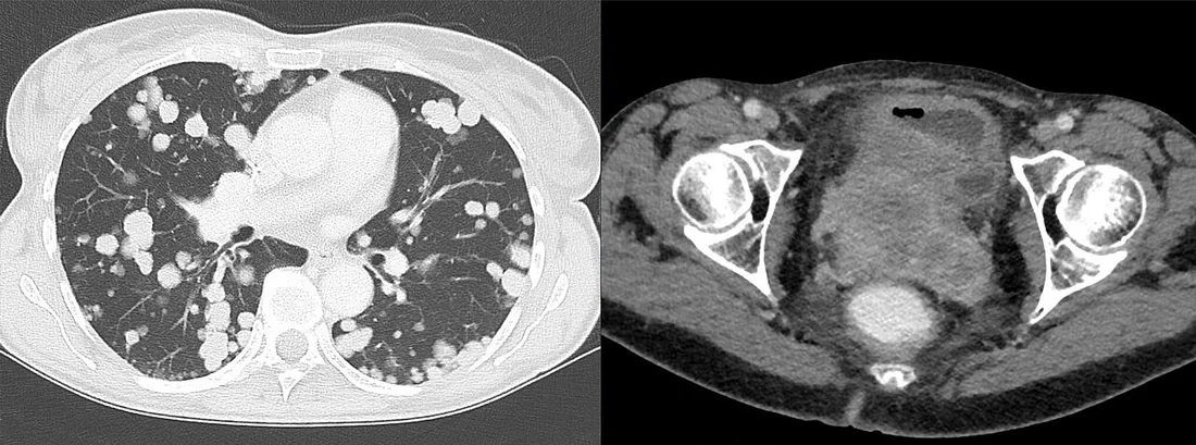
- Lokal ileri evre serviks kanserinde üreter tutulumu, hidroureteronefroz gelişsin ya da gelişmesin sık görülen bir bulgudur.
- Şüpheli pelvik veya paraaortik lenf nodlarında kısa eksenin ≥10 mm olması, yuvarlak şekil veya santral nekroz metastazla ilişkili olabilir; görüntüleme küçük hacimli metastazı dışlayamaz.
- Uzak yayılımda akciğer, karaciğer veya kemik metastazları görülebilir; PET-BT tek incelemede tüm vücudu değerlendirebilir.
Ölçütler ve sınıflamalar
- Şüpheli lenf nodu: kısa eksen boyutu (yuvarlak şekil, düzensiz kapsül, nekroz ve FDG tutulumuyla birlikte yorumlanır; normal boyut mikrometastazı dışlamaz)
- ≥10 mm
- FIGO 2018 serviks kanseri evrelemesi
- FIGO 2018, Evre IB tümörleri boyutlarına göre IB1, IB2 ve IB3 alt evrelerine ayırır. Evre IIB, pelvik yan duvar tutulumu olmaksızın parametrial tutulumu tanımlar. FIGO 2018'de IIIC1 pelvik, IIIC2 paraaortik lenf nodu tutulumu olarak sınıflandırılır. BT, lokal tümör değerlendirmesinde MRG'ye göre daha sınırlıdır; MRG lokal parametrial ve mesane invazyonunun değerlendirilmesinde üstündür.
Normalde
Metastazla ilişkili ölçütleri (kısa eksen 10 mm ve üzeri, yuvarlak şekil, santral nekroz) taşımayan pelvik nodları karşı taraftaki aynı istasyonla kıyaslayarak kaydedin.
Normal BT ile kıyasla
Aynı bölgenin, kaynak veri setinde patoloji içermediği belirtilen bir BT incelemesini kesit kesit kaydırın; organ sınırlarını açarak anatomiyi hasta görüntüsüyle karşılaştırın. Küçük rastlantısal bulgular tümüyle dışlanmamıştır.
Kaynak: TotalSegmentator v2.01 veri seti (Wasserthal ve ark., Radiology: Artificial Intelligence 2023), CC BY 4.0, vaka s0119; organ sınırları veri setinin otomatik segmentasyonundan, birleştirilerek sadeleştirildi. Görüntüler 2,5 mm kesit aralığına yeniden örneklendi.
Ayırıcı tanı
- Naboth kistleri
- servikste sıvı yoğunluklu, ince duvarlı kistik odaklar; solid kontrastlanan kitle ve infiltratif sınır oluşturmaz.
- Endometrium kaynaklı kanser
- miyometrial invazyon sonrası servikal stromaya uzanabilir; bu yayılım MRG ile saptanabilir ve yönetimi değiştirebilir.
- Radyasyon sonrası fibrozis
- tedavi öyküsü olan hastada pelvik dokularda hacim kaybı ve şerit tarzı yumuşak doku; yeni büyüyen nodüler kitle nüks kuşkusunu artırır.
Tuzaklar
- Parametrial bölgedeki değişiklikler, lokal tümör uzanımını değerlendirmek için MRG ile incelenebilir.
- Mesane veya rektum duvarına infiltrasyon derinliği MRG ile değerlendirilebilir; bu değerlendirme tek başına mukozal tutulumu doğrulamaz.
- Görüntüleme yöntemlerinin duyarlılığı küçük hacimli lenf nodu metastazlarını dışlamak için yetersiz olabilir; nodal boyut ve morfoloji ölçütleri birlikte değerlendirilmelidir.
- Lokal ileri evre serviks kanserinde üreter tutulumu, hidroureteronefroz gelişsin ya da gelişmesin sık görülür.
Kendini dene
Paraaortik lenf nodlarının BT değerlendirmesinde metastazla anlamlı ilişkili kısa eksen eşiği hangisidir?
Cevabı göster
≥10 mm. Paraaortik lenf nodlarında BT’de kısa eksenin ≥10 mm olması metastazla anlamlı ilişkilidir.
Serviks kanserli hastada paraaortik zincirde yuvarlak, santral nekrotik bir lenf nodu saptanıyor. Bu morfoloji hangi olasılığı artırır?
Cevabı göster
Paraaortik metastaz. Paraaortik zincirde yuvarlak veya santral nekrotik lenf nodu, serviks kanserinde metastaz olasılığını artırır. Bu morfolojik özellikler metastazla anlamlı biçimde ilişkilidir; lenf nodları birlikte değerlendirilen görüntüleme bulgularına göre yorumlanmalıdır.
İlgili konular
- Endometrium kanserinde yayılım değerlendirmesi
- Postpartum pelvik komplikasyonlar ve normal postpartum görünüm
Kaynaklar
Bu sayfadaki 39 cümle ve 2 quiz sorusu aşağıdaki kaynaklardan alıntılarla otomatik olarak karşılaştırıldı; 30 cümle bu karşılaştırmada düzeltildi (2026-10-09).
- Diagnostic accuracy of computed tomography alone versus computed tomography combined with ultrasound for diagnosing lymph node metastasis in thyroid cancerpmc.ncbi.nlm.nih.gov
- Advances in Positron Emission Tomography Combined With Computed Tomography (PET/CT) Imaging in Cervical Cancer: From Metabolic Assessment to Fibroblast Activation Protein-Targeted Imagingpmc.ncbi.nlm.nih.gov
- International Western Radiation Therapy Guidelines Applicability in Low- and Middle-Income Countries for Cervical Cancer Management: A Comparative Survey Example from South and North Border of the Mediterranean Area Countriespmc.ncbi.nlm.nih.gov
- FDG-PET/CT-based treatment response predicts recurrence and overall survival in locally advanced cervical cancerpmc.ncbi.nlm.nih.gov
- Laparoscopic Lymph Node Assessment Prior to Open Radical Hysterectomy—A Two-Center Retrospective Study on Individualized Surgical Management of Early-Stage Cervical Cancerpmc.ncbi.nlm.nih.gov
- The Role of Imaging in Cervical Cancer Staging: ESGO/ESTRO/ESP Guidelines (Update 2023).pubmed.ncbi.nlm.nih.gov
- Imaging in endometrial cancer.pubmed.ncbi.nlm.nih.gov
- [Cervical CUP syndrome].pubmed.ncbi.nlm.nih.gov
- Imaging to assist fertility-sparing surgery.pubmed.ncbi.nlm.nih.gov
- Minimally invasive mediastinal staging of non-small-cell lung cancer: emphasis on ultrasonography-guided fine-needle aspiration.pubmed.ncbi.nlm.nih.gov
- Staging, recurrence and follow-up of uterine cervical cancer using MRI: Updated Guidelines of the European Society of Urogenital Radiology after revised FIGO staging 2018.pubmed.ncbi.nlm.nih.gov
- Intraoral ultrasonography to measure tumor thickness of oral cancer: A systematic review and meta-analysis.pubmed.ncbi.nlm.nih.gov
- 68 Ga-FAPI-PET/CT in patients with various gynecological malignancies.pubmed.ncbi.nlm.nih.gov
- Gynecological cancers.pubmed.ncbi.nlm.nih.gov
- PET/CT-guided radiation therapy planning: from present to the future.pubmed.ncbi.nlm.nih.gov
- Comprehensive management of HPV-related squamous cell carcinoma of the head and neck of unknown primary.pubmed.ncbi.nlm.nih.gov
- PET/CT Variants and Pitfalls in Gynecological Cancers.pubmed.ncbi.nlm.nih.gov
- Quantitative Assessment of Gynecologic Malignancies.pubmed.ncbi.nlm.nih.gov
- Molecular imaging in cervical cancer.pubmed.ncbi.nlm.nih.gov
- Advances in diagnosis and treatment of metastatic cervical cancer.pubmed.ncbi.nlm.nih.gov
- Cervical tumors.pubmed.ncbi.nlm.nih.gov
- Endotracheal Leiomyoma Mimicking Malignancy on 18F-FDG PET/CT in a Patient With Papillary Thyroid Carcinoma.pubmed.ncbi.nlm.nih.gov
- Nabothian cyst a predominant cause of false-positive iodine uptake in uterus: comparison of SPECT/CT and pelvic MRI.pubmed.ncbi.nlm.nih.gov
- Imaging evaluation of para-aortic lymph nodes in cervical cancer.pubmed.ncbi.nlm.nih.gov
- Recommendations from Gynaecological (GYN) GEC-ESTRO Working Group (I): concepts and terms in 3D image based 3D treatment planning in cervix cancer brachytherapy with emphasis on MRI assessment of GTV and CTV.pubmed.ncbi.nlm.nih.gov
- Ureteral stenting in patients with locally advanced cervical cancer: Predictors of low success rate.pubmed.ncbi.nlm.nih.gov
- Management of stage I and II cervical cancer: a review.pubmed.ncbi.nlm.nih.gov
- Concordance between MRI and clinical examination in cervical cancer staging: a retrospective study.pubmed.ncbi.nlm.nih.gov
- Comparison of treatment outcomes of pelvis external radiotherapy with and without vaginal brachytherapy for cervical cancer patients with positive or close vaginal resected margins.pubmed.ncbi.nlm.nih.gov
- Survival outcomes of 2018 FIGO stage IIIC versus stages IIIA and IIIB in cervical cancer: A systematic review with meta-analysis.pubmed.ncbi.nlm.nih.gov
- Five-fraction HDR brachytherapy in locally advanced cervical cancer: A monocentric experience.pubmed.ncbi.nlm.nih.gov
- Prognostic Performance of the 2018 International Federation of Gynecology and Obstetrics Cervical Cancer Staging Guidelines.pubmed.ncbi.nlm.nih.gov
- An Unusual Presentation of a Cervical Paraspinal Leiomyoma in an Adolescent Female.pubmed.ncbi.nlm.nih.gov
- Angioleiomyomas of the Extremities and Trunk: An Observational Study.pubmed.ncbi.nlm.nih.gov
- Dual treatment approach for leiomyomas of urinary bladder: Experience from tertiary hospital.pubmed.ncbi.nlm.nih.gov
- Imaging and Staging of Endometrial Cancer.pubmed.ncbi.nlm.nih.gov
- Magnetic resonance imaging of endometrial carcinoma.pubmed.ncbi.nlm.nih.gov
- Clinical Utility of Diffusion-Weighted Imaging in Gynecological Imaging: Revisited.pubmed.ncbi.nlm.nih.gov
- Feasibility of radical hysterectomy in women with FIGO stage IIB cervical cancer: an observation study of 10-year experience in a tertiary center.pubmed.ncbi.nlm.nih.gov
- Validation of the 2018 FIGO Staging System of Cervical Cancer for Stage III Patients with a Cohort from China.pubmed.ncbi.nlm.nih.gov
- Magnetic resonance imaging-based validation of the 2018 FIGO staging system in patients treated with definitive radiotherapy for locally advanced cervix cancer.pubmed.ncbi.nlm.nih.gov
- Advances in Positron Emission Tomography Combined With Computed Tomography (PET/CT) Imaging in Cervical Cancer: From Metabolic Assessment to Fibroblast Activation Protein-Targeted Imaging.pubmed.ncbi.nlm.nih.gov
- HPV-associated Oropharyngeal Squamous Cell Carcinoma: An Imaging Framework for AJCC Version 9 TNM Staging.pubmed.ncbi.nlm.nih.gov
- The role of <sup>68</sup>Ga-FAPi PET/CT in Staging and Restaging of Breast Cancer Subtypes with Limited <sup>18</sup>F-FDG Uptake.pubmed.ncbi.nlm.nih.gov
- Simultaneous Occurrence of Breast Cancer Metastases and Cervical Cancer Metastases in the Same Lymph Node.pubmed.ncbi.nlm.nih.gov
- International Western Radiation Therapy Guidelines Applicability in Low- and Middle-Income Countries for Cervical Cancer Management: A Comparative Survey Example from South and North Border of the Mediterranean Area Countries.pubmed.ncbi.nlm.nih.gov
- Prospective comparison of diagnostic accuracy of ultrasound, PET/CT and DW-MRI for preoperative assessment of pelvic lymph nodes in cervical cancer patients: results of the CANNES trial.pubmed.ncbi.nlm.nih.gov
- Isolated ovarian metastasis in otherwise cervix-confined adenocarcinoma of the cervix: A rare clinical conundrum.pubmed.ncbi.nlm.nih.gov
- Endometrial carcinosarcoma with multi-classifier molecular signature (MMRd-p53abn): A case report.pubmed.ncbi.nlm.nih.gov
- Sentinel Lymph Node Mapping Without Intraoperative Frozen Section in Preoperative Endometrial Intraepithelial Neoplasia/Atypical Endometrial Hyperplasia: Feasibility, Diagnostic Yield, and the Estimated Consequence of Frozen-Section Triage.pubmed.ncbi.nlm.nih.gov
- Balancing Radicality and Function: Prospective Long-Term Outcomes of Muallem Nerve-Sparing Radical Hysterectomy Guided by an Anatomy-Oriented Classification.pubmed.ncbi.nlm.nih.gov
- Implementation of tumor-free and total Müllerian compartment resection techniques in robot-assisted radical hysterectomy: protocol for standardizing surgical procedures.pubmed.ncbi.nlm.nih.gov
- Prognostic Survival Model Following Primary Radical Surgery for Early-Stage Cervical Cancer.pubmed.ncbi.nlm.nih.gov
- Successful treatment of massive stage IIIB cervical cancer with a spindle cell component: A case report.pubmed.ncbi.nlm.nih.gov
- Disease laterality independently predicts survival in stage IIIB cervical cancer in a prospective cohort study from Northeast India.pubmed.ncbi.nlm.nih.gov
- Bone marrow-sparing radiotherapy in women with cervical cancer treated with radiochemotherapy.pubmed.ncbi.nlm.nih.gov
- HPV-associated moderately differentiated squamous cell carcinoma of the cervix with pathologically confirmed bladder invasion and radiologically presumed adrenal metastasis in a patient with prior lung adenocarcinoma resection: a rare case report and clinical discussion.pubmed.ncbi.nlm.nih.gov
- Fistula occurrence after definitive chemoradiotherapy in metastatic or persistent cervical cancer: a retrospective cohort study.pubmed.ncbi.nlm.nih.gov
- Decoding the 2023 FIGO Staging System in Endometrial Cancer: A Practical, Visually Guided Framework for Molecular Risk-Adapted Care Incorporating the 2025 Risk Group Update.pubmed.ncbi.nlm.nih.gov
- Laparoscopic Lymph Node Assessment Prior to Open Radical Hysterectomy-A Two-Center Retrospective Study on Individualized Surgical Management of Early-Stage Cervical Cancer.pubmed.ncbi.nlm.nih.gov
- Breast Metastasis From Cervical Small Cell Neuroendocrine Carcinoma Mimicking Primary Breast Cancer.pubmed.ncbi.nlm.nih.gov
- Leveraging artificial intelligence to streamline documentation and support patient-centered gynecologic oncology outpatient visits.pubmed.ncbi.nlm.nih.gov
- Granulomatosis with polyangiitis presenting with rare urogenital involvement: a case based review.pubmed.ncbi.nlm.nih.gov
- Contrast-enhanced MRI improves the diagnostic performance for vaginal fornix invasion in cervical carcinoma.pubmed.ncbi.nlm.nih.gov
- Early Rectal Cancer: Diagnostic Challenges and the Role of Endoscopic Intermuscular Dissection Within the Therapeutic Algorithm.pubmed.ncbi.nlm.nih.gov
- Gynecologic imaging errors and pitfalls: how to avoid misdiagnosis on pelvic MRI.pubmed.ncbi.nlm.nih.gov
- MRI in deciphering the superficial spread of cervical squamous cell carcinoma into the uterine corpus: A case report and literature review.pubmed.ncbi.nlm.nih.gov
- Lymphoma of the cervix invading the uterine body with distant lymphadenopathy: MRI and FDG-PET/CT findings.pubmed.ncbi.nlm.nih.gov
- Diagnostic accuracy of computed tomography alone versus computed tomography combined with ultrasound for diagnosing lymph node metastasis in thyroid cancer.pubmed.ncbi.nlm.nih.gov
- Impact of adjuvant therapy after radical hysterectomy for early-stage cervical cancer on mortality and recurrence: a systematic review and meta-analysis.pubmed.ncbi.nlm.nih.gov
- Evaluating Performance of Transvaginal Doppler Parameters in Differentiating Cervical Neoplastic Severity.pubmed.ncbi.nlm.nih.gov
- Validation of the 2023 International Federation of Gynecology and Obstetrics staging system in a large retrospective cohort.pubmed.ncbi.nlm.nih.gov
- The construction of a predictive model for postoperative bladder paralysis in patients with cervical cancer based on logistic regression and nomogram.pubmed.ncbi.nlm.nih.gov
- Stage Migration and Validity of the International Federation of Gynecology and Obstetrics (FIGO) 2018 Staging System for Cervical Cancer: A Retrospective Cohort Study From a Tertiary Cancer Center in Nepal.pubmed.ncbi.nlm.nih.gov
- Association of regional lymph node resection with survival in patients with locally advanced-stage cervical cancer: A retrospective SEER-based cohort study.pubmed.ncbi.nlm.nih.gov
- Nodal status in predicting the outcome of treatment with definitive chemoradiation in patients with cervical cancer in FIGO IIIC1 and IIIC2 stages.pubmed.ncbi.nlm.nih.gov
- Clinically Advanced Warty Invasive Squamous Cell Carcinoma of the Cervix with p16 Overexpression-Case Study and Literature Review.pubmed.ncbi.nlm.nih.gov
- Comparison of long-term prognoses between 3D-printed template-guided and conventional freehand interstitial brachytherapy for locally advanced cervical cancer: a single-center retrospective study.pubmed.ncbi.nlm.nih.gov
- Locally Advanced Cervical Cancer: Multiparametric MRI in Gynecologic Oncology and Precision Medicine.pubmed.ncbi.nlm.nih.gov
- Diagnostic value of routine endoscopic studies in staging locally advanced cervical cancer: a multicenter experience from a low and middle-income country.pubmed.ncbi.nlm.nih.gov
- Para-Aortic Lymph Node Staging and Oncologic Outcomes in Locally Advanced Cervical Cancer: A Narrative Review.pubmed.ncbi.nlm.nih.gov
- Treatment Pathways and Candidate Correlates of Surgical Intervention in Locally Advanced Cervical and Other Gynecological Cancers: A Hypothesis-Generating Single-Center Cohort Study.pubmed.ncbi.nlm.nih.gov
- Vaginal Adenocarcinoma: A Review of a Rare Gynecologic Cancer.pubmed.ncbi.nlm.nih.gov
- Diagnostic Imaging Performance of Dual-Energy Computed Tomography Compared with Conventional Computed Tomography and Magnetic Resonance Imaging for Uterine Cervical Cancer.pubmed.ncbi.nlm.nih.gov
- Conversion-oriented multimodal therapy enabling definitive surgery in a patient with FIGO stage IVA cervical cancer at high risk of fistula formation: a case report.pubmed.ncbi.nlm.nih.gov
- FIGO 2018 Versus Ontogenetic Staging for Locally Advanced Cervical Cancer: An International Multicenter Cohort Study Comparing the Two Classifications and Their Prognostic Implications.pubmed.ncbi.nlm.nih.gov
- Recent AJCC Version 9 Staging of Nasopharyngeal Cancer.pubmed.ncbi.nlm.nih.gov
- Feasibility of Different Tumor Delineation Approaches for 18 F-PSMA-1007 PET/CT Imaging in Prostate Cancer Patients.pubmed.ncbi.nlm.nih.gov
- Diagnostic Value of Pelvic MRI for Assessment of the Depth of Myometrial Invasion and Cervical Involvement in Endometrial Cancer: Comparison of New Versus Old FIGO Staging.pubmed.ncbi.nlm.nih.gov
- Magnetic resonance lymphography: a novel technique for lymph node assessment in gynecologic malignancies.pubmed.ncbi.nlm.nih.gov
- Radiomics in Oncological PET/CT: Clinical Applications.pubmed.ncbi.nlm.nih.gov
- Diagnostic Performance of Ultrasonography Compared to Cross-Sectional Imaging in Thyroid-Related Nodal Metastasis: A Retrospective Cohort Study from Saudi Arabia.pubmed.ncbi.nlm.nih.gov
- Gastric-Type Endocervical Adenocarcinoma with Ovarian and Lymph Node Metastases: A Rare Case Study.pubmed.ncbi.nlm.nih.gov
- The Prognostic Role of Magnetic-Resonance-Imaging-Detected Corpus Invasion in Patients with Cervical Carcinoma Who Underwent Definitive or Adjuvant Pelvic Radiotherapy.pubmed.ncbi.nlm.nih.gov
- Novel ultrasound features and diagnostic clues of gastric-type endocervical adenocarcinoma: a case series.pubmed.ncbi.nlm.nih.gov
- Metastasis of Breast Lobular Carcinoma to the Uterine Cervix: A Narrative Review.pubmed.ncbi.nlm.nih.gov
- Synchronous triple cancers of synchronous endometrial and ovarian carcinoma complicated by renal cancer: a case report.pubmed.ncbi.nlm.nih.gov
- Diagnostic value of magnetic resonance imaging in preoperative evaluation of cervical cancer and analysis of pathological features.pubmed.ncbi.nlm.nih.gov
- Diagnostic utility of apparent diffusion coefficient values in cervical cancer staging: A retrospective study.pubmed.ncbi.nlm.nih.gov
- Node-RADS in cervical cancer: a multi-reader agreement and diagnostic performance study.pubmed.ncbi.nlm.nih.gov
- Invasive Urothelial Carcinoma Presenting With Combined Obstructive Uropathy and Ischemic Small- and Large-Bowel Obstruction.pubmed.ncbi.nlm.nih.gov
- Laparoscopic Versus Robotic Lateral Pelvic Lymph Node Dissection in Locally-Advanced Rectal Cancer: A Cohort Study Comparing Perioperative Morbidity and Short-Term Oncological Outcomes.pubmed.ncbi.nlm.nih.gov
- Computer-assisted and image-guided surgery for surgical lymph-nodal staging of gynecologic cancer in the era of digital and robotic surgery: a review of current evidence.pubmed.ncbi.nlm.nih.gov
- Porphyrin-lipid nanotheranostics for multimodal imaging of nodal disease in preclinical oral cancers.pubmed.ncbi.nlm.nih.gov
- Decoding Occult Cervical Lymph Node Metastasis in Head and Neck Squamous Cell Carcinoma: From AI-Driven Multimodal Fusion to Clinical Translation.pubmed.ncbi.nlm.nih.gov
- Metastatic Breast Cancer to the Cervix and Endometrium Mimicking a Primary Gynecologic Malignancy.pubmed.ncbi.nlm.nih.gov
- <sup>68</sup>Ga-FAPI-PET/CT in extra-cervical CUP: Head-to-Head Comparison of <sup>68</sup>Ga-FAPI-46 with <sup>18</sup>F-FDG in 13 patients.pubmed.ncbi.nlm.nih.gov
- Diagnosis of Rare Cervical Tumors: IECC Classification, HPV Status, and Subtype-Directed Immunohistochemistry, with Treatment Outcomes from a Tertiary Center.pubmed.ncbi.nlm.nih.gov
- The Role of Interventional Radiology in the Diagnosis and Treatment of Gynecological Conditions.pubmed.ncbi.nlm.nih.gov
- First-trimester ovarian dysgerminoma in pregnancy: a case report highlighting the value of expert ultrasound.pubmed.ncbi.nlm.nih.gov
- Image-Guided Adaptive Brachytherapy for Uterine Cancer: A Comprehensive Review.pubmed.ncbi.nlm.nih.gov
- Integrated multiparametric ultrasound and biomarker assessment for clinical staging and treatment response prediction in cervical cancer: A single-center retrospective study.pubmed.ncbi.nlm.nih.gov
- Development and evaluation of a cervical cancer-specific restriction spectrum imaging (RSI) model.pubmed.ncbi.nlm.nih.gov
- Personalized brachytherapy in gynecological cancers: procedural narrative in patients with a poor response or challenging anatomy after chemoradiation.pubmed.ncbi.nlm.nih.gov
- Shaping surgical decisions: a practical MRI approach to cervical stromal invasion assessment in the post-SHAPE era.pubmed.ncbi.nlm.nih.gov
- Exploratory Analysis of Systemic Inflammatory Indices Across Locoregional Tumor Progression Parameters in Cervical Cancer: The CERTIFIKO Prospective Study.pubmed.ncbi.nlm.nih.gov
- Limited effectiveness of radioiodine therapy for residual cervical lymph node metastases in radioiodine-naïve papillary thyroid cancer.pubmed.ncbi.nlm.nih.gov
Eğitim amaçlıdır; hasta başında tanı ve tedavi kararının yerine geçmez, tıbbi tavsiye değildir. Metinler yapay zekâ desteğiyle yazıldı ve otomatik bir adımda kaynak alıntılarıyla karşılaştırıldı; bu bir tıbbi doğrulama değildir ve içerik uzman radyolog incelemesinden geçmedi. Klinik kararlarda güncel kılavuzları, kurum protokollerini ve radyoloji raporunu esas alın.