Özet
Üreter taşı, akut yan ağrısı ve hematüriyle başvuran erişkinlerde sık görülen, tıkanmaya yol açabilen bir nedendir. Kontrastsız BT taşı doğrudan gösterir; yerini, sayısını ve uzun eksendeki boyutunu belirleyerek tıkanma bulgularını ve alternatif ağrı nedenlerini ortaya koyar. Küçük distal taşlar pelvik flebolitle karışabilir; taşın atlanması devam eden obstrüksiyonun ve enfeksiyonun fark edilmesini geciktirebilir.
Faz ve pencere
- Kontrastsız tanısal
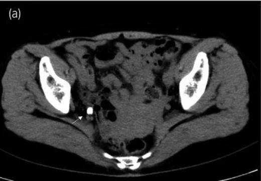
- Üreter lümenindeki taş, çevresindeki yumuşak dokudan belirgin yüksek atenüasyonlu odak olarak görünür; aksiyel görüntüde yerini bulup koronal planda uzun eksen boyutunu ölç. Üreter taşı araştırmasında kontrast verilmez; proksimal kaliks-pelvis ve üreter genişlemesi, çevre yağ dokusu çizgilenmesi de aynı seride aranır.
Önerilen pencereler: Batın (G 400 / M 50), Kemik (G 1800 / M 400).
BT bulguları
- Üreter lümeninde, böbrek pelvisinden üreterovezikal bileşkeye uzanan seyir üzerinde hiperdens kalkül; vertebra ve iliak damarlarla ilişkisine göre düzeyi belirt.
- Taşın kraniokaudal uzun eksen ölçümü ve aynı tarafta ek renal ya da üreteral taşların sayımı.
- Yumuşak doku rim işareti — kalkül çevresinde ödemli üreter duvarına ait ince halka, pelvik flebolitten ayrımı destekler.
- Taşın proksimalinde hidroureter ve kalikslerde genişleme; distal üreterin görece ince kalması geçiş seviyesini işaret eder.
- Periüreteral ve perinefrik yağ çizgilenmesi, böbrek boyutunda asimetri veya toplayıcı sistem çevresinde ödem gibi ikincil tıkanma bulguları.
- Üreter seyri dışındaki pelvik kalsifikasyon, özellikle damar boyunca uzanan kuyruk görünümüyle flebolit lehine olabilir.
Normalde
Taşın yerini ve boyutunu kontrastsız BT'de belirle, eşlik eden komplikasyonları da değerlendir.
Normal BT ile kıyasla
Aynı bölgenin, kaynak veri setinde patoloji içermediği belirtilen bir BT incelemesini kesit kesit kaydırın; organ sınırlarını açarak anatomiyi hasta görüntüsüyle karşılaştırın. Küçük rastlantısal bulgular tümüyle dışlanmamıştır.
Kaynak: TotalSegmentator v2.01 veri seti (Wasserthal ve ark., Radiology: Artificial Intelligence 2023), CC BY 4.0, vaka s0119; organ sınırları veri setinin otomatik segmentasyonundan, birleştirilerek sadeleştirildi. Görüntüler 2,5 mm kesit aralığına yeniden örneklendi.
Ayırıcı tanı
- Pelvik flebolit
- üreter dışı damar hattında yer alır; komet-kuyruk uzanımı flebolit lehine, proksimal üreter genişlemesi taş lehinedir.
- Üreter duvarı kalsifikasyonu
- lümen içinde ayrı kalkül yerine, üreter duvarında odak ya da diffüz biçimde izlenen duvar içi (intramural) kalsifikasyon görülür.
- Üst üriner sistem ürotelyal tümöründe BT ürografi, tanı ve evrelemede kullanılan görüntüleme yöntemidir.
- Papiller nekrozda papiller anormallikler, BT ürografisinin ekskretuar fazında değerlendirilebilir.
Tuzaklar
- Distal üreter taşları pelvik flebolitlerle karışabilir; rim ve comet-tail bulguları ayrımda yardımcı olsa da duyarlılıkları düşüktür ve ikincil tıkanma bulguları da değerlendirilmelidir.
- Yalnız rim işaretinin bulunmaması taşı dışlamaz; küçük taşlarda bu işaret silik olabilir ve ikincil bulgular daha yararlı olabilir.
- Taş geçtikten sonra kalan üreter genişlemesi ve yağ çizgilenmesi, görünür kalkül olmadan yakın dönem obstrüksiyonu düşündürebilir.
- Split-bolus dual-energy BT ürografide sanal kontrastsız ve postkontrast görüntülerin birlikte değerlendirilmesi, yalnız postkontrast görüntülere göre taş saptama duyarlılığını artırabilir; başlangıç kontrastsız BT belirsizse kontrastlı BT veya BT ürogram uygun olabilir.
Kendini dene
Kolik ağrılı hastanın distal üreter komşuluğunda kalsifikasyon var. Üstteki üreter geniş, odak ince yumuşak doku halkasıyla çevrili. En olası tanı nedir?
Cevabı göster
Üreter taşı. Lümen hattındaki kalsifik odağa eşlik eden proksimal dilatasyon ve rim işareti üreter taşını destekler. Flebolit genellikle üreter dışı venöz seyirdedir; tümör yumuşak doku lezyonudur, papiller nekroz ise kalsifik lümen taşı görünümü vermez.
Pelvik damar seyri üzerinde, üreter lümeni dışında bir kalsifikasyon var; aynı tarafta proksimal üreterde genişleme yok. En olası yorum hangisidir?
Cevabı göster
Pelvik flebolit. Üreter dışındaki venöz seyirde yerleşim ve üst sistemde tıkanma bulgusu olmaması fleboliti destekler. Taş lehine lümen içi konum ve proksimal dilatasyon aranır; belirsiz pelvik kalsifikasyon tek işaretle sınıflandırılmamalıdır.
İlgili konular
Kaynaklar
Bu sayfadaki 35 cümle ve 2 quiz sorusu aşağıdaki kaynaklardan alıntılarla tek tek karşılaştırıldı; 2 cümle bu karşılaştırmada düzeltildi (2026-10-09).
- Gastrointestinal perforation in anti-NXP2 antibody-associated juvenile dermatomyositis: case reports and a review of the literaturepmc.ncbi.nlm.nih.gov
- Ureteral calculus complicated by bladder malakoplakia: A case reportpmc.ncbi.nlm.nih.gov
- Residual Intravenous Contrast Mimicking a Ureteral Stent on Noncontrast Computed Tomography in a Patient With Ureterolithiasispmc.ncbi.nlm.nih.gov
- Adult primary obstructive megaureter revealed by a 2-cm ureterovesical junction stonepmc.ncbi.nlm.nih.gov
- A Pelvic Abscess and a Pelvic Stone Secondary to a Ureteral Calculuspmc.ncbi.nlm.nih.gov
- ACR Appropriateness Criteria® acute onset flank pain--suspicion of stone disease.pubmed.ncbi.nlm.nih.gov
- Incidental discovery of endourethral clip migration 10 years after radical prostatectomy: A case report and review of the literature.pubmed.ncbi.nlm.nih.gov
- ACR Appropriateness Criteria® Acute Onset Flank Pain-Suspicion of Stone Disease (Urolithiasis).pubmed.ncbi.nlm.nih.gov
- Calculus pyelonephritis associated with ureterocutaneostomy.pubmed.ncbi.nlm.nih.gov
- Recurrent nephrolithiasis associated with atazanavir use.pubmed.ncbi.nlm.nih.gov
- Double-J stent placement during laparoscopic ureterolithotomy: the "seagull" technique.pubmed.ncbi.nlm.nih.gov
- Spontaneous Large Urinoma Secondary to Obstructing Ureteral Calculus: A Kidney Pop-Off Mechanism.pubmed.ncbi.nlm.nih.gov
- Urinary stone size estimation: a new segmentation algorithm-based CT method.pubmed.ncbi.nlm.nih.gov
- Myopericytoma in urinary bladder: a case report.pubmed.ncbi.nlm.nih.gov
- Development and validation of a preoperative nomogram for predicting patients with impacted ureteral stone: a retrospective analysis.pubmed.ncbi.nlm.nih.gov
- Shockwave Lithotripsy Is an Efficacious Treatment Modality for Obese Patients with Upper Ureteral Calculi: Logistic Regression and Matched-Pair Analyses from a Dedicated Center Comparing Treatment Outcomes by Skin-to-Stone Distance.pubmed.ncbi.nlm.nih.gov
- Clinical effectiveness protocols for imaging in the management of ureteral calculous disease: AUA technology assessment.pubmed.ncbi.nlm.nih.gov
- Sonographic twinkling artifact for diagnosis of acute ureteral calculus.pubmed.ncbi.nlm.nih.gov
- Ultrasound-guided percutaneous antegrade pyelography for suspected ureteral obstruction in 6 pet guinea pigs ( Cavia porcellus ).pubmed.ncbi.nlm.nih.gov
- Management of pediatric stone disease.pubmed.ncbi.nlm.nih.gov
- Kidney Disease: Kidney Stones.pubmed.ncbi.nlm.nih.gov
- Symptomatic renal papillary varicosities and medullary nephrocalcinosis.pubmed.ncbi.nlm.nih.gov
- The natural history of asymptomatic calyceal stones.pubmed.ncbi.nlm.nih.gov
- Diagnostic Accuracy of Doppler Twinkling Artifact for Identifying Urinary Tract Calculi.pubmed.ncbi.nlm.nih.gov
- Side-by-side evaluation of virtual non-contrast and post-contrast images improves detection of clinically significant urolithiasis on single-phase split bolus dual-energy CT urography.pubmed.ncbi.nlm.nih.gov
- Could spot urine analysis of calcium and uric acid help predict density of urinary stone in computerized tomography? A preliminary study.pubmed.ncbi.nlm.nih.gov
- Imaging evaluation of amyloidosis of the urinary tract and retroperitoneum.pubmed.ncbi.nlm.nih.gov
- Pseudodistal ureteral stone resulting from calcified Deflux implantation.pubmed.ncbi.nlm.nih.gov
- Differentiation of distal ureteral stones and pelvic phleboliths using a convolutional neural network.pubmed.ncbi.nlm.nih.gov
- EAU Guidelines on Diagnosis and Conservative Management of Urolithiasis.pubmed.ncbi.nlm.nih.gov
- Beyond ureterolithiasis: gamut of abnormalities affecting the ureter.pubmed.ncbi.nlm.nih.gov
- [Renal colic].pubmed.ncbi.nlm.nih.gov
- Missed Diagnosis of Clear Cell Renal Cell Carcinoma on Noncontrast Computed Tomography: The Potential Role of Artificial Intelligence in Reducing Radiological Diagnostic Error.pubmed.ncbi.nlm.nih.gov
- Calculi in Three of the Four Ureters in a Case of Bilateral Complete Duplication, Complicated by Pyonephrosis in One Obstructed Moiety-A Rare Case Report.pubmed.ncbi.nlm.nih.gov
- Nontraumatic unilateral ureteral entrapment at the sacroiliac joint: A rare cause of obstructive uropathy.pubmed.ncbi.nlm.nih.gov
- Role of postoperative renal ultrasound in identifying ureteral injury despite normal intraoperative jet flow.pubmed.ncbi.nlm.nih.gov
- Modified Nondismembered Pyeloplasty for Ureteropelvic Junction Obstruction Using Axial Advancement of Renal Pelvic Flaps: Technique and Clinical Outcomes.pubmed.ncbi.nlm.nih.gov
- Three-dimensional reconstruction by a computer-assisted surgery system for pediatric hydronephrosis caused by accessory renal artery compression: a single-center retrospective observational comparative case series.pubmed.ncbi.nlm.nih.gov
- Fibroepithelial Polyp of the Proximal Ureter Presenting as Chronic Hematuria and Hydronephrosis in an Adolescent: A Case Report.pubmed.ncbi.nlm.nih.gov
- Eosinophilic vacuolated tumor of the kidney misdiagnosed as upper tract urothelial carcinoma: a case report and literature review.pubmed.ncbi.nlm.nih.gov
- Upper tract urothelial carcinoma mimicking emphysematous pyelonephritis: a diagnostic challenge case report.pubmed.ncbi.nlm.nih.gov
- Renal Papillary Necrosis Complicating Immune Checkpoint Inhibitor Therapy.pubmed.ncbi.nlm.nih.gov
- The Use of Prophylactic Ureteric Stents in Major Abdomino-Pelvic Sarcoma Surgery: Risks, Benefits, and Potential Complications.pubmed.ncbi.nlm.nih.gov
- A Rare Case of Emphysematous Pyelonephritis in a Non-diabetic Patient With Staghorn Calculus.pubmed.ncbi.nlm.nih.gov
- Risk factors and nomogram for predicting urinary tract infection in patients with ureterolithiasis complicated with hydronephrosis.pubmed.ncbi.nlm.nih.gov
- Is This Your Stone? Distinguishing Phleboliths and Nephroliths on Imaging in the Emergency Department Setting.pubmed.ncbi.nlm.nih.gov
- Clinical study on ureteritis observed in contrast-enhanced computed tomography in the emergency department.pubmed.ncbi.nlm.nih.gov
- Imaging in Upper Tract Urothelial Carcinoma: A Review.pubmed.ncbi.nlm.nih.gov
- Ureteral Dilatation with No Apparent Cause on Intravenous Urography: Normal or Abnormal? A Pilot Study.pubmed.ncbi.nlm.nih.gov
- Endoscopic combined intrarenal surgery for staghorn calculus in a horseshoe kidney with double collecting system: A case report.pubmed.ncbi.nlm.nih.gov
- Approach to the diagnosis of bilateral ureteral transitional cell carcinoma in a cat.pubmed.ncbi.nlm.nih.gov
- The Saudi urological association guidelines on urolithiasis.pubmed.ncbi.nlm.nih.gov
- Comparison between ultrasound and noncontrast helical computed tomography for identification of acute ureterolithiasis in a teaching hospital setting.pubmed.ncbi.nlm.nih.gov
- Goldenhar Syndrome Associated with Extensive Arterial Malformations.pubmed.ncbi.nlm.nih.gov
- MR Imaging of the Upper Urinary Tract: Techniques, Diagnostic Performance, and Imaging Biomarkers.pubmed.ncbi.nlm.nih.gov
- Pseudotumoral Bladder Schistosomiasis in a 16-Year-Old Adolescent: A Case Report.pubmed.ncbi.nlm.nih.gov
- Diagnostic Yield of Upper-Tract Imaging in Visible Haematuria: A Single-Centre Retrospective Clinical Audit.pubmed.ncbi.nlm.nih.gov
- CT Urography Findings of Upper Urinary Tract Carcinoma and Its Mimickers: A Pictorial Review.pubmed.ncbi.nlm.nih.gov
- Spilling the beans: an inside scoop on the imaging of renal parenchymal disease.pubmed.ncbi.nlm.nih.gov
- Conventional uroradiology with excretory urography: a forgotten art?pubmed.ncbi.nlm.nih.gov
- An overview of kidney stone imaging techniques.pubmed.ncbi.nlm.nih.gov
- Magnetic Resonance Imaging of Uterine Cervix: A Pictorial Essay.pubmed.ncbi.nlm.nih.gov
- Iatrogenic urinary tract injuries: etiology, diagnosis, and management.pubmed.ncbi.nlm.nih.gov
- CIRSE Standards of Practice on Nephrostomy and Ureteric Stent Placement and Exchange.pubmed.ncbi.nlm.nih.gov
- Three-dimensional image reconstruction technology in laparoscopic pyeloplasty for horseshoe kidney-associated ureteropelvic junction obstruction: a case series study.pubmed.ncbi.nlm.nih.gov
- MDCT-based Grading of Perirenal Changes Secondary to Acute Unilateral Upper Urinary Tract Obstruction.pubmed.ncbi.nlm.nih.gov
- Renal pelvic urothelial carcinoma with imaging features presenting as renal infarction 1 case report and literature review.pubmed.ncbi.nlm.nih.gov
- Complications after Nephron-sparing Interventions for Renal Tumors: Imaging Findings and Management.pubmed.ncbi.nlm.nih.gov
- Percutaneous nephrostomy: technical aspects and indications.pubmed.ncbi.nlm.nih.gov
- Urinary Ultrasound and Other Imaging for Ureteropelvic Junction Type Hydronephrosis (UPJHN).pubmed.ncbi.nlm.nih.gov
- Patterns of injury and violence in Yaoundé Cameroon: an analysis of hospital data.pubmed.ncbi.nlm.nih.gov
- What a difference a delay makes! CT urogram: a pictorial essay.pubmed.ncbi.nlm.nih.gov
- A Rare but Critical Presentation in Primary Care: A Case Report on Unilateral Ureterolithiasis Leading to Postrenal Acute Kidney Injury.pubmed.ncbi.nlm.nih.gov
- Assessment of anatomical outcomes in the upper urinary tract following flexible ureteroscopy with flexible and navigable suction ureteral access sheath: 1-year results from a multicentre study.pubmed.ncbi.nlm.nih.gov
- Ureteric squamous cell carcinoma in a duplex collecting system presenting with chronic obstructive uropathy: a rare case report.pubmed.ncbi.nlm.nih.gov
- Spontaneous Rupture of the Renal Pelvis Secondary to Upper Tract Urothelial Carcinoma.pubmed.ncbi.nlm.nih.gov
- Spectral CT findings of bladder urothelial carcinoma with gastric metastasis: a case report.pubmed.ncbi.nlm.nih.gov
- Investigating hematuria among young Indian adults using MDCT urography.pubmed.ncbi.nlm.nih.gov
- Pelvic Phlebolith: A Trivial Pursuit for the Urologist?pubmed.ncbi.nlm.nih.gov
- Low-Dose Unenhanced Computed Tomography with Iterative Reconstruction for Diagnosis of Ureter Stones.pubmed.ncbi.nlm.nih.gov
- Diagnostic performance and dose reduction of virtual noncontrast imaging in pediatric computed tomography urography.pubmed.ncbi.nlm.nih.gov
- Computed Tomography Urography: State of the Art and Beyond.pubmed.ncbi.nlm.nih.gov
- Dual-Energy Computed Tomography in Urological Diseases: A Narrative Review.pubmed.ncbi.nlm.nih.gov
Eğitim amaçlıdır; hasta başında tanı ve tedavi kararının yerine geçmez. İçerik yapay zekâ destekli bir süreçle güncel literatüre göre cümle cümle kaynaklarla karşılaştırıldı, ancak uzman radyolog incelemesinden geçmedi. Klinik kararlarda güncel kılavuzları, kurum protokollerini ve radyoloji raporunu esas alın.