
Özet
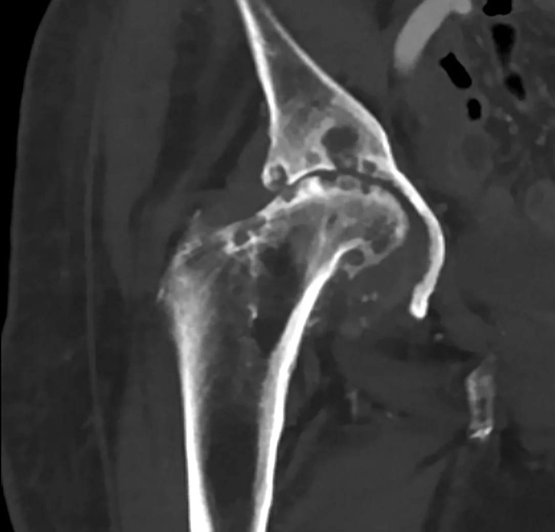
Kalça osteoartriti, eklem kıkırdağının kaybı ve buna eşlik eden subkondral kemik yeniden şekillenmesiyle ilerleyen dejeneratif eklem hastalığıdır; yaşla birlikte görülme olasılığı artar. BT, kalça eklemini çok düzlemli rekonstrüksiyonlarla ayrıntılı değerlendirir; yüksek çözünürlüklü kemik görüntülemesi osteofitleri, subkondral kistleri ve sklerozu ortaya koyar. Konvansiyonel BT eklem kıkırdağını doğrudan değerlendirmez; görüntüleme bulguları ağrı ve işlev kaybıyla iyi korelasyon göstermeyebilir, BT ise kalça eklemini çok düzlemli olarak ayrıntılı değerlendirebilir. BT ve radyografi, eklem aralığı genişliğinin yanı sıra osteofit oluşumu, subkondral kistler ve skleroz gibi dejeneratif kemik değişikliklerini de belirgin şekilde gösterir.
Faz ve pencere
- Kontrastsız tanısal
- Kontrastsız BT, eklem aralığı daralması, subkondral skleroz, kistik değişiklikler ve osteofit gibi dejeneratif kemik bulgularını yüksek çözünürlükle değerlendirmeyi sağlar. Dejeneratif kemik bulguları için kontrast gerekmez.
- Kemik algoritması tanısal
- Koronal ve sagittal kemik rekonstrüksiyonları eklem yüzü uyumunu, asetabular kenar osteofitlerini, femur başı osteofitlerini ve subkondral kistik boşlukların sklerotik kenarlarını gösterir; ince eklem aralığı değerlendirmesi kıkırdağı doğrudan göstermez.
Önerilen pencereler: Kemik (G 1800 / M 400).
BT bulguları
- Eklem aralığı daralması, kalça osteoartritinin yapısal görüntüleme özelliklerinden biridir.
- Asetabular osteofit — çatı kenarında marjinal kemik çıkıntısı oluşur.
- Femur başı osteofiti — kalça osteoartritinde osteofitler (eklem kenarından çıkan kemik çıkıntıları) temel dejeneratif kemik bulgularındandır ve BT bu çıkıntıları ayrıntılı gösterir.
- Subkondral skleroz — asetabular çatı ve femur başı eklem yüzlerinin altında kemik yoğunluğu artar.
- Subkondral kist — femur başı ya da asetabulumda sklerotik kenarlı, yuvarlak/oval kemik içi boşluklar görülür.
- Femur başı kontur kaybı — ileri kalça osteoartritinde femur başı ve asetabulumda deformite gelişir; bu bulgu radyografik evreleme sistemlerinde yer alır.
Ölçütler ve sınıflamalar
- Kellgren–Lawrence (kalça radyografisi)
- Kellgren–Lawrence sınıflaması; eklem aralığı daralması, osteofit, skleroz, kist ve femur başı–asetabulum deformitesinin derecesini yansıtan, 0-4 arasında derecelenen radyografik bir osteoartrit evreleme sistemidir. Bu radyografik evreleme BT'de doğrudan doğrulanmış ayrı bir eşik sistemi değildir.
Normalde
Normal kalça ile karşılaştırmada osteoartritin yapısal bulguları eklem aralığı daralması, osteofitler, subkondral kistler ve sklerozdur. Kalça BT'sinde eklem aralığı genişliği ile asetabular ve femoral osteofitler, subkondral skleroz ve kistler çok düzlemli olarak değerlendirilebilir.
Normal BT ile kıyasla
Aynı bölgenin, kaynak veri setinde patoloji içermediği belirtilen bir BT incelemesini kesit kesit kaydırın; organ sınırlarını açarak anatomiyi hasta görüntüsüyle karşılaştırın. Küçük rastlantısal bulgular tümüyle dışlanmamıştır.
Kaynak: TotalSegmentator v2.01 veri seti (Wasserthal ve ark., Radiology: Artificial Intelligence 2023), CC BY 4.0, vaka s1256; organ sınırları veri setinin otomatik segmentasyonundan, birleştirilerek sadeleştirildi. Görüntüler 2,5 mm kesit aralığına yeniden örneklendi.
Ayırıcı tanı
- Romatoid artrit
- inflamatuvar artritlerde agresif sinovit kalça ekleminde erozyon, eklem yıkımı ve protrusio yapabilir; bu tablo dejeneratif kemik bulgularından ayrışır.
- Septik artrit
- kısa sürede eklem yüzlerinde erozyon ve yıkım yapabilir; klinik enfeksiyon işaretleri ve çevre inflamasyon ayırıcıdır.
- Hızla yıkıcı kalça artropatisi
- eklem ve kemik kaybı seri görüntülerde hızlı ilerler, osteofitler beklenenden az olabilir.
- Femur başı osteonekrozu
- subkondral çökme ve baş deformitesi yapar; başlangıçta asetabular marjinal osteofit paterni baskın değildir.
- CPPD artropatisi
- kondrokalsinozis (eklem kıkırdağında kalsifikasyon) CPPD'nin başlıca radyolojik görüntüleme bulgusudur ve CPPD osteoartritle ilişkili olabilir.
Tuzaklar
- BT'de görülen eklem aralığı kıkırdak kalınlığının doğrudan ölçümü değildir; pozisyon ve kesit düzlemi alanın görünümünü değiştirir.
- Subkondral kistler tek başına OA kanıtı değildir; kenar sklerozu, osteofit ve eklem aralığı paterniyle birlikte yorumla.
- Vakum fenomeni — ileri dejeneratif disk ve faset eklem dejenerasyonuyla ilişkili gaz birikimidir ve radyografi ile MRI'da genellikle seçilmez.
- Kemik çıkıntıları asemptomatik morfolojik OA'yı gösterebilir; BT bulgularından ağrı şiddeti çıkarımı yapma.
Kendini dene
Kalça BT'sinde üst eklem aralığı daralmış; asetabular ve femoral osteofitler ile subkondral skleroz eşlik ediyor. En olası tanı nedir?
Cevabı göster
Kalça osteoartriti. Enfeksiyon ve romatolojik süreçler eklem yapısını değiştirebilir; osteonekroz ise femur başı çökmesiyle karakterizedir.
BT'de femur başı osteofitleri ve subkondral kistler görülüyor. Bu kemik bulgularından hangisi doğrudan çıkarılamaz?
Cevabı göster
Hastanın ağrı şiddeti. BT morfolojik değişiklikleri gösterir ancak yapısal OA şiddeti ile ağrı arasında bire bir ilişki yoktur. Osteofit, kist konumu ve kemik konturu görüntüde doğrudan değerlendirilebilir.
İlgili konular
Kaynaklar
Bu sayfadaki 39 cümle ve 2 quiz sorusu aşağıdaki kaynaklardan alıntılarla otomatik olarak karşılaştırıldı; 16 cümle bu karşılaştırmada düzeltildi (2026-10-09).
- The current state of computed tomography in osteoarthritis researchpmc.ncbi.nlm.nih.gov
- Advances in Imaging of Hip Osteoarthritis.pubmed.ncbi.nlm.nih.gov
- Novel cartilage imaging techniques for hip disorders.pubmed.ncbi.nlm.nih.gov
- Osteoarthritis of the hip: is radiography still needed?pubmed.ncbi.nlm.nih.gov
- Imaging of Osteoarthritis of the Hip.pubmed.ncbi.nlm.nih.gov
- Canine Hip Dysplasia: Diagnostic Imaging.pubmed.ncbi.nlm.nih.gov
- [Imaging in joint-preserving hip surgery].pubmed.ncbi.nlm.nih.gov
- Developmental Dysplasia of the Hip in Adolescents and Young Adults.pubmed.ncbi.nlm.nih.gov
- Standard and Advanced Imaging of Hip Osteoarthritis. What the Radiologist Should Know.pubmed.ncbi.nlm.nih.gov
- Surgical management of healed slipped capital femoral epiphysis.pubmed.ncbi.nlm.nih.gov
- MRI-based semiquantitative scoring of joint pathology in osteoarthritis.pubmed.ncbi.nlm.nih.gov
- The role of imaging in early hip OA.pubmed.ncbi.nlm.nih.gov
- Innovation in hip arthroscopy: is hip arthritis preventable in the athlete?pubmed.ncbi.nlm.nih.gov
- Current and Future Advanced Imaging Modalities for the Diagnosis of Early Osteoarthritis of the Hip.pubmed.ncbi.nlm.nih.gov
- Targeting subchondral osteoporosis in osteoarthritis: biomechanical crosstalk and the therapeutic rationale for inflammatory microenvironment remodeling.pubmed.ncbi.nlm.nih.gov
- Hip Osteoarthritis and the Human Microbiome: Current Concepts, Biological Insights and Future Directions.pubmed.ncbi.nlm.nih.gov
- MicroRNAs as Key Regulators of Degeneration and Inflammation in Osteoarthritis: A Narrative Review.pubmed.ncbi.nlm.nih.gov
- Current Landscape of Short-T2 Imaging Techniques in the Musculoskeletal System: The Past, Present and Future.pubmed.ncbi.nlm.nih.gov
- Review of CNN-Based Approaches for Preprocessing, Segmentation and Classification of Knee Osteoarthritis.pubmed.ncbi.nlm.nih.gov
- Is Ultrasonography an Effective Method for Diagnosing Degenerative Changes in the Temporomandibular Joint?pubmed.ncbi.nlm.nih.gov
- Osteoarthritis: Epidemiology, Diagnosis, and Treatment.pubmed.ncbi.nlm.nih.gov
- Intramuscular Fat Infiltration and Knee Osteoarthritis: A Systematic Review and Meta-Analysis.pubmed.ncbi.nlm.nih.gov
- The current state of computed tomography in osteoarthritis research.pubmed.ncbi.nlm.nih.gov
- Association of supine apical vertebral rotation with three-dimensional deformity and global spinal alignment in adult degenerative scoliosis: a retrospective study.pubmed.ncbi.nlm.nih.gov
- Guide to lower extremity radiologic measurements: part 1 hip.pubmed.ncbi.nlm.nih.gov
- Guidelines for the Design and Conduct of Clinical Studies in Knee Articular Cartilage Repair: International Cartilage Repair Society Recommendations Based on Current Scientific Evidence and Standards of Clinical Care.pubmed.ncbi.nlm.nih.gov
- Glossary of terms for musculoskeletal radiology.pubmed.ncbi.nlm.nih.gov
- Weight-bearing computed tomography in knee pathologies: current evidence and future perspectives.pubmed.ncbi.nlm.nih.gov
- Unveiling the Hidden Links: Anatomical and Radiological Insights into Primary Hip Osteoarthritis.pubmed.ncbi.nlm.nih.gov
- Surgical treatment of varus unicompartmental knee osteoarthritis: indications, trends, and outcomes-a narrative review.pubmed.ncbi.nlm.nih.gov
- Long-Term Results of Medial Fixed-Bearing Unicompartmental Knee Arthroplasty with Miller-Galante Prosthesis: A Minimum 10-Year Follow-Up Study.pubmed.ncbi.nlm.nih.gov
- Case Report: Combined posterolateral and direct anterior approaches for total hip arthroplasty in osteonecrosis of the femoral head with a rare massive osteophyte with literature review.pubmed.ncbi.nlm.nih.gov
- Where is acetabular reaming concentrated during total hip arthroplasty for hip osteoarthritis? A computed tomography-based simulation analysis.pubmed.ncbi.nlm.nih.gov
- Hip Osteoarthritis Associated with Prominent Synovitis Revealed by <sup>68</sup>Ga-FAPI PET/CT.pubmed.ncbi.nlm.nih.gov
- Radiological pattern of hip osteoarthritis is associated with one-year patient-reported outcomes but not long-term mortality or revision after total hip arthroplasty : a retrospective case series.pubmed.ncbi.nlm.nih.gov
- Case Report of Rapidly Destructive Osteoarthritis of the Hip following Multiple High-dose Intra-articular Steroid Injections.pubmed.ncbi.nlm.nih.gov
- Adipose-derived mesenchymal stem cells for early hip osteoarthritis: mechanistic insights and possible clinical evidence.pubmed.ncbi.nlm.nih.gov
- Optimizing diagnosis and management of ONFH, FNSF, and SIF.pubmed.ncbi.nlm.nih.gov
- Intraosseous tumours of the knee.pubmed.ncbi.nlm.nih.gov
- Wrist-arthroscopy treatment of carpal bone cysts.pubmed.ncbi.nlm.nih.gov
- Chondroblastoma of the femoral head: curettage without dislocation.pubmed.ncbi.nlm.nih.gov
- Ultrasonographic Evaluation of Canine Hip Dysplasia: Comparison with FCI Radiographic Scoring System.pubmed.ncbi.nlm.nih.gov
- Legg-Calvé-Perthes disease- surgical treatment options.pubmed.ncbi.nlm.nih.gov
- Conservative Management of Late-Onset Legg-Calvé-Perthes Disease in a 10-Year-Old Child: A Case Report.pubmed.ncbi.nlm.nih.gov
- Total hip arthroplasty in patients with alkaptonuria: a case report.pubmed.ncbi.nlm.nih.gov
- Case Report: Ochronotic arthropathy mimicking spondyloarthritis: a case-based review of diagnostic pitfalls and a novel likely pathogenic HGD variant.pubmed.ncbi.nlm.nih.gov
- The evolving landscape of minimally invasive procedures in musculoskeletal diseases-Part II.pubmed.ncbi.nlm.nih.gov
- Hydrogels for Bone Repair: Construction Strategies and Applications.pubmed.ncbi.nlm.nih.gov
- Three-dimensional computed tomographic angular measurements of the canine tibia using a bone-centered coordinate system.pubmed.ncbi.nlm.nih.gov
- Radiographic Analysis of Lower Limb Alignment in Patients with Knee Osteoarthritis.pubmed.ncbi.nlm.nih.gov
- Characterizing symptom burden in knee osteoarthritis: the predominant role of Kellgren-Lawrence grade 4 in functional decline and pain severity.pubmed.ncbi.nlm.nih.gov
- The clinical utility of imaging in osteoarthritis and its importance in future prediction of total hip replacement; a nested case-control study within the AGES-Reykjavik cohort.pubmed.ncbi.nlm.nih.gov
- Magnetic resonance imaging of knee arthropathies: A pictorial review.pubmed.ncbi.nlm.nih.gov
- Treatment of Posterior Hip Dislocation due to Juvenile Idiopathic Arthritis. Report of Two Cases.pubmed.ncbi.nlm.nih.gov
- Idiopathic chondrolysis of hip in children: New proposal and implication for radiological staging.pubmed.ncbi.nlm.nih.gov
- Methicillin-Resistant Staphylococcus aureus Iliac Osteomyelitis with Cortical Breach Mimicking Septic Arthritis of the Hip in a Child: A Diagnostic Pitfall.pubmed.ncbi.nlm.nih.gov
- From Physicochemical Properties to Rehabilitation Outcomes: Understanding Corticosteroid Injection Adverse Effects.pubmed.ncbi.nlm.nih.gov
- Bilateral Knee Infectious Arthritis Caused by <i>Proteus mirabilis</i> Mimicking Reactive Arthritis in an Elderly Patient with Metastatic Cancer: A Case Report.pubmed.ncbi.nlm.nih.gov
- Characterization of pain-related mechanisms and bone destruction in mild and severe monoiodoacetate-induced hip arthropathy in rats.pubmed.ncbi.nlm.nih.gov
- Impact of Chronic Opioid Use in Recurrent Dislocations following Revision Total Hip Arthroplasty: An Iatrogenic Charcot-Like Arthropathy Case Report.pubmed.ncbi.nlm.nih.gov
- Beyond Cartilage Regeneration: Targeting the Subchondral Bone with Extracorporeal Shock Wave Therapy and Regenerative Medicine in Knee Osteoarthritis.pubmed.ncbi.nlm.nih.gov
- Artificial Intelligence Applications in MRI for the Diagnosis and Management of Osteonecrosis of the Femoral Head: A Comprehensive Review.pubmed.ncbi.nlm.nih.gov
- Crowned Dens Syndrome: A Case Report Describing Refractory Symptoms.pubmed.ncbi.nlm.nih.gov
- Concurrence of Calcium Pyrophosphate Deposition Disease in Patients Undergoing Surgery for Degenerative Spine Conditions: A Case Series and Systematic Review.pubmed.ncbi.nlm.nih.gov
- Zinc Deficiency in Older Adults Undergoing Rehabilitation With Calcium Pyrophosphate Deposition Disease: A Case Series.pubmed.ncbi.nlm.nih.gov
- Update of the articular representation of the TLEM 2.0 musculoskeletal model.pubmed.ncbi.nlm.nih.gov
- Repeatability of objective bone and joint measures in the knee using weight-bearing computed tomography: three-dimensional quantification of bone mineral density, joint space width, and subchondral bone plate thickness.pubmed.ncbi.nlm.nih.gov
- Accuracy of Computed Tomography in Diagnosing Temporomandibular Joint Osteoarthritis Relative to Histopathological Findings-An Ex Vivo Study of 41 Horses.pubmed.ncbi.nlm.nih.gov
- Can Deep Learning Methods Differentiate Temporomandibular Joint Disorders From Healthy Joints? A 3D Artificial Intelligence Algorithm Study Based on CBCT Images.pubmed.ncbi.nlm.nih.gov
- The evolving landscape of minimally invasive procedures in musculoskeletal diseases-part I.pubmed.ncbi.nlm.nih.gov
- Porous Ti-6Al-4V Architectures in Load-Bearing Orthopedic Reconstruction: A Critical Narrative Review of the Translational Gap Across Microstructure, Fatigue, Surface Function, and Clinical Failure Modes.pubmed.ncbi.nlm.nih.gov
- Quantitative Imaging of the Mandibular Condyle: Radiomics and Microarchitectural Biomarkers.pubmed.ncbi.nlm.nih.gov
- Multi-Detector Helical Computer Tomography, Transrectal Ultrasonography and Histology of the Lumbosacral Area: A Comparative Study in Warmblood Horse Cadavers.pubmed.ncbi.nlm.nih.gov
- Diagnostic Utility of CT-Based Acetabular Sector Angles in Pincer-Type Femoroacetabular Impingement: Limitations and Clinical Integration.pubmed.ncbi.nlm.nih.gov
- Radiographic evaluation of the painful adolescent and young adult hip.pubmed.ncbi.nlm.nih.gov
- Hip preservation.pubmed.ncbi.nlm.nih.gov
- Comprehensive Clinical Evaluation of Femoroacetabular Impingement: Part 2, Plain Radiography.pubmed.ncbi.nlm.nih.gov
- Glenohumeral cartilage lesions: reviewing diagnostic imaging strategies and preoperative evaluation.pubmed.ncbi.nlm.nih.gov
- Rapidly destructive hip osteoarthritis: a diagnosis not to miss.pubmed.ncbi.nlm.nih.gov
- Hip Arthroscopy With Triangular Drilling and Suction Decompression for Osteonecrosis of the Femoral Head: A Retrospective Study.pubmed.ncbi.nlm.nih.gov
- Development of osteoarthritis in adults with untreated hip dysplasia: a longitudinal observational study.pubmed.ncbi.nlm.nih.gov
- Posterior Acetabular Wall Fracture Mimicking a Subcapital Femoral Neck Fracture on Plain Radiographs: A Diagnostic Pitfall and the Role of Intraoperative Fluoroscopy: A Case Report.pubmed.ncbi.nlm.nih.gov
- Patterns of intra-discal vacuum phenomenon in adult degenerative scoliosis.pubmed.ncbi.nlm.nih.gov
- Minimally invasive prone transpsoas lateral lumbar interbody fusion using navigation.pubmed.ncbi.nlm.nih.gov
- Non-Infectious Spinal Diseases Mimicking Infection: A Pictorial Essay.pubmed.ncbi.nlm.nih.gov
- Rapidly progressive arthropathy identified on imaging of patients treated with Crizotinib for ALK-rearranged/ROS1-positive non small cell lung cancer: A retrospective single-center study.pubmed.ncbi.nlm.nih.gov
- Radiation Decreases Bone Density while Diabetes Increases Osteoarthritis in Non-Human Primates.pubmed.ncbi.nlm.nih.gov
- Non-neoplastic Orthopedic Pathology - A Systematic Review.pubmed.ncbi.nlm.nih.gov
- Musculoskeletal tumors and tumor-like lesions with "dark" signal intensity on T2-weighted MR images: A pictorial review.pubmed.ncbi.nlm.nih.gov
- The state of trial registrations in the field of Orthopaedics in years 2015-2020. A meta-epidemiological study.pubmed.ncbi.nlm.nih.gov
- Arthroplasties for hip fracture in adults.pubmed.ncbi.nlm.nih.gov
- Radiologic Mimics of Osteomyelitis and Septic Arthritis: A Pictorial Essay.pubmed.ncbi.nlm.nih.gov
- Axial Spondyloarthritis: Mimics and Pitfalls of Imaging Assessment.pubmed.ncbi.nlm.nih.gov
- Do Not Throw Out the Radiographs! Radiographic Disease Severity as a Prognostic Biomarker for the Onset of Frequent Knee Pain.pubmed.ncbi.nlm.nih.gov
- What is end-stage knee osteoarthritis? A scoping and narrative review.pubmed.ncbi.nlm.nih.gov
- Association between radial meniscal tears with and without posterior root involvement and subchondral insufficiency fracture of the knee on MRI: The mediating role of meniscal extrusion.pubmed.ncbi.nlm.nih.gov
- Deep learning-based multiclass classification for the diagnosis of hand arthritis on digital radiography: a multicenter study.pubmed.ncbi.nlm.nih.gov
- Central Sensitization and Nociplastic Pain: Shared Mechanisms in Fibromyalgia, Osteoarthritis, and Inflammatory Arthritis.pubmed.ncbi.nlm.nih.gov
- Imaging and management of calcium pyrophosphate deposition disease.pubmed.ncbi.nlm.nih.gov
- Crystal deposition disease of the shoulder at uncommon sites: Diagnostic challenges and nomenclature issues in the absence of synovial fluid analysis.pubmed.ncbi.nlm.nih.gov
- Association of Intra-articular Ozone/Prolozone Injections With Cutibacterium acnes Infection in a Shoulder With Pseudogout and No Prior Surgery: Management Principles and Literature Review.pubmed.ncbi.nlm.nih.gov
- Robot-Assisted Hip Arthroscopy for the Treatment of Femoroacetabular Impingement Syndrome Combined With Labral Injury: Surgical Technique.pubmed.ncbi.nlm.nih.gov
- Eight months of misdiagnosis: Intra-articular osteoid osteoma in a 48-year-old woman.pubmed.ncbi.nlm.nih.gov
- Osteocyte and vascular network in hip osteoarthritis: A multimodal imaging and histological analysis.pubmed.ncbi.nlm.nih.gov
- Super-acute massive osteolysis of the femoral head after acetabular fracture fixation : a comparative study with conventional post-traumatic osteonecrosis requiring total hip arthroplasty.pubmed.ncbi.nlm.nih.gov
- Musculoskeletal Manifestations of Alkaptonuria: A Case-Based Review of Literature.pubmed.ncbi.nlm.nih.gov
- Computed tomography of the equine temporohyoid joint: Association between imaging changes and potential risk factors.pubmed.ncbi.nlm.nih.gov
- Impaired Preoperative Spinopelvic Function Does Not Compromise the Patient-Reported Outcome at 2-Years After Total Hip Arthroplasty.pubmed.ncbi.nlm.nih.gov
- Diagnostics of Sacroiliac Joint Differentials to Axial Spondyloarthritis Changes by Magnetic Resonance Imaging.pubmed.ncbi.nlm.nih.gov
- Preoperative lower radiographic osteoarthritis severity in primary total knee arthroplasty increases revision risk by tenfold compared to severe osteoarthritis.pubmed.ncbi.nlm.nih.gov
- Imaging findings of intra-articular tumor/tumor-like lesions based on pathologic correlation.pubmed.ncbi.nlm.nih.gov
Eğitim amaçlıdır; hasta başında tanı ve tedavi kararının yerine geçmez, tıbbi tavsiye değildir. Metinler yapay zekâ desteğiyle yazıldı ve otomatik bir adımda kaynak alıntılarıyla karşılaştırıldı; bu bir tıbbi doğrulama değildir ve içerik uzman radyolog incelemesinden geçmedi. Klinik kararlarda güncel kılavuzları, kurum protokollerini ve radyoloji raporunu esas alın.