Özet
Kronik böbrek hastalığına bağlı mineral-kemik bozukluğunda sekonder hiperparatiroidi kemik rezorpsiyonuna, osteomalazi ise yetersiz mineralizasyona yol açabilen farklı mekanizmalardır. BT kortikal bütünlüğü, fokal litik lezyonu ve kırık hattını gösterir; tek başına altta yatan metabolik nedeni belirlemez. Karşı femur ve iskeletin diğer bölgelerindeki benzer bulgular, PTH ve mineral metabolizması laboratuvarlarıyla birlikte yorumlandığında olası paterni güçlendirir. Brown tümörü tek başına litik morfolojiyle metastazdan kesin ayırt edilemez; Looser bölgesi osteomalaziye eşlik edebilen bir psödofraktürdür.
Okuma sırası
- Femur medial korteksi. Femurun medial korteksinde kemiğe dik yönde geniş radyolüsent bant (Looser bölgesi) ve kortikal incelme varlığını değerlendir.
- Femur medullası. Medüller kemikte ekspansil, litik odak varsa sınırını ve korteksi ne kadar incelttiğini takip et.
- Medial femur korteksi. Medial femur korteksinde kemiğe dik yönde radyolüsent bant ve çevresinde minimal sklerotik kenar varlığını araştırmak gerekir.
- Femur ve karşı taraf. Femurda psödofraktür olup olmadığını kontrol et; saptarsan karşı femuru da incele.
Faz ve pencere
- Kontrastsız tanısal
- Kontrastsız BT’nin kemik penceresinde diffüz osteopeni, kortikal incelme, subperiostal veya intrakortikal rezorpsiyon, litik Brown tümörü ve medial kortikal Looser bölgesi araştırılır.
Önerilen pencereler: Kemik (G 1800 / M 400).
BT bulguları
- Hiperparatiroidide femur korteksi incelmiş, dış kortikal yüz boyunca subperiostal rezorpsiyon çentikleri gelişmiş olabilir.
- İntrakortikal rezorpsiyon, korteks içinde tünel veya oval lüsensi biçiminde görülebilir.
- Brown tümörü, uzun kemik medullasında ekspansil litik odak ve kortikal incelme oluşturabilir.
- Osteomalazide Looser bölgesi medial femurda kortekse dik transvers radyolüsensi şeklinde görülebilir ve kemiğin yalnızca bir kısmından geçebilir.
- Yetersiz mineralizasyon genel osteopeni ve trabeküler yapının belirsizleşmesiyle birlikte görülebilir.
- Kemik zayıflığında femurun medial korteksi boyunca transvers Looser bölgeleri ve subkondral yetersizlik kırıkları aranmalıdır.
Normalde
Proksimal femurde primer bası trabekülleri dikey, primer gerilme trabekülleri daha yatay, sekonder bası trabekülleri ise oblik yönlenir. Psödofraktür saptandığında karşı femur da incelenmelidir.
Normal BT ile kıyasla
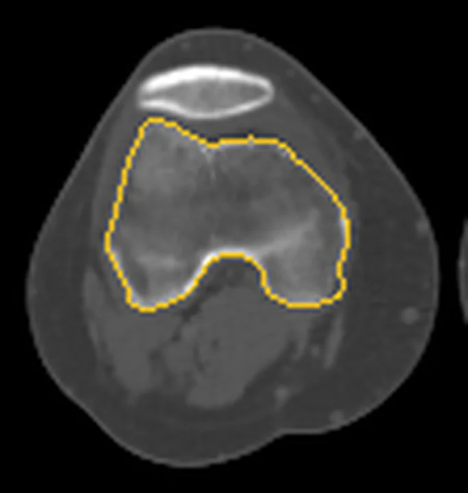
Aynı bölgenin, kaynak veri setinde patoloji içermediği belirtilen bir BT incelemesini kesit kesit kaydırın; organ sınırlarını açarak anatomiyi hasta görüntüsüyle karşılaştırın. Küçük rastlantısal bulgular tümüyle dışlanmamıştır.
Kaynak: TotalSegmentator v2.01 veri seti (Wasserthal ve ark., Radiology: Artificial Intelligence 2023), CC BY 4.0, vaka s0685; organ sınırları veri setinin otomatik segmentasyonundan, birleştirilerek sadeleştirildi. Görüntüler 2,5 mm kesit aralığına yeniden örneklendi.
Ayırıcı tanı
- Metastaz
- medüller osteoliz yapabilir; Brown tümörüyle görüntüleme bulguları örtüşebildiğinden klinik, biyokimya ve iskelet dağılımıyla birlikte değerlendirilir.
- Multipl miyelom
- birden fazla osteolitik odak yapabilir; eşlik eden Looser bölgesi osteomalaziyi düşündürür ancak miyelomu tek başına dışlamaz.
- Osteoporotik yetersizlik kırığı
- trabeküler kırık hattı veya impaksiyon görülebilir; Looser bölgesi ise medial kortekste kemiğin uzun eksenine dik psödofraktür bandıdır.
- Paget hastalığı
- kemik genişlemesiyle birlikte kortikal ve trabeküler kalınlaşma, mikst litik-sklerotik yeniden yapılanma gösterebilir; hiperparatiroidide kortikal rezorpsiyon, incelme ve tünelleşme görülebilir.
Tuzaklar
- Tek bir litik femur odağını biyokimyasal bağlam olmadan brown tümör diye etiketleme; hiperparatiroidi ve diğer iskelet bulguları ile korelasyon gerekir.
- Medial korteksteki Looser bölgesini lateral korteksten başlayan atipik femur kırığıyla karıştırma; yerleşim ve eşlik eden osteomalazi bulguları ayırıcı tanıya katkı verir.
- Korteks içindeki tünel benzeri rezorpsiyonları küçük tümör odağı sanma; çevre korteks boyunca benzer rezorpsiyon alanlarını ve karşı femuru incele.
Kendini dene
Kronik böbrek hastalığı olan kişide ekspansil litik femur odağı, kortikal incelme ve başka kemik rezorpsiyonları var. En olası lezyon nedir?
Cevabı göster
Brown tümörü. Kronik böbrek hastalığına eşlik eden sekonder hiperparatiroidi ve ekspansil litik odak Brown tümörünü düşündürür; ancak görüntüleme tek başına tanıyı kesinleştirmez. Brown tümörün görüntüleme ayırıcı tanısında multipl miyelom ve metastaz gibi diğer litik kemik lezyonları da yer alır.
Osteomalazide femurda medial kortekse dik, kemiğin yalnız bir bölümünü kat eden radyolüsent bant izleniyor. En uygun adlandırma hangisidir?
Cevabı göster
Looser zonu. Medial femurda kortekse dik, kemiğin yalnız bir bölümünü kat eden radyolüsent bant Looser bölgesi morfolojisiyle uyumludur. Paget çatlağı kemiğin konveks yüzünde görülür; atipik femur kırığı tipik olarak diyafizin lateral korteksinden başlar. Osteoid osteom ise kısa bant değil, fokal niduslu yuvarlak veya oval lezyondur.
Kaynaklar
Bu sayfadaki 34 cümle ve 2 quiz sorusu aşağıdaki kaynaklardan alıntılarla otomatik olarak karşılaştırıldı; 11 cümle bu karşılaştırmada düzeltildi (2026-10-09).
- Osteomalacia Is Not a Single Diseasepmc.ncbi.nlm.nih.gov
- Atraumatic fractures of the femurpmc.ncbi.nlm.nih.gov
- Osseous Manifestations of Primary Hyperparathyroidism: Imaging Findingspmc.ncbi.nlm.nih.gov
- Osteoporosis and Chronic Kidney Disease-Mineral and Bone Disorder (CKD-MBD): Back to Basics.pubmed.ncbi.nlm.nih.gov
- Osteo-renal cross-talk and phosphate metabolism by the FGF23-Klotho system.pubmed.ncbi.nlm.nih.gov
- Osteomalacia and Renal Osteodystrophy.pubmed.ncbi.nlm.nih.gov
- Osteoporosis and adynamic bone in chronic kidney disease.pubmed.ncbi.nlm.nih.gov
- Bone microarchitecture in patients undergoing parathyroidectomy for management of secondary hyperparathyroidism.pubmed.ncbi.nlm.nih.gov
- The Non-invasive Diagnosis of Bone Disorders in CKD.pubmed.ncbi.nlm.nih.gov
- Multiple Brown tumors in a Case of Primary Hyperparathyroidism with Pathological Fracture in Femur.pubmed.ncbi.nlm.nih.gov
- Brown Tumor as an Index Presentation of Severe Vitamin D Deficiency in a Teenage Girl.pubmed.ncbi.nlm.nih.gov
- Occult renal cell carcinoma with acrometastasis and ipsilateral juxta-articular knee lesions mimicking acute inflammation.pubmed.ncbi.nlm.nih.gov
- Proximal Femoral Fractures: What the Orthopedic Surgeon Wants to Know.pubmed.ncbi.nlm.nih.gov
- Peri-implant atypical femoral fracture after nail or plate osteosynthesis.pubmed.ncbi.nlm.nih.gov
- Case series of patients with pathological dyaphiseal fractures from metastatic bone disease.pubmed.ncbi.nlm.nih.gov
- Proximal femoral density distribution and structure in relation to age and hip fracture risk in women.pubmed.ncbi.nlm.nih.gov
- Longitudinal Effects of Single Hindlimb Radiation Therapy on Bone Strength and Morphology at Local and Contralateral Sites.pubmed.ncbi.nlm.nih.gov
- Challenges in the treatment of parathyroid carcinoma: a case report.pubmed.ncbi.nlm.nih.gov
- Case report: Intraosseous hibernoma (IOH) mimics osseous metastasis: another rare pitfall in FDG-PET-CT.pubmed.ncbi.nlm.nih.gov
- Giant Cell Lesions Associated with Primary Hyperparathyroidism.pubmed.ncbi.nlm.nih.gov
- Primary Hyperparathyroidism With Brown Tumor-A Case Mistaken as Non-Ossifying Fibroma Resolved by Biochemical and Radiological Correlation.pubmed.ncbi.nlm.nih.gov
- Oro-mandibular manifestations of primary hyperparathyroidism.pubmed.ncbi.nlm.nih.gov
- Atypical femoral fracture as the cause of greater trochanteric pain syndrome - a case report.pubmed.ncbi.nlm.nih.gov
- The effects of dual plating on the vascularity of the distal femur.pubmed.ncbi.nlm.nih.gov
- Bone histomorphometry and indicators of bone and mineral metabolism in wait-listed dialysis patients.pubmed.ncbi.nlm.nih.gov
- Prevalence of radiological findings among cases of severe secondary hyperparathyroidism.pubmed.ncbi.nlm.nih.gov
- Brown Tumor With Spine Involvement at Multiple Levels in a Hemodialysis Patient.pubmed.ncbi.nlm.nih.gov
- Multisystem Imaging Manifestations of Kidney Failure.pubmed.ncbi.nlm.nih.gov
- Primary hyperparathyroidism presenting with sudden onset paraplegia.pubmed.ncbi.nlm.nih.gov
- Brown tumour mimicking skeletal metastasis.pubmed.ncbi.nlm.nih.gov
- Osteoid osteoma - radiofrequency ablation treatment guided by computed tomography: a case series.pubmed.ncbi.nlm.nih.gov
- Intractable Scapular Pain Due to Undiagnosed Osteoid Osteoma: A Case Report.pubmed.ncbi.nlm.nih.gov
- Percutaneous Radiofrequency Ablation for the Treatment of Osteoid Osteoma in Children and Adults: A Comparative Analysis in 92 Patients.pubmed.ncbi.nlm.nih.gov
- Melatonin Accelerates Osteoporotic Bone Defect Repair by Promoting Osteogenesis-Angiogenesis Coupling.pubmed.ncbi.nlm.nih.gov
- OTUD1 inhibits osteoclast differentiation and osteoclastic bone loss through deubiquitinating and stabilizing PRDX1.pubmed.ncbi.nlm.nih.gov
- Genotype-phenotype relationship and comparison between eastern and western patients with osteogenesis imperfecta.pubmed.ncbi.nlm.nih.gov
- Endocrine-related osteoporosis: bone quality, clinical phenotype, and targeted management.pubmed.ncbi.nlm.nih.gov
- Phosphate: An Anion Controlling Metabolic Functions.pubmed.ncbi.nlm.nih.gov
- Endocrine-related osteoporosis: the state of the art.pubmed.ncbi.nlm.nih.gov
- Sequential stress fractures of the ulna and radius in a patient with neurofibromatosis type 1: A case report.pubmed.ncbi.nlm.nih.gov
- Brown tumors presenting with pathological fractures: case series focused on metabolic management.pubmed.ncbi.nlm.nih.gov
- Pathological fracture of primary lymphoma of bone: A case report on diagnosis and novel surgical management.pubmed.ncbi.nlm.nih.gov
- International Harmonization of Nomenclature and Diagnostic Criteria (INHAND): Non-proliferative and Proliferative Lesions of the Non-human Primate ( M. fascicularis ).pubmed.ncbi.nlm.nih.gov
- Patterns of injury and violence in Yaoundé Cameroon: an analysis of hospital data.pubmed.ncbi.nlm.nih.gov
- A Modern-Day Captain Charles Martell: Catastrophic Skeletal Manifestations of Primary Hyperparathyroidism.pubmed.ncbi.nlm.nih.gov
- Osteitis fibrosa cystica recovery following parathyroidectomy for primary hyperparathyroidism: a case series and review of literature.pubmed.ncbi.nlm.nih.gov
- Surgically Treated Benign Bone Tumors and Tumor-like Conditions in the Pediatric Population-A 10-Year Institutional Experience.pubmed.ncbi.nlm.nih.gov
- Bone in Parathyroid Diseases Revisited: Evidence From Epidemiological, Surgical and New Drug Outcomes.pubmed.ncbi.nlm.nih.gov
- Solitary phalangeal brown tumour in primary hyperparathyroidism: Report of a rare presentation.pubmed.ncbi.nlm.nih.gov
- Giant cell reparative granuloma of the calcaneus in a 32-year-old man without recurrence at 7-year follow-up: a case report.pubmed.ncbi.nlm.nih.gov
- Brown's Tumor Presenting as a Pathological Humerus Fracture: A Rare Diagnostic Pitfall.pubmed.ncbi.nlm.nih.gov
- Proximal Tibial Giant Cell Tumor in a Skeletally Immature Child: A Diagnostic Dilemma and Review of the Literature.pubmed.ncbi.nlm.nih.gov
- Bone disease in kidney transplant: don't forget about osteomalacia: a case report and literature review.pubmed.ncbi.nlm.nih.gov
- Metabolic Bone Disease in Captive Flying Foxes: A Conceptual Framework and Future Perspectives.pubmed.ncbi.nlm.nih.gov
- Approach to a Child with Hypophosphatemia.pubmed.ncbi.nlm.nih.gov
- Asian Federation of Osteoporosis Societies 2025 consensus on atypical femoral fractures in patients with osteoporosis.pubmed.ncbi.nlm.nih.gov
- Comparing incomplete atypical femur fractures in patients with or without bisphosphonate treatment: radiography and bone morphology in a retrospective study of 19 cases.pubmed.ncbi.nlm.nih.gov
- Management of postmenopausal osteoporosis.pubmed.ncbi.nlm.nih.gov
- Patient-specific osteoarticular reconstruction of the proximal tibia for giant cell tumor using a 3D-printed trabecular implant and hinged knee arthroplasty: a case report.pubmed.ncbi.nlm.nih.gov
- New Insight in Pediatric Orthopedic Oncology: The Use of a Xeno-Hybrid Bone Substitute in Loss of Bone Tissue After Oncological Resections, a Case Series.pubmed.ncbi.nlm.nih.gov
- Extraosseous osteochondroma of the hypothenar region: a case report and literature review.pubmed.ncbi.nlm.nih.gov
- Intraosseous tumours of the knee.pubmed.ncbi.nlm.nih.gov
- Dialysis-related amyloidosis mimicking metastatic bone disease: a scintigraphic pitfall of non-MDP-avid osteolytic lesions on bone SPECT/CT.pubmed.ncbi.nlm.nih.gov
- Hungry bone syndrome-like state following denosumab in a patient with multiple myeloma.pubmed.ncbi.nlm.nih.gov
- Metachronous multicentric giant cell tumor of bone treated with denosumab and mega-prosthesis revision: a rare case report.pubmed.ncbi.nlm.nih.gov
- Glossary of terms for musculoskeletal radiology.pubmed.ncbi.nlm.nih.gov
- Postmenopausal Osteoporosis: From Molecular Pathways to Therapeutic Targets-A Mechanism-to-Practice Framework Integrating Pharmacotherapy, Fall Prevention, and Adherence into Patient-Centered Care.pubmed.ncbi.nlm.nih.gov
- Review of imaging modalities and radiological findings of calvarial lesions.pubmed.ncbi.nlm.nih.gov
- Craniofacial disorders and dysplasias: Molecular, clinical, and management perspectives.pubmed.ncbi.nlm.nih.gov
- Brown tumor mimicking giant cell tumor in primary hyperparathyroidism: a case report.pubmed.ncbi.nlm.nih.gov
- Osseous Manifestations of Primary Hyperparathyroidism: Imaging Findings.pubmed.ncbi.nlm.nih.gov
- Nonproliferative and Proliferative Lesions of the Rat and Mouse Skeletal Tissues (Bones, Joints, and Teeth).pubmed.ncbi.nlm.nih.gov
- Prediction of incident hip fracture risk by femur geometry variables measured by hip structural analysis in the study of osteoporotic fractures.pubmed.ncbi.nlm.nih.gov
- Unveiling Primary Bone Tumors of the Spine: A Review of Essential Imaging Clues.pubmed.ncbi.nlm.nih.gov
- Check the Calcium first in any Bone Lesion: Brown Tumour of the Talus Misinterpreted as Giant Cell Tumour.pubmed.ncbi.nlm.nih.gov
- Radiological Assessment of Atypical Femoral Fractures: A 10-Year Experience Across Two Centres.pubmed.ncbi.nlm.nih.gov
- Bisphosphonate-Associated Atypical Femoral Fractures: A Description of Surgical Techniques in the Revision Fixation Setting With an Emphasis on Avoiding Varus Malalignment: A Two-Case Report.pubmed.ncbi.nlm.nih.gov
- Femoral neck-specific cortical thickness predicts internal fixation complications across adults with implications for elderly surgical decision-making.pubmed.ncbi.nlm.nih.gov
- Malignancy-mimicking osteitis fibrosa cystica secondary to primary hyperparathyroidism.pubmed.ncbi.nlm.nih.gov
- Brown tumours of a rib initially detected by ultrasound: a case report.pubmed.ncbi.nlm.nih.gov
- Multifocal osseous and cutaneous Rosai-Dorfman-Destombes disease presenting as an aggressive intramedullary femoral lesion.pubmed.ncbi.nlm.nih.gov
- Disorganized bone collagen fibers in early-stage type 2 diabetes mellitus and its association with adipose tissue redistribution.pubmed.ncbi.nlm.nih.gov
- Systemic lupus erythematosus complicated by refractory <i>Mycobacterium avium complex</i> infection involving bilateral lower extremities: a case report and literature review.pubmed.ncbi.nlm.nih.gov
- Conventional radiography for the assessment of focal bone lesions of the appendicular skeleton: fundamental concepts in the modern imaging era.pubmed.ncbi.nlm.nih.gov
- Benign incidental do-not-touch bone lesions.pubmed.ncbi.nlm.nih.gov
- Dedifferentiated Central Chondrosarcoma: A Clinical, Histopathological, and Immunohistochemical Analysis of 57 Cases.pubmed.ncbi.nlm.nih.gov
- Osteoid Osteoma, a Diagnostic Problem: A Series of Atypical and Mimicking Presentations and Review of the Recent Literature.pubmed.ncbi.nlm.nih.gov
- Positron emission tomography/computed tomography imaging appearance of benign and classic "do not touch" osseous lesions.pubmed.ncbi.nlm.nih.gov
- Lymphoma masquerading as Paget's disease of bone: a rare diagnostic challenge.pubmed.ncbi.nlm.nih.gov
- Dedifferentiation of Metastatic Recurrent Chondrosarcoma of the Right Distal Femur Into High-Grade Sarcoma Favouring Leiomyosarcoma: A Report of a Rare Case With Review of Literature.pubmed.ncbi.nlm.nih.gov
- HR-pQCT measures of bone in autosomal dominant osteopetrosis highly correlate with fractures and inversely correlate with bone resorption markers.pubmed.ncbi.nlm.nih.gov
- Musculoskeletal tumors and tumor-like lesions with "dark" signal intensity on T2-weighted MR images: A pictorial review.pubmed.ncbi.nlm.nih.gov
- Anatomy-driven automated subsegmentation of pelvic and proximal femoral CT into clinically relevant subregions and landmarks.pubmed.ncbi.nlm.nih.gov
- Single-baseline virtual progression modeling of Crowe type I-III developmental dysplasia of the hip: a finite element study of stress-location migration.pubmed.ncbi.nlm.nih.gov
- Finite Element Evaluation of Biomimetic Porous Ti6Al4V Implants for Femoral Reconstruction: Mechanical Performance of Mono-Block and Modular Designs.pubmed.ncbi.nlm.nih.gov
- Limb Salvage in Myxoid Liposarcoma With Tumor Prosthetic Reconstruction: A Case Report and Literature Review.pubmed.ncbi.nlm.nih.gov
- Delayed Diagnosis of an Occult Femoral Neck Fracture Undetected by MRI and Dual-Energy CT: A Case Report.pubmed.ncbi.nlm.nih.gov
- How do forelimb long bones adapt in rhinoceroses? An in-depth examination of their microanatomy.pubmed.ncbi.nlm.nih.gov
- Abnormal mechanical stress due to excessive femoral torsion is associated with dysplasia of the distal femoral epiphyseal plate and trochlea.pubmed.ncbi.nlm.nih.gov
Eğitim amaçlıdır; hasta başında tanı ve tedavi kararının yerine geçmez, tıbbi tavsiye değildir. Metinler yapay zekâ desteğiyle yazıldı ve otomatik bir adımda kaynak alıntılarıyla karşılaştırıldı; bu bir tıbbi doğrulama değildir ve içerik uzman radyolog incelemesinden geçmedi. Klinik kararlarda güncel kılavuzları, kurum protokollerini ve radyoloji raporunu esas alın.