Özet
SCFE, büyüme çağındaki çocuk ve ergenlerde proksimal femur fizisinin yetmezliğiyle epifizin metafize göre yer değiştirmesidir. Ağrının dizde hissedilmesi ve başlangıçta ince olan kayma tanıyı geciktirebilir; instabil kaymada femur başı kanlanması tehlikeye girebilir. Tanıda ilk tercih radyografidir; BT, belirgin kaymanın üç boyutlu yönünü, deformiteyi ve cerrahi planlamayı kemik ayrıntısıyla gösterir, erken kayma şüphesinde MR daha duyarlıdır. Kayma ve eşlik eden baş-boyun deformitesi fark edilmezse ilerleyici deformite, femoroasetabular sıkışma ve femur başı osteonekrozu gözden kaçabilir.
Faz ve pencere
- Kontrastsız tanısal
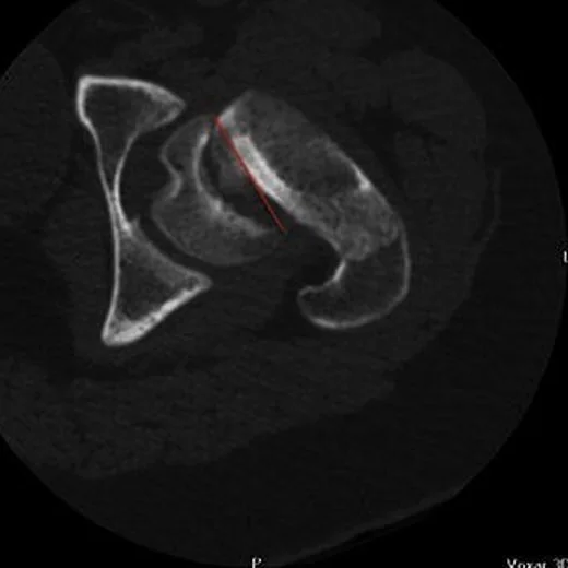
- Kontrast kullanılmadan epifiz-metafiz ilişkisi değerlendirilir: epifiz, metafize göre çoğunlukla posteromediale kaymış görünür ve fizis genişlemiş ya da düzensiz olabilir. Erken/pre-slip evrede BT bulgular sınırlı olabilir; bu aşamada tanı için en duyarlı yöntem manyetik rezonans görüntülemedir.
- Kemik algoritması tanısal
- Aksiyel oblik ve sagittal düzlemler BT'de fizis yer değiştirmesinin en belirgin olduğu düzlemlerdir; ameliyat öncesi BT kayma şiddetini değerlendirmeye ve fizis boyunca kırık hattı ya da oluşmuş kallus bulgularıyla instabiliteyi öngörmeye yardımcı olabilir.
Önerilen pencereler: Kemik (G 1800 / M 400).
BT bulguları
- İnce kaymalarda Klein çizgisi femur başını kesmeyebilir veya iki taraf arasında 2 mm'den fazla baş-boyun genişliği farkı görülebilir.
- Epifiz-metafiz basamaklanması — femur başı epifizinin metafize göre posterior ve medial konumu değişmiştir.
- Fizis boyunca kırık hattı — BT'de fizis boyunca uzanan kırık hattı, özellikle kallus yokluğunda intraoperatif instabiliteyi düşündürebilir.
- Metafizer çıkıntı — kronik kaymada metafizin anterosuperior kenarı belirginleşerek baş-boyun bileşkesini bozar.
- Femur boynu kısalığı ve varus — proksimal femur geometrisi karşı tarafa kıyasla değişmiştir.
- Femur başı kontur değişikliği — uzun süren deformite veya eklem içi temas femur başı sferisitesini bozabilir.
- Karşı taraf fizis değişikliği — eşzamanlı ya da gelişmekte olan karşı kalça kayması için aynı kemik düzlemlerinde taranır.
Ölçütler ve sınıflamalar
- Southwick kayma şiddeti (lateral grafide)
- hafif <30°; orta 30–50°; ağır >50°
- Southwick
- Frog-leg lateral grafide ölçülen Southwick açısı ile kaymanın şiddeti hafif (<30°), orta (30–50°) ve ağır (>50°) olarak sınıflandırılır. BT deformitenin uzaysal yönünü tamamlar.
- Loder stabilite sınıflaması
- Yürüme yeteneğine göre yapılır: koltuk değneğiyle veya değneksiz yük taşıyabiliyorsa stabil, hiç yük taşıyamıyorsa instabil kabul edilir.
Normalde
Normal kalçada femur başı epifizi proksimal metafizle fizis boyunca hizalıdır; fizis düzgün ve iki taraf arasında benzer genişliktedir. Aynı aksiyel oblik düzeyde epifiz merkezinin metafiz merkezine göre posterior-medial konumunu ve baş-boyun bileşkesindeki kemik çıkıntısını karşı tarafla kıyaslayın.
Normal BT ile kıyasla
Aynı bölgenin, kaynak veri setinde patoloji içermediği belirtilen bir BT incelemesini kesit kesit kaydırın; organ sınırlarını açarak anatomiyi hasta görüntüsüyle karşılaştırın. Küçük rastlantısal bulgular tümüyle dışlanmamıştır.
Kaynak: TotalSegmentator v2.01 veri seti (Wasserthal ve ark., Radiology: Artificial Intelligence 2023), CC BY 4.0, vaka s1256; organ sınırları veri setinin otomatik segmentasyonundan, birleştirilerek sadeleştirildi. Görüntüler 2,5 mm kesit aralığına yeniden örneklendi.
Ayırıcı tanı
- Femur boynu kırığı
- çocuklarda femur boynu kırığı ile SCFE ayırıcı tanıda birlikte düşünülmelidir.
- Geçici sinovitte görülebilen eklem sıvısı ve sinovit bulguları özgül değildir; SCFE şüphesinde fizis ve epifiz-metafiz ilişkisi de değerlendirilmelidir.
- Femur başı osteonekrozu
- femur başının kanlanmasının bozulmasıyla ilişkilidir ve ilerlediğinde başta çökme ile eklem dejenerasyonuna yol açabilir.
- Femoroasetabular sıkışma, cam tipi
- baş-boyun bileşkesindeki çıkıntı izole olabilir; epifiz-metafiz hizası korunur.
- Proksimal femur gelişimsel varyasyonu (primer cam morfolojisi)
- femur baş-boyun bileşkesindeki kemik veya kıkırdak çıkıntı başın sferik şeklini bozabilir ve asemptomatik erkek sporcularda iki taraflı görülebilir.
Tuzaklar
- Tek aksiyel kesitte epifizin posterior görünmesi fizis düzleminin oblikliğinden kaynaklanabilir; koronal ve sagittal rekonstrüksiyonlarda gerçek basamaklanmayı doğrula.
- Erken kayma BT'de belirgin kemik basamağı oluşturmadan önce başlayabilir; normal BT, klinik şüphede pre-slip hastalığı dışlamaz.
- Metafizer çıkıntıyı akut kayma sanma; düzgün kortikal yeniden şekillenme ve baş-boyun deformitesi kronikliği destekler.
- Southwick açısı lateral radyografilerde kayma şiddetini değerlendirmek için kullanılır; BT'de fizis yer değiştirmesi aksiyel oblik ve sagittal düzlemlerde daha belirgin görülür.
Kendini dene
Ergende kalça ağrısı var. BT'de femur başı epifizinin metafize göre posteriora ve mediale kaydığı görülüyor. En olası tanı nedir?
Cevabı göster
Femur başı epifiz kayması. SCFE, büyüme sırasında femur başı epifizinin posteriora ve mediale kaymasıyla tanımlanır.
BT'de belirgin kemik basamağı olmayan ancak fizis çevresinde erken hastalık şüphesi bulunan çocukta en duyarlı ek inceleme hangisidir?
Cevabı göster
Kalça MR. MR erken/pre-slip fizis ve kemik iliği değişikliklerini BT'den önce gösterebilir. Kontrastlı BT rutin erken tanı yöntemi değildir; diğer seçenekler fizis morfolojisini aynı duyarlılıkla değerlendirmez.
Kaynaklar
Bu sayfadaki 43 cümle ve 2 quiz sorusu aşağıdaki kaynaklardan alıntılarla otomatik olarak karşılaştırıldı; 5 cümle bu karşılaştırmada düzeltildi (2026-10-09).
- Comparison of Management Strategies and Associated Complications With Slipped Capital Femoral Epiphysispmc.ncbi.nlm.nih.gov
- Novelties in slipped capital femoral epiphysis imaging: A narrative reviewpmc.ncbi.nlm.nih.gov
- Angular and Rotational Lower Limb Alignment Variations in Children: Practical Guidance for Paediatricianspmc.ncbi.nlm.nih.gov
- A Review of Pediatric Orthopedic Disorders: Diagnosis and Treatment Updatespmc.ncbi.nlm.nih.gov
- Slipped capital femoral epiphysis with hypopituitarism in adultspmc.ncbi.nlm.nih.gov
- Hip preservation.pubmed.ncbi.nlm.nih.gov
- Hip Pain in Children.pubmed.ncbi.nlm.nih.gov
- Imaging SCFE: diagnosis, treatment and complications.pubmed.ncbi.nlm.nih.gov
- Applications of musculoskeletal ultrasonography in pediatric patients.pubmed.ncbi.nlm.nih.gov
- [Femoroacetabular impingement syndrome in adolescents-How to adivse? How to treat?].pubmed.ncbi.nlm.nih.gov
- Adolescent primary hyperparathyroidism.pubmed.ncbi.nlm.nih.gov
- Surgery for slipped capital femoral epiphysis in adolescents.pubmed.ncbi.nlm.nih.gov
- Surgical management of healed slipped capital femoral epiphysis.pubmed.ncbi.nlm.nih.gov
- [Slipped capital femoral epiphysis: clinical presentation, diagnostic procedure and classification].pubmed.ncbi.nlm.nih.gov
- Predicting epiphyseal stability of slipped capital femoral epiphysis with preoperative CT imaging.pubmed.ncbi.nlm.nih.gov
- Radiography, CT, and MRI of Hip and Lower Limb Disorders in Children and Adolescents.pubmed.ncbi.nlm.nih.gov
- Novelties in slipped capital femoral epiphysis imaging: A narrative review.pubmed.ncbi.nlm.nih.gov
- Increased body mass index percentile is associated with decreased epiphyseal tubercle size in asymptomatic children and adolescents with healthy hips.pubmed.ncbi.nlm.nih.gov
- Mechanical failure of single cannulated screw fixation of unstable slipped capital femoral epiphysis.pubmed.ncbi.nlm.nih.gov
- A 5-Year Outcome of Closed Reduction and Screw Fixation for an Atypical Slipped Capital Femoral Epiphysis Associated with Hypogonadism: A Case Report.pubmed.ncbi.nlm.nih.gov
- Direct anterior approach for open correction of Slipped Capital Femoral Epiphysiolysis: surgical technique and clinical outcome.pubmed.ncbi.nlm.nih.gov
- Identification and management of unrecognized femoral head epiphysiolysis: a case report.pubmed.ncbi.nlm.nih.gov
- Diagnosis of Slipped Capital Femoral Epiphysis: How to Stay out of Trouble?pubmed.ncbi.nlm.nih.gov
- A Retrospective Study of Risk Factors and Outcomes in the Surgical Management of Slipped Capital Femoral Epiphysis.pubmed.ncbi.nlm.nih.gov
- Delayed Slipped Capital Femoral Epiphysis Following Surgical Fixation of a Femoral Neck Fracture in a Child: A Rare Case Report.pubmed.ncbi.nlm.nih.gov
- Evaluation of the Reliability of Radiographic and MRI Angles in Superior Femoral Epiphysiolysis: A Comparative Study.pubmed.ncbi.nlm.nih.gov
- Imaging of pediatric musculoskeletal trauma: Age-specific considerations and challenges.pubmed.ncbi.nlm.nih.gov
- Sequential Traumatic and Non-traumatic Slipped Upper Femoral Epiphysis in a Young Girl: A Case Report and Review of Diagnostic Challenges.pubmed.ncbi.nlm.nih.gov
- The Unique Features of Pediatric Sports Injuries: The Hip.pubmed.ncbi.nlm.nih.gov
- Bilateral Total Hip Replacement for Slipped Capital Femoral Epiphysis in a Young Adult After Growth Hormone Therapy: A Case Report.pubmed.ncbi.nlm.nih.gov
- A Review of Pediatric Orthopedic Disorders: Diagnosis and Treatment Updates.pubmed.ncbi.nlm.nih.gov
- Ipsilateral Knee Pain After Total Hip Arthroplasty: Pathogenesis and Intervention Strategies-A Narrative Review.pubmed.ncbi.nlm.nih.gov
- Virtual Reality Simulation for Orthopedic Surgical Training: A Narrative Review of Current Evidence and Educational Impact.pubmed.ncbi.nlm.nih.gov
- Recent Advances in the Diagnosis and Management of Pediatric Orthopedic Disorders: A Comprehensive Review.pubmed.ncbi.nlm.nih.gov
- Total hip arthroplasty preoperative planning for childhood hip disorders' sequelae: Focus on developmental dysplasia of the hip.pubmed.ncbi.nlm.nih.gov
- What hip and spinopelvic characteristics are associated with ischiofemoral impingement?pubmed.ncbi.nlm.nih.gov
- Fast Field Echo Magnetic Resonance Imaging For Quantifying Acetabular Wall Coverage: A Validation Study With Computed Tomography.pubmed.ncbi.nlm.nih.gov
- Not All Cams Are Equal: MRI-Based Statistical Shape Modeling Reveals High Variability in Cam Shape in a Large FAI Cohort.pubmed.ncbi.nlm.nih.gov
- Evolving Understanding of and Treatment Approaches to Slipped Capital Femoral Epiphysis.pubmed.ncbi.nlm.nih.gov
- Combined Imhauser osteotomy and osteochondroplasty in slipped capital femoral epiphysis through surgical hip dislocation approach.pubmed.ncbi.nlm.nih.gov
- Indications and techniques of proximal femoral osteotomies in 2026.pubmed.ncbi.nlm.nih.gov
- The Etiology of Neuromuscular Hip Dysplasia and Implications for Management: A Narrative Review.pubmed.ncbi.nlm.nih.gov
- Do osteotomies of the proximal femur still have a role?pubmed.ncbi.nlm.nih.gov
- Association Between Body Weight and Clinical Characteristics of Slipped Capital Femoral Epiphysis in Switzerland: A 21-Year Single-Centre Retrospective Analysis (2005-2025).pubmed.ncbi.nlm.nih.gov
- Pain Sensitivity, Psychological Factors, and Muscle Function in Male Athletes With Long-Standing Groin Pain and Matched Controls.pubmed.ncbi.nlm.nih.gov
- Between Consecutive Fractures: Time and Sex as Dominant Factors in Type and Severity Concordance of Contralateral Hip Injuries.pubmed.ncbi.nlm.nih.gov
- Diagnostic Delay in Slipped Capital Femoral Epiphysis: A Single-Centre Study in Switzerland.pubmed.ncbi.nlm.nih.gov
- Bony adaptation signs are predictive of anterior head-neck offset remodeling after internal fixation for slipped capital femoral epiphysis: a multicenter study on 217 patients (228 hips) with follow-up until end of growth.pubmed.ncbi.nlm.nih.gov
- Invited Perspective: It is Time to Rename SCFE.pubmed.ncbi.nlm.nih.gov
- Angular and Rotational Lower Limb Alignment Variations in Children: Practical Guidance for Paediatricians.pubmed.ncbi.nlm.nih.gov
- Long-term outcomes of the modified Dunn procedure in moderate and severe slipped capital femoral epiphysis: a prospective case series with 7-year follow-up.pubmed.ncbi.nlm.nih.gov
- Immediate Versus Semi-Elective Treatment of Stable Slipped Capital Femoral Epiphyses (SCFE).pubmed.ncbi.nlm.nih.gov
- Favourable outcome of severe, unstable grade III slipped capital femoral epiphysis managed by closed reduction percutaneous pinning with mid-term follow up: A case report and literature review.pubmed.ncbi.nlm.nih.gov
- Application of biodegradable implants in pediatric orthopedics: shifting from absorbable polymers to biodegradable metals.pubmed.ncbi.nlm.nih.gov
- Precision stratified treatment of primary hyperparathyroidism based on multilevel emerging biomarkers.pubmed.ncbi.nlm.nih.gov
- Stop Searching under the Streetlight! A Primer and Practical Guide to the Diagnosis of Joint Pain and Inflammation.pubmed.ncbi.nlm.nih.gov
- Idiopathic chondrolysis of hip in children: New proposal and implication for radiological staging.pubmed.ncbi.nlm.nih.gov
- Magnetic resonance imaging of the hip: anatomy and pathology.pubmed.ncbi.nlm.nih.gov
- Hypoparathyroidism and Avascular Necrosis of the Hip Joint: A Case Report.pubmed.ncbi.nlm.nih.gov
- What is the prevalence of extra-articular and intra-articular magnetic resonance imaging findings in football players with and without hip and/or groin pain? A cross-sectional study of 166 football players.pubmed.ncbi.nlm.nih.gov
- [Pediatric Hip Disorders].pubmed.ncbi.nlm.nih.gov
- Ischiofemoral impingement in joint preserving hip surgery: prevalence and imaging predictors.pubmed.ncbi.nlm.nih.gov
- Hip Arthroscopy for Labral Tears and FAI is Associated with a High Rate of Return to Play for Professional American Football Athletes, A Literature Review.pubmed.ncbi.nlm.nih.gov
- Current Trends and Future Directions in the Diagnosis and Management of Pediatric Orthopedic Disorders.pubmed.ncbi.nlm.nih.gov
- The Swedish experience of capital realignment surgery for slipped capital femoral epiphysis: results from a nationwide retrospective cohort of 28 patients.pubmed.ncbi.nlm.nih.gov
- Remodelling After Percutaneous Pinning for Slipped Capital Femoral Epiphysis: The Influence of Transphyseal Screw Position.pubmed.ncbi.nlm.nih.gov
- Primary cam morphology; bump, burden or bog-standard? A concept analysis.pubmed.ncbi.nlm.nih.gov
- Comparison of Management Strategies and Associated Complications With Slipped Capital Femoral Epiphysis.pubmed.ncbi.nlm.nih.gov
- The Rising Popularity of Growth Hormone Therapy and Ensuing Orthopedic Complications in the Pediatric Population: A Review.pubmed.ncbi.nlm.nih.gov
- Slipped capital femoral epiphysis: a review of management in the hip impingement era.pubmed.ncbi.nlm.nih.gov
- Recognition and management of traumatic sports injuries in the skeletally immature athlete.pubmed.ncbi.nlm.nih.gov
- Improved Midterm Outcomes After Hip Arthroscopy for Femoroacetabular Impingement Syndrome in the Setting of a Previous Slipped Capital Femoral Epiphysis.pubmed.ncbi.nlm.nih.gov
- Treatment of Displaced Femoral Neck Fractures in Children: Mid-Term Outcomes and Predictors of Failure.pubmed.ncbi.nlm.nih.gov
- Finite element analysis of the proximal femoral growth plate biomechanics: insights into cam deformity risk of adolescent athletes.pubmed.ncbi.nlm.nih.gov
- Clinical Diagnosis of Slipped Capital Femoral Epiphysis in a Child With Negative Radiological Findings: A Case Report.pubmed.ncbi.nlm.nih.gov
- Imaging of Hip Pain: From Radiography to Cross-Sectional Imaging Techniques.pubmed.ncbi.nlm.nih.gov
- A Race against Time: Managing Acute Unstable Slipped Capital Femoral Epiphysis in a 15-year old with Normal Body Mass Index using the Modified Dunn's Procedure - A Case Report.pubmed.ncbi.nlm.nih.gov
- Review for the generalist: evaluation of pediatric hip pain.pubmed.ncbi.nlm.nih.gov
- The Genetic Epidemiology of Joint Shape and the Development of Osteoarthritis.pubmed.ncbi.nlm.nih.gov
- Comparison of different radiographic methods to measure the slip angle in children with slipped capital femoral epiphysis (SCFE).pubmed.ncbi.nlm.nih.gov
- Changing Treatment Philosophy of Slipped Capital Femoral Epiphysis (SCFE) after Introduction of the Modified Dunn Procedure (MDP): Our Experience with MDP and Its Complications.pubmed.ncbi.nlm.nih.gov
- Ischiopubic Osteochondrosis (Van Neck-Odelberg Disease) as a Rare Cause of Pediatric Hip Pain: A Systematic Review.pubmed.ncbi.nlm.nih.gov
- Double versus single fluoroscopy for screw fixation of slipped capital femoral epiphysis: does double fluoroscopy improve outcomes? A retrospective comparative study.pubmed.ncbi.nlm.nih.gov
- Outcomes by BMI Following Arthroscopic Management of Symptomatic Discoid Lateral Meniscus in Children and Adolescents: A Retrospective Cohort Study With a Minimum 5-Year Follow-Up.pubmed.ncbi.nlm.nih.gov
- Capital Femoral Epiphysis with Acute Unstable Valgus Type Slip Managed with Closed Reduction and Percutaneous Fixation: A Case Report.pubmed.ncbi.nlm.nih.gov
- Outcomes in Pediatric Hip FAI Surgery: a Scoping Review.pubmed.ncbi.nlm.nih.gov
- Corticosteroid-associated osteonecrosis in paediatric compared with adult patients: are they the same disease?pubmed.ncbi.nlm.nih.gov
- Intraosseous bone marrow concentrate delays total hip arthroplasty in osteoarthritis: A fifteen year matched cohort study with dose-response analysis.pubmed.ncbi.nlm.nih.gov
- Does the Capital Femoral Physis Bony MorphologyDiffer in Children with Symptomatic Cam-type Femoroacetabular Impingement.pubmed.ncbi.nlm.nih.gov
- The etiology of primary femoroacetabular impingement: genetics or acquired deformity?pubmed.ncbi.nlm.nih.gov
Eğitim amaçlıdır; hasta başında tanı ve tedavi kararının yerine geçmez, tıbbi tavsiye değildir. Metinler yapay zekâ desteğiyle yazıldı ve otomatik bir adımda kaynak alıntılarıyla karşılaştırıldı; bu bir tıbbi doğrulama değildir ve içerik uzman radyolog incelemesinden geçmedi. Klinik kararlarda güncel kılavuzları, kurum protokollerini ve radyoloji raporunu esas alın.