Özet
Yüksek enerjili künt travmada veya osteoporotik kemikte düşük enerjili düşme sonrasında sakrum, iki innominat kemik ve bunları birleştiren eklemler birlikte yaralanabilir. BT, özellikle direkt grafide örtüşen posterior halka kırıklarını ve yer değiştirmeyi göstererek yaralanma örüntüsünü tanımlar. Kırıkların halka boyunca tek tek sayılması, ikinci bir kopma noktasının ve mekanik instabilitenin gözden kaçmasını önler. Pelvik hematom, damar kanaması ve komşu mesane, üreter, bağırsak ya da organ yaralanması atlanırsa yaşamı tehdit eden eşlikçi hasar raporda görünmeyebilir.
Faz ve pencere
- Kontrastsız tanısal
- Kemik penceresinde pubik ramuslar, asetabulum komşuluğu, sakrum ve iliak kemiklerde kortikal kesinti, trabeküler impaksiyon ve kırık fragmanlarının konumu seçilir; damar kanaması bu fazda doğrulanamaz.
- Portal tanısal
- Kontrastlı travma incelemesinde pelvik/retroperitoneal hematom, serbest sıvı ve eşlik eden viseral yaralanmalar değerlendirilir; hematom içindeki kontrast odağı kanama şüphesi doğurur. Mesane rüptürü intravenöz kontrastlı portal fazdan tek başına dışlanamaz; gerektiğinde retrograd BT sistografi gerekir.
- Arteriyel tanısal
- Kontrastlı BT'de aktif arteriyel ekstravazasyon gözlenmesi, transkateter arteriyel embolizasyon için temel görüntüleme tetikleyicisidir.
- Kemik algoritması tanısal
- İnce kesit aksiyel görüntülerden koronal ve sagittal rekonstrüksiyonlar sakral kırık yönünü, foraminal uzanımı, sakroiliak eklem uyumunu ve halka boyunca ikinci kırılma noktasını daha iyi ortaya koyar.
Önerilen pencereler: Kemik (G 1800 / M 400), Yumuşak doku (G 400 / M 40), Anjiyo (G 600 / M 150).
BT bulguları
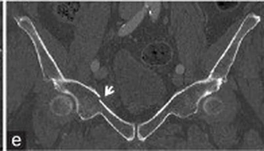
- Ön halka kırığı — en sık görülen ön halka yaralanması olan pubis kolu korteksinde süreksizlik; straddle örüntülerinde her iki kolun tutulması beklenir.
- Posterior halka kırığı — sakral ala, sakral foramen, sakral kanal veya iliak kanatta kırık; nöral foramen ve kanal uzanımı ayrıca gösterilir.
- Sakroiliak eklem bozulması — eklem aralığında asimetri, açılma, basamaklanma veya eklem yüzlerinde translasyon.
- Halka bütünlüğünün iki noktada kesilmesi — ön ramus hasarıyla birlikte sakrum/SI eklemi/iliak kemikte ikinci lezyon aranır; tek kırık hattı varsayılmaz.
- Hemipelvis rotasyonu ya da kraniyal kayması — obturator foramen ve iliak kanat konturları karşı tarafla kıyaslandığında yönlü deformasyon belirginleşir.
- Pelvik retroperitoneal hematom — presakral, perivezikal veya iliopsoas komşuluğunda ekstraperitoneal yumuşak doku dansitesi.
- Aktif kontrast ekstravazasyonu — hematom içinde damar dışı kontrast odağı arteriyel fazda belirginleşir ve gecikmiş/portal görüntüde yayılabilir.
- Eşlik eden yumuşak doku hasarı — mesane çevresi, bağırsak, üreter ve sakral sinir foraminaları travma bağlamında taranır.
Ölçütler ve sınıflamalar
- Young–Burgess
- LC: iç rotasyon ve kompresyon; APC: dış rotasyon/açılma; VS: kraniyal hemipelvis yer değiştirmesi; CM: birden fazla kuvvet örüntüsünün birlikteliği. LC-I sakral kompresyon, LC-II ipsilateral iliak crescent, LC-III karşı tarafta APC bileşeni; APC-I hafif simfizis açılması, APC-II anterior SI kompleks hasarı, APC-III anterior ve posterior SI kompleks ayrışmasıdır.
- Tile
- Tip A posterior kompleks korunmuş ve mekanik olarak stabil; Tip B rotasyonel instabil fakat vertikal stabil; Tip C hem rotasyonel hem vertikal instabildir.
Normalde
Normal pelvis BT’sinde pubik simfizis eklem yüzleri karşılıklı ve hizalı, simfizis aralığı düzenli; her iki sakroiliak eklem benzer genişlikte, sakral ala ve pubik ramus korteksleri kesintisiz izlenir. Aynı aksiyel ve koronal seviyelerde karşı taraf eklem aralığını, sakral korteksi ve obturator halka konturunu kıyaslayarak kırık, dönme veya basamaklanmayı ayırt edin.
Normal BT ile kıyasla
Aynı bölgenin, kaynak veri setinde patoloji içermediği belirtilen bir BT incelemesini kesit kesit kaydırın; organ sınırlarını açarak anatomiyi hasta görüntüsüyle karşılaştırın. Küçük rastlantısal bulgular tümüyle dışlanmamıştır.
Kaynak: TotalSegmentator v2.01 veri seti (Wasserthal ve ark., Radiology: Artificial Intelligence 2023), CC BY 4.0, vaka s1256; organ sınırları veri setinin otomatik segmentasyonundan, birleştirilerek sadeleştirildi. Görüntüler 2,5 mm kesit aralığına yeniden örneklendi.
Ayırıcı tanı
- İzole pubik ramus kırığı
- posterior halka, sakrum ve SI eklemlerinde eşlik eden lezyon yoktur; ince kesitlerde posterior halkayı özellikle kontrol edin.
- Asetabulum kırığı
- hat asetabular kolon veya duvarı izleyip kalça eklem yüzüne ulaşır; halka bağlantısındaki ikinci lezyonla karıştırmayın.
- Sakral yetersizlik kırığı
- çoğunlukla alar bölgede dikey sklerotik/kırık çizgileri ve düşük enerjili klinik bağlam görülür; akut deplase travma örüntüsü beklenmez.
- İliak kemik kontüzyonu
- BT'de belirsiz kalan travmatik ödem veya kırık çizgileri, MRG ve çift enerjili BT ile saptanan ek lezyonlara yol açabilir; sadece BT'ye güvenilirse ark halka yaralanmaları kaçırılabilir.
- Pelvik hematom veya kitle
- yumuşak doku dansitesi tek başına tanı koydurucu değildir; kontrastlı BT'de damar dışına çıkmış kontrast odağı arteriyel kaçağı gösterirken, kesintisiz korteks kırığı dışlar.
Tuzaklar
- Ön ramus kırığını tek başına raporlayıp posterior halkayı atlamak, stabiliteyi yanlış gösterir; sakrum ve iki SI eklemini koronal/sagittal rekonstrüksiyonlarda tamamlayın.
- Sakral foramendeki çizgiyi damar oluğu veya kısmi hacim etkisi sanabilirsiniz; komşu kesitlerde korteks sürekliliğini ve kemik algoritmasını izleyin.
- Aktif arteriyel ekstravazasyonun görülmemesi kanamanın olmadığını göstermez; pelvik kanamanın büyük kısmı venöz ve gubrenin yüzeylerinden kaynaklanır ve mekanik tamponaj gerektirir.
Kendini dene
Yüksek enerjili travmada pubik ramus kırığına aynı tarafta sakral ala impaksiyonu eşlik ediyor. En uygun örüntü hangisidir?
Cevabı göster
LC-I yaralanması. LC-I, ramus kırığıyla ipsilateral sakral kompresyon/impaksiyon örüntüsüdür. APC-II'de dış rotasyon ve anterior SI kompleks açılması; dikey makaslamada kraniyal hemipelvis kayması beklenir. Asetabular arka duvar kırığı kalça eklem yuvasını tutar.
Ramus kırığı görülen travma BT'sinde halka yaralanmasının tamamını göstermek için hangi bölge özellikle incelenmelidir?
Cevabı göster
Sakrum ve SI eklemleri. Pelvik halka ön ve arka bağlantıların ikisi birlikte değerlendirilerek tamamlanır; sakrum ve SI eklemi ikinci kopma noktasını gösterebilir. Diğer seçenekler halka sürekliliğini tamamlayan posterior bağlantılar değildir.
İlgili konular
Kaynaklar
Bu sayfadaki 45 cümle ve 2 quiz sorusu aşağıdaki kaynaklardan alıntılarla tek tek karşılaştırıldı; 6 cümle bu karşılaştırmada düzeltildi (2026-10-09).
- Haemorrhage control and definitive fixation strategies in unstable pelvic ring fractures: Integrating damage control and endovascular techniques A narrative reviewpmc.ncbi.nlm.nih.gov
- Evolution of the reduction technique for unstable pelvic ring fractures: a narrative reviewpmc.ncbi.nlm.nih.gov
- Early vs. late definitive fixation of pelvic ring fractures in resuscitated polytraumatized patients: a systematic review and meta-analysispmc.ncbi.nlm.nih.gov
- Artificial Intelligence in Pelvic Fracture Diagnosis and Outcome Prediction: A Systematic Review and Meta-analysispmc.ncbi.nlm.nih.gov
- Robotisch assistierte Schraubenplatzierung einer Kriechschraube und einer SI-S1-Schraubepmc.ncbi.nlm.nih.gov
- Osteoporotic Pelvic Fractures.pubmed.ncbi.nlm.nih.gov
- [Osteoporotic fractures of the pelvis].pubmed.ncbi.nlm.nih.gov
- Evaluation and Treatment of Blunt Pelvic Trauma.pubmed.ncbi.nlm.nih.gov
- [Pelvic Ring Fractures in the Elderly].pubmed.ncbi.nlm.nih.gov
- Methodology and interpretation of radiographic outcomes in surgically treated pelvic fractures: a systematic review.pubmed.ncbi.nlm.nih.gov
- [Radiological diagnosis of pelvic ring fractures].pubmed.ncbi.nlm.nih.gov
- [Current treatment of pelvic ring fractures].pubmed.ncbi.nlm.nih.gov
- Pelvic fractures: experience of pelvic ring fractures at a major trauma centre.pubmed.ncbi.nlm.nih.gov
- [Recommendations for Releasing the Pelvic Binder After a Non-Invasive Pelvic Stabilisation Procedure Under Emergency Room Conditions].pubmed.ncbi.nlm.nih.gov
- Imaging Review of Pelvic Ring Fractures and Its Complications in High-Energy Trauma.pubmed.ncbi.nlm.nih.gov
- [Pelvic ring fractures in the elderly. Underestimated osteoporotic fracture].pubmed.ncbi.nlm.nih.gov
- Hemorrhage in Pelvic Ring Fractures After Low-Energy Trauma: A Systematic Review.pubmed.ncbi.nlm.nih.gov
- Imaging of pelvic ring fractures in older adults and its clinical implications-a systematic review.pubmed.ncbi.nlm.nih.gov
- Intelligent robot-assisted minimally invasive reduction system for reduction of unstable pelvic fractures.pubmed.ncbi.nlm.nih.gov
- The 'nightstick' ischial fracture: a unique oddity of the pelvic injury family.pubmed.ncbi.nlm.nih.gov
- Long-term safety and effectiveness of the "OptEase" vena cava filter.pubmed.ncbi.nlm.nih.gov
- Clinical Outcomes of Sacral Alar-Iliac Screw Fixation for Pelvic Ring Fractures: A Retrospective Case Series.pubmed.ncbi.nlm.nih.gov
- Robot-assisted closed reduction and navigated fixation for pelvic ring injuries: a focused narrative review of a reduction-fixation-audit framework.pubmed.ncbi.nlm.nih.gov
- Functional Outcomes of Surgical Management of Posterior Wall Acetabular Fractures: A Prospective Study.pubmed.ncbi.nlm.nih.gov
- Transarterial embolization in musculoskeletal care: an indication-stratified narrative review.pubmed.ncbi.nlm.nih.gov
- Prognostic model and U-shaped SBP-risk correlation in unstable pelvic fractures with TBI.pubmed.ncbi.nlm.nih.gov
- Analysis of risk factors affecting the prognosis of external fixation in the treatment of unstable pelvic fractures in children: a retrospective study of 96 patients.pubmed.ncbi.nlm.nih.gov
- Guidance for clinical practice using emergency and point-of-care ultrasonography.pubmed.ncbi.nlm.nih.gov
- Evaluating the Effectiveness of Yttrium-90 Glass Microspheres in the Treatment of Hepatocellular Carcinoma, Intrahepatic Cholangiocarcinoma, and Metastatic Colorectal Cancer in Practice: Protocol for the Prospective PROACTIF Phase IV Registry Study in France.pubmed.ncbi.nlm.nih.gov
- Computed tomography of pediatric abdominal trauma: optimizing utilization and enhancing diagnostic interpretation.pubmed.ncbi.nlm.nih.gov
- Radiologic Imaging of Traumatic Bowel and Mesenteric Injuries: A Comprehensive Up-to-Date Review.pubmed.ncbi.nlm.nih.gov
- 2022 ACC/AHA guideline for the diagnosis and management of aortic disease: A report of the American Heart Association/American College of Cardiology Joint Committee on Clinical Practice Guidelines.pubmed.ncbi.nlm.nih.gov
- Clinical Characteristics and In-Hospital Outcomes of Traumatic Bladder Rupture: An 11-Year Retrospective Cohort Study at a Single Regional Trauma Center.pubmed.ncbi.nlm.nih.gov
- Acute and chronic complications of cesarean delivery: An update for the general radiologist.pubmed.ncbi.nlm.nih.gov
- Isolated Lymphatic Injury Mimicking Bladder Rupture in a Young Male Following Blunt Trauma: A Case Report.pubmed.ncbi.nlm.nih.gov
- Collateral-pathway-oriented clinico-angiographic framework for inferior epigastric artery-related hemorrhage: embolization planning and clinical outcomes.pubmed.ncbi.nlm.nih.gov
- Beyond Chordoma: A Comprehensive Review of Sacral Lesions.pubmed.ncbi.nlm.nih.gov
- Open and Percutaneous Fixation of Traumatic Sacral Fracture-Dislocation with Spinopelvic Dissociation: Two Adolescent Cases and a Systematic Literature Review.pubmed.ncbi.nlm.nih.gov
- CT of Sacral Fractures: Classification Systems and Management.pubmed.ncbi.nlm.nih.gov
- Staged management of a Tile C3 pelvic ring injury with bilateral sacroiliac disruption and T-shaped sacral fracture.pubmed.ncbi.nlm.nih.gov
- Fatal Injuries Caused by Falling Trees: Correlation Between Postmortem Computed Tomography and Forensic Autopsy Findings in Eight Cases.pubmed.ncbi.nlm.nih.gov
- Evaluation of cannulated compression headless screws as an alternative implant for superior pubic ramus fracture fixation: a biomechanical study.pubmed.ncbi.nlm.nih.gov
- Sacral Insufficiency Fractures : How to Classify?pubmed.ncbi.nlm.nih.gov
- Fragility fractures of the pelvis in the elderly: protocol for an international prospective multicentre observational cohort study.pubmed.ncbi.nlm.nih.gov
- Gorham-Stout disease with thoracic involvement: pathogenic mechanisms, respiratory complications, and multimodal therapies.pubmed.ncbi.nlm.nih.gov
- Decoding triple negative breast cancer bone metastasis: from 3D bioprinted models to clinical translation.pubmed.ncbi.nlm.nih.gov
- Special screw corridors and imaging in pelvic ring trauma.pubmed.ncbi.nlm.nih.gov
- Quadrilateral plate fractures.pubmed.ncbi.nlm.nih.gov
- Successful Management of a Delayed Traumatic Hemothorax by Timely Imaging Re-evaluation in a Hybrid Emergency Room System.pubmed.ncbi.nlm.nih.gov
- Incidental findings in female pelvis MRI performed for gynaecological malignancies.pubmed.ncbi.nlm.nih.gov
- Beyond the gut: spectrum of magnetic surgery devices.pubmed.ncbi.nlm.nih.gov
- Anatomy-driven automated subsegmentation of pelvic and proximal femoral CT into clinically relevant subregions and landmarks.pubmed.ncbi.nlm.nih.gov
- A novel internal fixation system for sacral fractures: a finite element biomechanical study.pubmed.ncbi.nlm.nih.gov
- Total Hip Arthroplasty for Post-Traumatic Hip Arthritis in Chronic Pubic Diastasis: A Case Report.pubmed.ncbi.nlm.nih.gov
- The Pelvic Bridge: A Subcutaneous Approach for Anterior Pelvic Fixation.pubmed.ncbi.nlm.nih.gov
- International Society of Paediatric Surgical Oncology (IPSO) Surgical Practice Guidelines.pubmed.ncbi.nlm.nih.gov
- Trunk training following stroke.pubmed.ncbi.nlm.nih.gov
- Epidemiology and injury patterns of high-energy pediatric pelvic and acetabular fractures: A retrospective study with comparative outcomes.pubmed.ncbi.nlm.nih.gov
- Safe definitive surgery in unstable pelvic ring injuries - a retrospective cohort study.pubmed.ncbi.nlm.nih.gov
- Minimally invasive circumferential fixation using TIFI and INFIX for unstable pelvic ring injuries: a prospective evaluation of clinical and radiological outcomes.pubmed.ncbi.nlm.nih.gov
- Surgical Management of a Lateral Compression (LC)III Pelvic Ring Fracture With Type I Crescent Fracture-Dislocation: A Case Report.pubmed.ncbi.nlm.nih.gov
- Rethinking LC-type in Young-Burgess classification of pelvic fracture: A retrospective study.pubmed.ncbi.nlm.nih.gov
- Surgical management of severely displaced IPRF: operative technique, indications, and preliminary clinical outcomes.pubmed.ncbi.nlm.nih.gov
- Minimally invasive percutaneous cannulated screw fixation for pelvic fractures: a retrospective case cohort study of clinical and radiological outcomes.pubmed.ncbi.nlm.nih.gov
- Clinical and Patient-Focused Outcomes After Percutaneous Screw Fixation of Pelvic Ring Fractures in Older Adults.pubmed.ncbi.nlm.nih.gov
- Chronic pelvic insufficiency fractures and their treatment.pubmed.ncbi.nlm.nih.gov
- Pelvic insufficiency fractures in the elderly - what insights has the past year of research brought? A narrative review.pubmed.ncbi.nlm.nih.gov
- Long-Term Outcome of Patients with a Floating Hip Injury of Müller Type A: An Analysis of Prognostic Factors Linked to Functional Outcomes.pubmed.ncbi.nlm.nih.gov
- Algorithms in acetabular fracture classifications.pubmed.ncbi.nlm.nih.gov
- Traumatic Bilateral Asymmetrical Hip Dislocation with Acetabular Fracture: A Case Report and Review of Current Evidence.pubmed.ncbi.nlm.nih.gov
- Safety and Early Clinical Outcomes of Sacroiliac Fusion for Sacral Fractures via a Modified S1 Corridor: An Exploratory Comparison of Triangular and Threaded 3D-Printed Titanium Implants.pubmed.ncbi.nlm.nih.gov
- Preoperative 3D-Planned S1 Corridors Transferred into 2D Fluoroscopy Allow for Safe Intraoperative Large-Diameter Implant Placement: Description of a Novel Sacroiliac Fixation Technique and Proof of Concept in 137 Implantations.pubmed.ncbi.nlm.nih.gov
- Sacral and Pelvic Fractures: Historical Systems and Advancements with the AO Spine Classification.pubmed.ncbi.nlm.nih.gov
- Integrated Clinical Workflow for Preoperative Planning and Resection of Giant Iliofemoral Heterotopic Ossification Using Three-Dimensional Technologies.pubmed.ncbi.nlm.nih.gov
- Current State-of-the-Art 3D MRI Sequences for Assessing Bone Morphology with Emphasis on Cranial and Spinal Imaging: A Narrative Review.pubmed.ncbi.nlm.nih.gov
- Clinical applications of portable CT scanners.pubmed.ncbi.nlm.nih.gov
- Contralateral screw loosening of through-and-through fixation following unilateral sacroiliac joint fusion: illustrative case.pubmed.ncbi.nlm.nih.gov
- Assessing Pediatric Pelvic Fracture Patterns: New Insights and Unique Fracture Characteristics.pubmed.ncbi.nlm.nih.gov
- Fragility Fractures of the Pelvis-Current Understanding and Open Questions.pubmed.ncbi.nlm.nih.gov
- Multidisciplinary management of spinal metastases: what the radiologist needs to know.pubmed.ncbi.nlm.nih.gov
- In the shadow of stability lies ruin: Occult vascular injuries in geriatric pelvic trauma.pubmed.ncbi.nlm.nih.gov
- Transcatheter Arterial Embolization for Bleeding Related to Pelvic Trauma: Comparison of Technical and Clinical Results between Hemodynamically Stable and Unstable Patients.pubmed.ncbi.nlm.nih.gov
- Clinical Outcomes of Straddle Fracture Patterns: A Retrospective Multicenter Analysis Based on a New Descriptive Classification Framework.pubmed.ncbi.nlm.nih.gov
- Risser growth plate injury in unstable paediatric pelvic fractures: a multicentre retrospective study.pubmed.ncbi.nlm.nih.gov
- Biomechanical stability evaluation of isolated straddle fracture using finite element analysis.pubmed.ncbi.nlm.nih.gov
- Emergency radiology of severe acute injury in extreme alpine summer sports: a pictorial essay.pubmed.ncbi.nlm.nih.gov
- Association of prophylactic transcatheter arterial embolization with perioperative blood loss and operative time in complex acetabular fractures: a propensity score-matched retrospective study.pubmed.ncbi.nlm.nih.gov
- Machine Learning-based Radiomic Model for Early Diagnosis of Male Urethral Injury in Pelvic Fracture Patients.pubmed.ncbi.nlm.nih.gov
- Clinico-demographical Profile of Pelvis and Acetabular Fracture Presenting in Tertiary Care Center of Nepal: An Observational Study.pubmed.ncbi.nlm.nih.gov
- Fetal radiation during acetabular and pelvic ring fixation: Which injury patterns dictate dangerous levels of fluoroscopic cumulative dose.pubmed.ncbi.nlm.nih.gov
- Isolated Unilateral Posterior Sacroiliac Dislocation With an Intact Anterior Pelvic Ring: Case Report of an Unusual Injury Pattern.pubmed.ncbi.nlm.nih.gov
- Examination under anesthesia in anterior posterior compression pelvic ring injuries; additional assessment of the posterior pelvic ring reveals occult sacroiliac joint instability.pubmed.ncbi.nlm.nih.gov
- Lateral rectus abdominis approach fixation of a high-energy both-column acetabular fracture after total hip arthroplasty with retention of a stable acetabular cup: a case report.pubmed.ncbi.nlm.nih.gov
- Specifying the Entry Point for a Transiliac-Transsacral Screw in Posterior Pelvic Ring Fixation.pubmed.ncbi.nlm.nih.gov
- Association between sacroiliac joint reduction quality and acetabular fracture alignment: a comparative study of the lateral window and pararectus approaches.pubmed.ncbi.nlm.nih.gov
- CT-based three-dimensional fracture mapping reveals characteristic patterns in pelvic insufficiency fractures.pubmed.ncbi.nlm.nih.gov
- Examination under anesthesia imaging changes surgeons' classification and treatment decisions of anterior posterior compression pelvic ring injuries.pubmed.ncbi.nlm.nih.gov
- Persistent impairment after surgically treated lateral compression pelvic injury.pubmed.ncbi.nlm.nih.gov
- Bilateral traumatic testicular dislocation associated with an unstable pelvic ring injury: a rare case report and literature review.pubmed.ncbi.nlm.nih.gov
- Bladder Traumapubmed.ncbi.nlm.nih.gov
- Bladder Rupturepubmed.ncbi.nlm.nih.gov
- Case Report: Life-saving external hemipelvectomy after missed external iliac artery occlusion in severe pelvic trauma.pubmed.ncbi.nlm.nih.gov
- Pelvic heterotopic ossification causing bladder compression: a rare post-traumatic case report.pubmed.ncbi.nlm.nih.gov
- Radiographic Changes After Pubic Symphysis Plating and Their Clinical Relevance: An Exploratory Longitudinal Cohort Study.pubmed.ncbi.nlm.nih.gov
- Pneumothorax and Hemothorax: Practical Approaches for Radiologists.pubmed.ncbi.nlm.nih.gov
- Point-of-Care Ultrasound in Blunt Chest Trauma: Diagnostic Performance and Clinical Relevance.pubmed.ncbi.nlm.nih.gov
- Sacroiliac Joint Ankylosis in Pelvic Ring Injuries with Posterior Ilium Fractures.pubmed.ncbi.nlm.nih.gov
- osteopoikilosis and fracture healing: the bone quantity-quality paradox-a case report and systematic review with synthesis without meta-analysis.pubmed.ncbi.nlm.nih.gov
- A Primer on Anatomy, Biophysics, Pathology, Imaging and Treatment of the Intervertebral Disc and the Anterior Spinal Column: The Discogenic, Intervertebral, Spinal Column (DISC) ASPN Workgroup.pubmed.ncbi.nlm.nih.gov
- Point-of-care ultrasound in trauma: a systematic review of recent literature.pubmed.ncbi.nlm.nih.gov
- Diagnostic performance of an artificial intelligence algorithm for detecting pneumoperitoneum on abdominal CT scans.pubmed.ncbi.nlm.nih.gov
- Thoracic Endometriosis and Catamenial Pneumothorax: Imaging Pitfalls and an Integrated Diagnostic Approach.pubmed.ncbi.nlm.nih.gov
- Understanding a mass in the paraspinal region: an anatomical approach.pubmed.ncbi.nlm.nih.gov
- Glossary of terms for musculoskeletal radiology.pubmed.ncbi.nlm.nih.gov
- Commentary: an eye on PET quantification.pubmed.ncbi.nlm.nih.gov
Eğitim amaçlıdır; hasta başında tanı ve tedavi kararının yerine geçmez. İçerik yapay zekâ destekli bir süreçle güncel literatüre göre cümle cümle kaynaklarla karşılaştırıldı, ancak uzman radyolog incelemesinden geçmedi. Klinik kararlarda güncel kılavuzları, kurum protokollerini ve radyoloji raporunu esas alın.