Özet
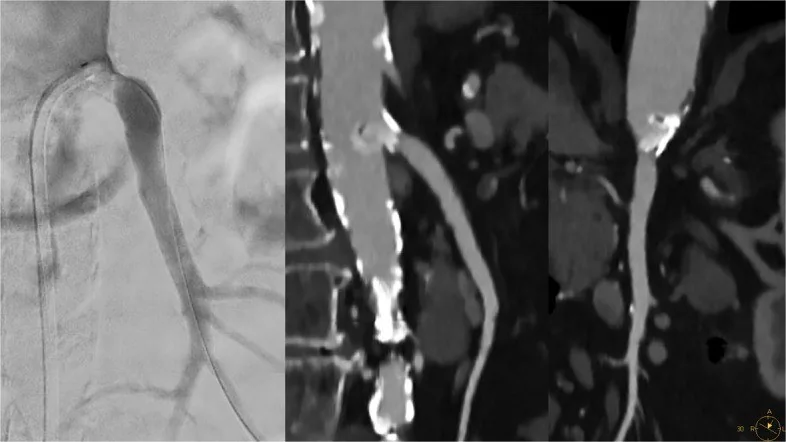
Ostial kalsifik plakta kalsiyum blooming’i BT anjiyografide stenoz derecesinin olduğundan fazla değerlendirilmesine yol açabilir. Ostial darlığın değerlendirilmesinde ince kesitli BT anjiyografi ile merkez çizgisi ve eğrisel MPR üzerinden damar çapı ölçümleri kullanılabilir.
Faz ve pencere
- BTA tanısal
- Kalsifik damarların BT anjiyografik değerlendirmesinde daha yüksek uzaysal çözünürlük kalsiyum blooming’ini azaltabilir ve stenoz ölçümünü etkileyebilir.
Önerilen pencereler: Anjiyo (G 600 / M 150), Batın (G 400 / M 50).
BT bulguları
- Yoğun ostial kalsifik plakta kalsiyum blooming’i BT anjiyografide stenoz derecesinin olduğundan fazla değerlendirilmesine yol açabilir.
- Renal arter ostiumu ve çevresindeki damar anatomisi, BT’de multiplanar ve 3D rekonstrüksiyonlarla değerlendirilir.
Normalde
Ostial kalsifik plak değerlendirilirken kalsiyum blooming’inin stenoz derecesinin olduğundan yüksek ölçülmesine yol açabileceği göz önünde bulundurulmalıdır.
Normal BT ile kıyasla
Aynı bölgenin, kaynak veri setinde patoloji içermediği belirtilen bir BT incelemesini kesit kesit kaydırın; organ sınırlarını açarak anatomiyi hasta görüntüsüyle karşılaştırın. Küçük rastlantısal bulgular tümüyle dışlanmamıştır.
Kaynak: TotalSegmentator v2.01 veri seti (Wasserthal ve ark., Radiology: Artificial Intelligence 2023), CC BY 4.0, vaka s0115; organ sınırları veri setinin otomatik segmentasyonundan, birleştirilerek sadeleştirildi. Görüntüler 2,5 mm kesit aralığına yeniden örneklendi.
Ayırıcı tanı
- Renal arter fibromüsküler displazisinde klasik “boncuk dizisi” örüntüsü görülebilir; ostial aterosklerotik darlık farklı bir örüntüdür.
- Hareket artefaktı
- damar konturunu kesitler arasında bulanıklaştırabilir; darlığın anatomik sürekliliğini başka düzlemlerde kontrol edin.
Tuzaklar
- Kalsifikasyonu değerlendirmek için önce aksiyel kaynak görüntüleri incele; damar çapı ve uzunluğunu merkez çizgisi ve eğrisel MPR ile değerlendir.
- Kalsiyum blooming’i BT anjiyografide kalsifik damar darlığının olduğundan fazla ölçülmesine yol açabilir.
- Ostial kalsifik plakta kalsiyum blooming’i BT anjiyografide stenoz derecesinin fazla değerlendirilmesine yol açabilir; uzaysal çözünürlüğün artırılması blooming etkisini azaltabilir.
Kendini dene
BT anjiyografide damar çapı ve uzunluğunu ölçmek için hangi yaklaşım kaynakta önerilmektedir?
Cevabı göster
Önce aksiyel kaynak görüntüleri inceleyip ardından merkez çizgisi ve eğrisel MPR kullanmak. Aksiyel kaynak görüntülerinin ardından merkez çizgisi ve eğrisel MPR ile damar ölçümleri yapılabilir.
BT anjiyografide damar çapı ve uzunluğunu ölçmek için hangi yaklaşım kaynakta önerilmektedir?
Cevabı göster
Önce aksiyel kaynak görüntülerini inceleyip ardından merkez çizgisi ve eğrisel MPR kullanmak. Aksiyel kaynak görüntülerinin ardından merkez çizgisi ve eğrisel MPR ile damar ölçümleri yapılabilir.
Kaynaklar
Bu sayfadaki 20 cümle ve 2 quiz sorusu aşağıdaki kaynaklardan alıntılarla otomatik olarak karşılaştırıldı; 12 cümle bu karşılaştırmada düzeltildi (2026-10-09).
- Assessment of Coronary Stenosis Using Coronary CT Angiography in Patients with High Calcium Scores: Current Limitations and Future Perspectiveskjronline.org
- Multisystem Imaging Manifestations of Kidney Failurepubs.rsna.org
- Cardiac CT blooming artifacts: clinical significance, root causes and potential solutionspubmed.ncbi.nlm.nih.gov
- Chronic mesenteric ischemia diagnosed via incidental CT findings with gastroenterologist perspective: Two case reportsf6publishing.blob.core.windows.net
- Multisystem Imaging Manifestations of Fibromuscular Dysplasiapubs.rsna.org
- Role of imaging in visceral vascular emergencieslink.springer.com
- Global Vascular Guidelines on the Management of Chronic Limb-Threatening Ischemia.pubmed.ncbi.nlm.nih.gov
- Global vascular guidelines on the management of chronic limb-threatening ischemia.pubmed.ncbi.nlm.nih.gov
- Plaque-Adapted Virtual Monoenergetic Image Selection for Carotid Plaque Visualization Using Photon-Counting CT.pubmed.ncbi.nlm.nih.gov
- Prospective comparison of ultra-high-resolution photon-counting detector CT coronary angiography versus conventional CT.pubmed.ncbi.nlm.nih.gov
- Understanding Photon-Counting CT: Physics, Detector Technology, and Image Reconstructions.pubmed.ncbi.nlm.nih.gov
- Carotid plaque characteristics by computed Tomography: A diagnostic accuracy systematic review.pubmed.ncbi.nlm.nih.gov
- Cardiovascular Applications of Photon-Counting CT Technology: A Revolutionary New Diagnostic Step.pubmed.ncbi.nlm.nih.gov
- CT imaging post-TAVI: Murphy's first law in action-preparing to recognize the unexpected.pubmed.ncbi.nlm.nih.gov
- A scoping review of photon-counting detector computed tomography in cardiovascular imaging.pubmed.ncbi.nlm.nih.gov
- Use of Calcium Modification in Percutaneous Coronary Intervention: A Comprehensive Review.pubmed.ncbi.nlm.nih.gov
- Current Trends and Future Challenges in Transcatheter Aortic Valve Replacement: Utility of Cardiac Computed Tomography Angiography.pubmed.ncbi.nlm.nih.gov
- The Emerging Role of Computed Tomography Coronary Angiography in the Left Main Stem Percutaneous Coronary Intervention.pubmed.ncbi.nlm.nih.gov
- Technical principles, benefits, challenges, and applications of photon counting computed tomography in coronary imaging: a narrative review.pubmed.ncbi.nlm.nih.gov
- Photon-Counting Computed Tomography Angiography of Carotid Arteries: A Topical Narrative Review with Case Examples.pubmed.ncbi.nlm.nih.gov
- Coronary CT Angiography for PCI Planning and Guidance: A Comprehensive Narrative Review.pubmed.ncbi.nlm.nih.gov
- Beyond Angiography: Cardiac CT for Planning Complex PCI in Calcified Coronary Lesions.pubmed.ncbi.nlm.nih.gov
- High-pitch photon-counting detector computed tomography angiography of the coronary arteries: Qualitative and quantitative evaluation of monoenergetic image reconstructions.pubmed.ncbi.nlm.nih.gov
- Noninvasive aortic imaging.pubmed.ncbi.nlm.nih.gov
- Myocardial blood flow quantification for evaluation of coronary artery disease by computed tomography.pubmed.ncbi.nlm.nih.gov
- Advanced quantitative CT for coronary artery disease: integrating stenosis quantification, plaque quantification, and fractional flow reserve-CT.pubmed.ncbi.nlm.nih.gov
- Association of Composite Metabolic Indices and Oxidative Stress Biomarkers with Coronary Atherosclerosis Detected by CT Angiography in Premenopausal Women with Metabolic Syndrome.pubmed.ncbi.nlm.nih.gov
- Myval Beyond the Aortic Valve: Device-Anatomy Interaction, Procedural Strategy, and Clinical Evidence in Mitral, Tricuspid, and Pulmonary Positions.pubmed.ncbi.nlm.nih.gov
- Preoperative planning for endovascular aortic arch repair.pubmed.ncbi.nlm.nih.gov
- Deep learning-based assessment of coronary artery disease using curved-multiplanar reconstruction: comprehensive evaluation of stenosis, calcification, and plaque.pubmed.ncbi.nlm.nih.gov
- Application of postmortem imaging modalities in cases of sudden death due to cardiovascular diseases-current achievements and limitations from a pathology perspective : Endorsed by the Association for European Cardiovascular Pathology and by the International Society of Forensic Radiology and Imaging.pubmed.ncbi.nlm.nih.gov
- The Role of Multimodality Imaging for Percutaneous Coronary Intervention in Patients With Chronic Total Occlusions.pubmed.ncbi.nlm.nih.gov
- Ultrasound Methods in the Evaluation of Atherosclerosis: From Pathophysiology to Clinic.pubmed.ncbi.nlm.nih.gov
- Multislice CT angiography of fenestrated endovascular stent grafting for treating abdominal aortic aneurysms: a pictorial review of the 2D/3D visualizations.pubmed.ncbi.nlm.nih.gov
- Endovascular revascularisation in chronic occlusive mesenteric ischaemia: safety and efficacy of intravascular lithotripsy.pubmed.ncbi.nlm.nih.gov
- Aortoiliac calcification hindering cardiac interventions: A case for intravascular lithotripsy.pubmed.ncbi.nlm.nih.gov
- Perioperative mortality in patients with acute aortic dissection and coronary artery problems: report of 3 cases and literature review.pubmed.ncbi.nlm.nih.gov
- Impact of Multidetector CT on 3D CT Angiography.pubmed.ncbi.nlm.nih.gov
- Spectral computed tomography characterization of coronary atherosclerotic plaque: principles, imaging biomarkers, and clinical significance.pubmed.ncbi.nlm.nih.gov
- Clinical benefits and current challenges of photon-counting detector CT in vascular imaging.pubmed.ncbi.nlm.nih.gov
- Study on the combined application of selected virtual monoenergetic imaging, virtual non-contrast imaging, and adaptive statistical iterative reconstruction-V in head and neck CT angiography using 256-slice wide-body CT.pubmed.ncbi.nlm.nih.gov
- The Human Hyoid Body-Greater Cornu Junction: Development, Histological Diversity, and Imaging Correlates.pubmed.ncbi.nlm.nih.gov
- Coronary Computed Tomography Angiography for the Diagnosis and Revascularization Guidance of Coronary Bifurcation Lesions: A Contemporary Review.pubmed.ncbi.nlm.nih.gov
- Coronary CT Angiography in PCI Planning: Advances, Clinical Applications, and Challenges.pubmed.ncbi.nlm.nih.gov
- Coronary Computed Tomography Angiography to Guide Percutaneous Coronary Intervention: Expert Opinion from a SCAI/SCCT Roundtable.pubmed.ncbi.nlm.nih.gov
- Computed Tomography Evaluation of Coronary Atherosclerosis: The Road Travelled, and What Lies Ahead.pubmed.ncbi.nlm.nih.gov
- Noncontrast MR angiography: An update.pubmed.ncbi.nlm.nih.gov
- Radiologic evaluation of the kidney transplant donor and recipient.pubmed.ncbi.nlm.nih.gov
- Diagnostic performance of dual-energy CT stress myocardial perfusion imaging: direct comparison with cardiovascular MRI.pubmed.ncbi.nlm.nih.gov
- Cardiac Computed Tomography in Structural Heart Interventions: From Preprocedural Planning to Procedural Strategy.pubmed.ncbi.nlm.nih.gov
- aCTA structured reporting of aortic dissection and updated knowledge required by radiologists.pubmed.ncbi.nlm.nih.gov
- Pre-Procedural Assessment of the Femoral Access Route for Transcatheter Aortic Valve Implantation: Comparison of a Non-Contrast Time-of-Flight Magnetic Resonance Angiography Protocol with Contrast-Enhanced Dual-Source Computed Tomography Angiography.pubmed.ncbi.nlm.nih.gov
- MDCT Imaging of Non-Traumatic Thoracic Aortic Emergencies and Its Impact on Diagnosis and Management-A Reappraisal.pubmed.ncbi.nlm.nih.gov
- 2022 ACC/AHA guideline for the diagnosis and management of aortic disease: A report of the American Heart Association/American College of Cardiology Joint Committee on Clinical Practice Guidelines.pubmed.ncbi.nlm.nih.gov
- 2022 ACC/AHA Guideline for the Diagnosis and Management of Aortic Disease: A Report of the American Heart Association/American College of Cardiology Joint Committee on Clinical Practice Guidelines.pubmed.ncbi.nlm.nih.gov
- Computed Tomographic Evaluation of Congenital Left Ventricular Outflow Obstruction.pubmed.ncbi.nlm.nih.gov
- Left Atrial Appendage: Mere Spectator or Protagonist? A Narrative Review of Its Multimodal Imaging Assessment and Its Closure Techniques.pubmed.ncbi.nlm.nih.gov
- Intraprocedural echocardiographic guidance in transcatheter aortic valve implantation.pubmed.ncbi.nlm.nih.gov
- Bilateral internal carotid artery fibromuscular dysplasia identified during acute ischemic stroke evaluation: A case report.pubmed.ncbi.nlm.nih.gov
- Role of 99mTc-DTPA Captopril Renal Scintigraphy in the Diagnosis of Renal Fibromuscular Dysplasia: A Case Report.pubmed.ncbi.nlm.nih.gov
- Fibromuscular Dysplasia and Cerebrovascular Dissection: Insights From a Multicenter Cohort.pubmed.ncbi.nlm.nih.gov
- Radiation dose optimization in coronary CT angiography clinical application of absolute phase gating in patients with atrial fibrillation.pubmed.ncbi.nlm.nih.gov
- Technical Recommendations for Computed Tomography Guidance of Intervention in the Right Ventricular Outflow Tract: Native RVOT, Conduits and Bioprosthetic Valves: A White Paper of the Society of Cardiovascular Computed Tomography (SCCT), Congenital Heart Surgeons' Society (CHSS), and Society for Cardiovascular Angiography & Interventions (SCAI).pubmed.ncbi.nlm.nih.gov
- Photon-counting computed tomography in coronary artery bypass grafting planning and follow-up: a narrative review of a high-resolution adjunct to invasive angiography.pubmed.ncbi.nlm.nih.gov
- Photon-Counting CT in Cardiovascular Imaging: Clinical Applications.pubmed.ncbi.nlm.nih.gov
- State-of-the-art CT and MR imaging and assessment of atherosclerotic carotid artery disease: standardization of scanning protocols and measurements-a consensus document by the European Society of Cardiovascular Radiology (ESCR).pubmed.ncbi.nlm.nih.gov
- Coronary Artery Calcium Score and Carotid Intima-Media Thickness As Markers of Computed Tomography Angiographic Severity in Coronary Artery Disease: A Cross-Sectional Study.pubmed.ncbi.nlm.nih.gov
- Acute Traumatic Aortic Injury: What the Radiologist Needs to Know.pubmed.ncbi.nlm.nih.gov
- Operative Procedures for Ultrasound Assessment of Extracranial Artery Disease: A Narrative Review by the Italian Society for Vascular Investigation (SIDV).pubmed.ncbi.nlm.nih.gov
- Percutaneous Transluminal Renal Angioplasty for Fibromuscular Dysplasia and Prognostic Risk Factors: A Retrospective Chinese Cohort Study.pubmed.ncbi.nlm.nih.gov
- Clinical Characteristics and Outcomes of Aortic Arch Emergencies: Takayasu Disease, Fibromuscular Dysplasia, and Aortic Arch Pathologies: A Retrospective Study and Review of the Literature.pubmed.ncbi.nlm.nih.gov
- Intraoperative direct coronary angiography clarifying a misleading coronary stenosis caused by an ascending aortic floating thrombus: a case report.pubmed.ncbi.nlm.nih.gov
- Vascular imaging: the evolving role of the multidisciplinary team meeting in peripheral vascular disease.pubmed.ncbi.nlm.nih.gov
- Renovascular Hypertension with Progressive Atherosclerotic Renal Artery Stenosis in an Elderly Patient with Known Fibromuscular Dysplasia: A Case Report.pubmed.ncbi.nlm.nih.gov
- Dual-Source Photon-Counting Computed Tomography-Part III: Clinical Overview of Vascular Applications beyond Cardiac and Neuro Imaging.pubmed.ncbi.nlm.nih.gov
- The Shifting Boundary Between Invasive and Non-Invasive Angiographic Investigation in Contemporary Cardiology and Cardiac Surgery: An Up-to-Date Narrative Review.pubmed.ncbi.nlm.nih.gov
- Practical guide on the use of the PulseSelect pulsed field ablation system for atrial fibrillation ablation procedures.pubmed.ncbi.nlm.nih.gov
- Atherosclerotic Abdominal Aortic Aneurysms on Computed Tomography Angiography: A Narrative Review on Spectrum of Findings, Structured Reporting, Treatment, Secondary Complications and Differential Diagnosis.pubmed.ncbi.nlm.nih.gov
- Clinical physiology: the crucial role of MRI in evaluation of peripheral artery disease.pubmed.ncbi.nlm.nih.gov
- Lung imaging methods: indications, strengths and limitations.pubmed.ncbi.nlm.nih.gov
- Imaging assessment of acute ischaemic stroke: a review of radiological methods.pubmed.ncbi.nlm.nih.gov
- Accuracy of dual-source CT coronary angiography: First experience in a high pre-test probability population without heart rate control.pubmed.ncbi.nlm.nih.gov
- 2022 ACC/AHA Guideline for the Diagnosis and Management of Aortic Disease: A Report of the American Heart Association/American College of Cardiology Joint Committee on Clinical Practice Guidelines.pubmed.ncbi.nlm.nih.gov
- Long-term safety and effectiveness of the "OptEase" vena cava filter.pubmed.ncbi.nlm.nih.gov
- Quantitative coronary CT angiography: absolute lumen sizing rather than %stenosis predicts hemodynamically relevant stenosis.pubmed.ncbi.nlm.nih.gov
- Visualization of the renal artery in kidney transplant patients using time-resolved computed tomography angiography.pubmed.ncbi.nlm.nih.gov
- Imaging and Surveillance of Chronic Aortic Dissection: A Scientific Statement From the American Heart Association.pubmed.ncbi.nlm.nih.gov
- 2.5-mm articulated endoluminal forceps using a compliant mechanism.pubmed.ncbi.nlm.nih.gov
- From intravascular imaging to adaptive vascular care: intelligent photonics and digital twins in panvascular disease.pubmed.ncbi.nlm.nih.gov
- Chest pain: coronary CT in the ER.pubmed.ncbi.nlm.nih.gov
- Multimodal imaging in neuro-ophthalmology.pubmed.ncbi.nlm.nih.gov
- Evaluation of Extracellular Volume and Coronary Artery Disease in Cardiac Amyloidosis Using Photon-Counting CT.pubmed.ncbi.nlm.nih.gov
- Coronary computed tomography and magnetic resonance imaging.pubmed.ncbi.nlm.nih.gov
- Intravascular ultrasound-mediated stent-assisted coil embolization and follow-up of a large renal artery aneurysm with ostial stenosis in a solitary kidney.pubmed.ncbi.nlm.nih.gov
- Computed Tomography Coronary Angiography as a Gatekeeper for Invasive Coronary Assessment Before Transcatheter Aortic Valve Implantation.pubmed.ncbi.nlm.nih.gov
- Fractional Flow Reserve from Coronary CT: Evidence, Applications, and Future Directions.pubmed.ncbi.nlm.nih.gov
- SCMR Position Paper (2020) on clinical indications for cardiovascular magnetic resonance.pubmed.ncbi.nlm.nih.gov
Eğitim amaçlıdır; hasta başında tanı ve tedavi kararının yerine geçmez, tıbbi tavsiye değildir. Metinler yapay zekâ desteğiyle yazıldı ve otomatik bir adımda kaynak alıntılarıyla karşılaştırıldı; bu bir tıbbi doğrulama değildir ve içerik uzman radyolog incelemesinden geçmedi. Klinik kararlarda güncel kılavuzları, kurum protokollerini ve radyoloji raporunu esas alın.