Özet
Koroner venler bazı koroner arterlere komşu seyreder: büyük kardiyak ven LAD'ye, koroner sinüs LCx'ye, orta kardiyak ven ise posterior interventriküler oluğa yakındır. Konus dalı, sağ koroner sinüs düzeyindeki ayrı bir ostiumdan çıkabilir. Koroner BT anjiyografi, koroner venöz sistemin ve komşu yapıların BT ile değerlendirilmesine olanak verir. Koroner CTA, anomal koroner damarların kökenini ve üç boyutlu seyrini kesitsel ve çok düzlemli gösterir.
Okuma sırası
- sağ koroner sinüs ve RCA ostiumu. Aort kökünün sağ koroner sinüsünde RCA ostiumunu bul; bu seviyede komşu küçük bir açıklık varsa ayrı konus dalı olasılığını akılda tut.
- konus dalı. Aday dalı ostiumdan distal ucuna kadar ardışık kesitlerde izleyin; sağ ventrikül çıkış bölgesine yönelen rotayı farklı düzlemlerde doğrulayın.
- koroner sinüs, büyük kardiyak ven ve sirkumfleks. Sol atriyoventriküler olukta sirkumfleks arter ile komşu koroner venin ilişkisi değişken olabilir; bireysel anatomiyi BT görüntülerinde değerlendirin.
- ana pulmoner arter ve olası fistül. Pulmoner trunkusun ana gövde ve dallara devamlılığını, şüpheli koroner dalın ise aort kökü veya koroner ağaca bağlantısını karşılaştır; fistül şüphesinde pulmoner artere boşalma noktasını ara.
Faz ve pencere
- BTA tanısal
- Koroner venler koroner arterlere yakın seyredebilir; bu yakınlığın yeri ve derecesi değişkendir. Koroner venlerin arterlerle komşuluğu bireyler arasında değişkenlik gösterir; bu ilişkilerin BT’de bireysel anatomiye göre değerlendirilmesi önemlidir.
Önerilen pencereler: Anjiyo (G 600 / M 150).
BT bulguları
- Büyük kardiyak ven, ön interventriküler olukta LAD'ye eşlik eder ve sol atriyoventriküler oluğa yönelerek koroner sinüse katılır.
- Koroner sinüs, posterior atriyoventriküler olukta LCx’nin distal bölümüne yakın seyreder; bu iki yapının göreli konumu değişken olabilir ve kesitlerde ayrı izlenmelidir.
- Orta kardiyak venin yakınında seyreden arter bireysel anatomiye göre değişebilir; incelenen kalplerde bu vene en sık yakın seyreden arter RCA’dır.
- Konus dalı ayrı bir ostiumla sağ koroner sinüsten çıkabilir.
- Konus dalı, sağ koroner sinüs düzeyindeki ayrı bir ostiumdan çıkabilir.
- Koroner arter fistülünde besleyici koroner dalın ana pulmoner artere gerçek devamlılığı ve boşalma noktası izlenir.
- Ana pulmoner arter sağ ventrikül çıkış yolundan ana gövde ve dallanmaya devam eder; pulmoner venler sol atriyuma bağlanır ve şüpheli damarların devamlılığı kesitlerde izlenir.
Normalde
Konus dalı, sağ koroner sinüsten ayrı bir ostiumla çıkabilir. Koroner venlerin arterlerle komşuluğu değişkenlik gösterir; sağ koroner arter orta kardiyak venin yakınında seyredebilir. Şüpheli koroner damarın başlangıcı ve komşu koroner venlerle ilişkisi, bireysel koroner anatomiye göre birlikte değerlendirilir.
Normal BT ile kıyasla
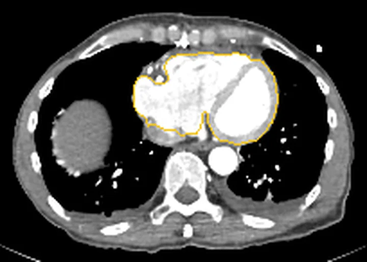
Aynı bölgenin, kaynak veri setinde patoloji içermediği belirtilen bir BT incelemesini kesit kesit kaydırın; organ sınırlarını açarak anatomiyi hasta görüntüsüyle karşılaştırın. Küçük rastlantısal bulgular tümüyle dışlanmamıştır.
Kaynak: TotalSegmentator v2.01 veri seti (Wasserthal ve ark., Radiology: Artificial Intelligence 2023), CC BY 4.0, vaka s0977; organ sınırları veri setinin otomatik segmentasyonundan, birleştirilerek sadeleştirildi. Görüntüler 2,5 mm kesit aralığına yeniden örneklendi.
Ayırıcı tanı
- Sirkumfleks arter
- sol atriyoventriküler olukta koroner arter ağacına bağlıdır; koroner sinüse venöz devamlılığı yoktur.
- Büyük kardiyak ven
- ön interventriküler olukta LAD'ye yakın seyreder, ardından koroner sinüse katılır.
- Konus dalı
- sağ koroner sinüsten ayrı bir ostiumla çıkabilir.
- Koroner-pulmoner arter fistülü
- koroner besleyici damar ile pulmoner arter arasında izlenebilir anormal bağlantı bulunur.
- Ana pulmoner arter
- sağ ventrikül çıkış yolundan ana gövdeye ve pulmoner bifurkasyona devam eder.
- Orta kardiyak ven ile komşu arterlerin ilişkisi bireysel anatomide değişkenlik gösterir; incelenen kalplerde RCA bu vene en sık yakın seyreden arterdir. Damarın kökenini ve devamlılığını bireysel kardiyak anatomi ve komşu damar ilişkilerini dikkate alarak değerlendirin.
Tuzaklar
- Koroner sinüs veya büyük kardiyak venin koroner arterlere komşuluğu değişken olduğundan, bu damarların ilişkisini koroner BT’de bireysel anatomiye göre değerlendirin.
- Ayrı ostiumlu konus dalı RCA ostiumuna komşu olabilir; başlangıcını aort kökünden ardışık kesitlerde izleyerek ayrı kökeni doğrulayın.
- Aortik kök veya ana pulmoner arter çevresindeki şüpheli damarların kökeni ve seyri, koroner CTA'nın çok düzlemli rekonstrüksiyonlarıyla değerlendirilebilir. Koroner sinüs venöz bir yapıdır ve miyokard kanını sağ atriyuma taşır.
- MIP ve hacimsel gösterim tek başına damar kökenini kesinleştirmeyebilir; kaynak aksiyel kesit ve eğrisel düzlemsel rekonstrüksiyonla doğrulayın.
Kendini dene
İncelenen kalplerde orta kardiyak venin yakınında en sık seyreden koroner arter hangisidir?
Cevabı göster
Sağ koroner arter (RCA). S6’daki anatomik incelemede sağ koroner arter, orta kardiyak venin yakınında en sık seyreden arter olarak bildirilmiştir.
Sağ koroner sinüsten ayrı bir ostiumla çıktığı bildirilen bağımsız koroner dal hangisidir?
Cevabı göster
Konus dalı. Kaynakta sağ koroner sinüsten çıkan ayrı ostiumlar arasında bağımsız konus dalı tanımlanmıştır.
Kaynaklar
Bu sayfadaki 37 cümle ve 2 quiz sorusu aşağıdaki kaynaklardan alıntılarla otomatik olarak karşılaştırıldı; 21 cümle bu karşılaştırmada düzeltildi (2026-10-09).
- Coronary Veins: Comprehensive CT-Anatomic Classification and Review of Variants and Clinical Implicationspubs.rsna.org
- Coronary venous system in cardiac computer tomography: Visualization, classification and rolewjgnet.com
- Atypical Chest Pain: Coronary, Aortic, and Pulmonary Vasculature Enhancement at Biphasic Single-Injection 64-Section CT Angiographypubs.rsna.org
- Coronary artery anomalies overview: The normal and the abnormalpmc.ncbi.nlm.nih.gov
- Conus artery in coronary CT angiographyelektrokardiologia.com
- CT of Congenital Heart Disease: Normal Anatomy and Typical Pathologic Conditionspubs.rsna.org
- Revisiting Coronary Artery Anomaliespubs.rsna.org
- Coronary-to-pulmonary artery fistula in a patient with atypical chest pain: case presentation and literature reviewlink.springer.com
- Surgical treatment of giant diagonal branch aneurysms accompanied by obstructive coronary artery disease with guidance from 3D printed models: a case reportpmc.ncbi.nlm.nih.gov
- Segmental Arterial Mediolysis and Other Mimics of Medium Vessel Vasculitis: A Case and Reviewpmc.ncbi.nlm.nih.gov
- Traumatic intracranial pseudoaneurysms of cortical middle cerebral artery and posterior cerebral artery branches presenting as subdural hematomas in elderly patients: A two-case reportpmc.ncbi.nlm.nih.gov
- Cinematic rendering in CT angiography: a pictorial review of clinical applicationspmc.ncbi.nlm.nih.gov
- Intestinal necrosis in a patient with Behçet's disease and infective endocarditis: a case reportpmc.ncbi.nlm.nih.gov
- The relative anatomy of the coronary arterial and venous systems: implications for coronary interventions.pubmed.ncbi.nlm.nih.gov
- Coronary Arterial Anomalies in Adults with Congenital Heart Disease: From Detection to Management Strategies.pubmed.ncbi.nlm.nih.gov
- Arterial Supply to the Atrioventricular Node: Anatomical Variability and Clinical Implications.pubmed.ncbi.nlm.nih.gov
- All three coronary arteries and a separate conus branch arising from the right sinus of Valsalva in an octogenarian with an acute myocardial infarction: a case report.pubmed.ncbi.nlm.nih.gov
- Assessment of Coronary Arteries during the Prenatal and Postnatal Periods using Two- and Three-dimensional Ultrasound.pubmed.ncbi.nlm.nih.gov
- Catheter ablation of ventricular tachycardia via the coronary veins.pubmed.ncbi.nlm.nih.gov
- Navigation of the Clinical Implications, Interventional Challenges, and Complexities of the Circumflex Coronary Artery: A Comprehensive Review.pubmed.ncbi.nlm.nih.gov
- Optimized contrast enhancement and homogeneity in aortic CT angiography: bolus tracking with personalized post-trigger delay.pubmed.ncbi.nlm.nih.gov
- Role of CT in the Pre- and Postoperative Assessment of Conotruncal Anomalies.pubmed.ncbi.nlm.nih.gov
- Thoracic Complications in Behçet's Disease: Imaging Findings.pubmed.ncbi.nlm.nih.gov
- Sudden Cardiac Arrest in a Pregnant Woman with Wolff-Parkinson-White Syndrome with Fractionated Delta Wave Due to Middle Cardiac Vein Diverticulum.pubmed.ncbi.nlm.nih.gov
- Angiographic Anatomy of the Left Coronary Veins: Beyond Conventional Cardiac Resynchronization Therapy.pubmed.ncbi.nlm.nih.gov
- Preclinical modeling of Loeys-Dietz syndrome: insights into mechanisms and therapy.pubmed.ncbi.nlm.nih.gov
- Essentials of Imaging the Repaired Tetralogy of Fallot Patient.pubmed.ncbi.nlm.nih.gov
- Detection of Coronary Artery and Aortic Arch Anomalies in Patients with Tetralogy of Fallot Using CT Angiography.pubmed.ncbi.nlm.nih.gov
- Prevalence and characteristics of coronary anomalies in Bulgarian patients undergoing coronary angiography.pubmed.ncbi.nlm.nih.gov
- Aortic Valve Leaflet Modification: A Working Group Position Statement on Best Practices and Step-by-Step Guide.pubmed.ncbi.nlm.nih.gov
- Anatomical characterisation of an additional atrioventricular node artery contributing to the arterial complex of proximal conduction components: Bonapace's septal branch or Kugel's collateral artery.pubmed.ncbi.nlm.nih.gov
- A Cadaveric Case Report of a Double Right Coronary Artery and Its Clinical Implications.pubmed.ncbi.nlm.nih.gov
- Coronary sinus and cardiac venous anatomy for cardiac resynchronization therapy: A Clinician's view.pubmed.ncbi.nlm.nih.gov
- A Rare Vascular Tangle Unraveled: Multimodal Imaging in Giant Coronary Arteriovenous Fistula.pubmed.ncbi.nlm.nih.gov
- Cardiovascular Computed Tomography Angiographic Assessment of Simple Cardiac Shunts in Adults.pubmed.ncbi.nlm.nih.gov
- Computed tomography and magnetic resonance imaging findings in congenital cardiovascular anomalies.pubmed.ncbi.nlm.nih.gov
- Society for Cardiovascular Magnetic Resonance 2024 Cases of SCMR case series.pubmed.ncbi.nlm.nih.gov
- Imaging considerations in tetralogy of Fallot: A comprehensive review.pubmed.ncbi.nlm.nih.gov
- Cardiac Development and Factors Influencing the Development of Congenital Heart Defects (CHDs): Part I.pubmed.ncbi.nlm.nih.gov
- Long-term safety and effectiveness of the "OptEase" vena cava filter.pubmed.ncbi.nlm.nih.gov
- Cardiac Computed Tomography in Structural Heart Interventions: From Preprocedural Planning to Procedural Strategy.pubmed.ncbi.nlm.nih.gov
- Pathogenesis and Surgical Treatment of Dextro-Transposition of the Great Arteries (D-TGA): Part II.pubmed.ncbi.nlm.nih.gov
- Segmental Arterial Mediolysis and Other Mimics of Medium Vessel Vasculitis: A Case and Review.pubmed.ncbi.nlm.nih.gov
- Intestinal necrosis in a patient with Behçet's disease and infective endocarditis: a case report.pubmed.ncbi.nlm.nih.gov
- Traumatic intracranial pseudoaneurysms of cortical middle cerebral artery and posterior cerebral artery branches presenting as subdural hematomas in elderly patients: A two-case report.pubmed.ncbi.nlm.nih.gov
- Cinematic rendering in CT angiography: a pictorial review of clinical applications.pubmed.ncbi.nlm.nih.gov
- Point-of-Care Agitated Saline Contrast Echocardiography in the Diagnosis of Respiratory Failure: A Case of Hepatopulmonary Syndrome With Decompensated Heart Failure.pubmed.ncbi.nlm.nih.gov
- Anomalous Coronary Artery Origin from Pulmonary Artery and Coronary Fistulas: A Review About Coronary Steal Phenomenon.pubmed.ncbi.nlm.nih.gov
- Single Coronary Artery in the Context of Vascular Disease: Anatomy, Development, Multimodality Imaging, and Clinical Interpretation.pubmed.ncbi.nlm.nih.gov
- State-of-the-Art Review on Finite Element Analysis Applied to Ultrathin-Strut Drug-Eluting Stents: Examining the Present and Looking Ahead to the Future.pubmed.ncbi.nlm.nih.gov
- Concomitant Aortic Valve Neocuspidization and Resection of a Cardiac Calcified Amorphous Tumor in a Hemodialysis Patient: A Case Report.pubmed.ncbi.nlm.nih.gov
- "Left Main" Pattern Electrocardiogram: A Diagnostic Trap for Cardiologists.pubmed.ncbi.nlm.nih.gov
- Common Clinical Scenarios in Adult Congenital Heart Disease: A Practical Guide for General Cardiologists.pubmed.ncbi.nlm.nih.gov
- A rare invader on a cardiac stent: <i>Mycobacterium abscessus</i> mediastinitis managed with tedizolid after linezolid intolerance.pubmed.ncbi.nlm.nih.gov
- Adult presentation of cor triatriatum sinister with anomalous coronary venous drainage into the left atrium: A rare congenital abnormality.pubmed.ncbi.nlm.nih.gov
- Coronary Artery Origin Anomalies on Coronary CT Angiography: A Single-Center Tertiary-Care Cohort.pubmed.ncbi.nlm.nih.gov
- Anomalous Coronary Venous Drainage to the Right Atrium: A Case Report.pubmed.ncbi.nlm.nih.gov
- Idiopathic IgG4-negative perivascular fibrosis with extensive thoracic aortic, coronary artery, and supra-aortic branch involvement: A case report.pubmed.ncbi.nlm.nih.gov
- Multimodal Imaging of a Post-Traumatic Cervical Arteriovenous Fistula with Concomitant Pseudoaneurysm After a Gunshot Wound Treated with a Covered Stent-Graft.pubmed.ncbi.nlm.nih.gov
- Mediastinal adipose tissue as an active player in cardiovascular disease: a multimodality imaging narrative review.pubmed.ncbi.nlm.nih.gov
- Interventricular septal dissection secondary to acute inferior myocardial infarction: case series and literature review.pubmed.ncbi.nlm.nih.gov
- Left Atrial Appendage: Mere Spectator or Protagonist? A Narrative Review of Its Multimodal Imaging Assessment and Its Closure Techniques.pubmed.ncbi.nlm.nih.gov
- When Cancer Clots: An Extensive Radiologic Analysis of Cancer-Associated Thromboembolism.pubmed.ncbi.nlm.nih.gov
- Bronchial Arteriovenous Malformation Detection Prior to Catheter Ablation for Paroxysmal Atrial Flutter and Fibrillation: A Case Report.pubmed.ncbi.nlm.nih.gov
- Unfavorable venous outflow correlates with poor prognosis in acute ischemic stroke due to large vessel occlusion (AIS-LVO) patients assessed dynamically and quantitatively based on four-dimensional computed tomography angiography/perfusion (4D-CTA/CTP).pubmed.ncbi.nlm.nih.gov
- Urgent and emergent pediatric cardiovascular imaging.pubmed.ncbi.nlm.nih.gov
- Technical Recommendations for Computed Tomography Guidance of Intervention in the Right Ventricular Outflow Tract: Native RVOT, Conduits, and Bioprosthetic Valves.pubmed.ncbi.nlm.nih.gov
- Contrast medium dose optimization in the era of multi-energy CT.pubmed.ncbi.nlm.nih.gov
- The Marshall Complex in the Human Heart: Embryology, Microanatomy, Autonomic Features and Clinical Implications for Atrial Fibrillation-A State-of-the-Art Narrative Review.pubmed.ncbi.nlm.nih.gov
- Intramural left ventricular posterior superior process premature ventricular complex ablation with intracardiac echocardiography.pubmed.ncbi.nlm.nih.gov
- Terminal S-Wave Amplitude in Lead V<sub>2</sub> With Monophasic R Waves in Other Precordial Leads: A Novel ECG Marker for Predicting Acute Successful Endocardial Ablation of a Subtype of Left Ventricular Outflow Tract Idiopathic Ventricular Arrhythmias.pubmed.ncbi.nlm.nih.gov
- Coronary Artery Bypass Grafting for Anomalous RCA From LAD With Proximal LAD Stenosis.pubmed.ncbi.nlm.nih.gov
- Presumed Primary Cardiac Angiosarcoma With Atrioventricular Groove Rupture and Right Coronary Artery Compromise: A Case Report and Literature Review.pubmed.ncbi.nlm.nih.gov
- Protocol for Post-Mortem Micro-CT Imaging of Coronary Arteries in Low-Mass Neonatal Puppy Hearts Using Barium-Based Contrast.pubmed.ncbi.nlm.nih.gov
- Visualization of coronary small branch at ultra-high field 5T Noncontrast Dixon Whole-Heart Coronary MR Angiography: compared with 3T MRI.pubmed.ncbi.nlm.nih.gov
- Coronary computed tomography angiography findings of anomalous left hepatic vein drainage into the coronary sinus with complex anomalies: a case report.pubmed.ncbi.nlm.nih.gov
- Case Report: Imaging findings of neonatal horseshoe lung with multiple system malformations.pubmed.ncbi.nlm.nih.gov
- Delayed Anterior Interventricular Vein Opacification Guides True-Lumen Wiring in Stump-Less LAD Occlusion.pubmed.ncbi.nlm.nih.gov
- Comprehensive Review of Coronary Artery Anatomy Relevant to Cardiac Surgery.pubmed.ncbi.nlm.nih.gov
- The Role of MDCT Coronary Angiography in the Detection of Benign Varieties and Anomalies of Coronary Blood Vessels-A Narrative Review.pubmed.ncbi.nlm.nih.gov
- Comprehensive macroscopic living anatomy of the swine heart: comparative visual approach with virtual dissection.pubmed.ncbi.nlm.nih.gov
- Obesity and Cardiovascular Disease: An Obesity Medicine Association (OMA) Clinical Practice Statement (CPS) 2026.pubmed.ncbi.nlm.nih.gov
- Echocardiography in the cardiac assessment of young athletes: a 2025 guideline from the British Society of Echocardiography (endorsed by Cardiac Risk in the Young).pubmed.ncbi.nlm.nih.gov
- Trifurcation of the Right Coronary Artery With Myocardial Bridging of the Posterior Interventricular Artery and the Absence of the Left Marginal Artery: A Report of a Cadaveric Case.pubmed.ncbi.nlm.nih.gov
- Anatomical Study of the Triangle of Brocq and Mouchet and Its Clinical Implications for Coronary Interventions in Human Cadaveric Hearts.pubmed.ncbi.nlm.nih.gov
- Guidewire Ablation via Coronary Venous System for Frequent VPBs: A Case Report.pubmed.ncbi.nlm.nih.gov
- Open Surgical Management of Renal Cell Carcinoma with Infradiaphragmatic Venous Tumor Thrombus (Mayo Levels 0-III): The Epitome of Surgical Self-Reliance in Urology.pubmed.ncbi.nlm.nih.gov
- Commentary: an eye on PET quantification.pubmed.ncbi.nlm.nih.gov
- High-affinity alphavbeta3 integrin targeted optical probe as a new imaging biomarker for early atherosclerosis: initial studies in Watanabe rabbits.pubmed.ncbi.nlm.nih.gov
- 2019 HRS/EHRA/APHRS/LAHRS expert consensus statement on catheter ablation of ventricular arrhythmias.pubmed.ncbi.nlm.nih.gov
- 2019 HRS/EHRA/APHRS/LAHRS expert consensus statement on catheter ablation of ventricular arrhythmias.pubmed.ncbi.nlm.nih.gov
- Extra-Carotid Arterial Webs: A Single-Center Series and Systematic Review.pubmed.ncbi.nlm.nih.gov
- Non-contrast enhanced visualization of the equine foot vasculature in a cadaver model using time-of-flight sequence.pubmed.ncbi.nlm.nih.gov
Eğitim amaçlıdır; hasta başında tanı ve tedavi kararının yerine geçmez, tıbbi tavsiye değildir. Metinler yapay zekâ desteğiyle yazıldı ve otomatik bir adımda kaynak alıntılarıyla karşılaştırıldı; bu bir tıbbi doğrulama değildir ve içerik uzman radyolog incelemesinden geçmedi. Klinik kararlarda güncel kılavuzları, kurum protokollerini ve radyoloji raporunu esas alın.