Özet
Koroner BT anjiyografide koroner arterlerin ostiumlarını, seyirlerini ve dominans örüntülerini belirlemek, anomali ve varyantları doğru sınıflandırmak için temel adımdır. Koroner dominans, anatomik olarak arka interventriküler oluğa uzanan posterior inen arterin (PDA) hangi sistemden çıktığını gösterir; posterior yüz dallarının kaynağını da rapora eklemek, eş-dominans teriminin farklı kullanımlarını açıklığa kavuşturur. EKG eşzamanlı koroner BT anjiyografi (CTCA), konuslu verileri 3B yeniden yapılandırma ve eğri çok düzlemli yeniden formatlama (cMPR) ile damarların kökeni, seyri ve komşu yapılarla ilişkilerini yüksek çözünürlükte gösterir. PDA kökeninin ve dominans örüntüsünün doğru belirlenmesi, alt duvar iskemi değerlendirmesi, kronik total oklüzyonda kollateral kullanımı ve girişimsel planlama açısından kritiktir.
Faz ve pencere
- BTA tanısal
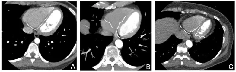
- EKG eşzamanlı, koroner arteriyel kontrastlanmalı seride aort sinüslerinden çıkan lümenler; sol ana arterin dallanması; RCA ile LCx'in atriyoventriküler oluklardaki devamı ve kruksu geçerek posterior interventriküler oluğa giren PDA izlenir.
Önerilen pencereler: Anjiyo (G 600 / M 150).
BT bulguları
- Sol ana koroner arter sol koroner sinüsten çıkar; çoğunlukla LAD ve LCx olarak ikiye ayrılır, bazı kalplerde ayrım noktasından ramus intermedius da doğar.
- LAD ön interventriküler olukta apekse doğru ilerler; diagonal dallar sol ventrikülün ön-yan yüzüne, septal perforatörler interventriküler septuma yönelir.
- LCx sol atriyoventriküler olukta arkaya ilerler ve obtus marginal dallar verir; distal uzanımı ve arka yüzü beslemesi dominans örüntüsüne göre değişir.
- RCA sağ koroner sinüsten çıkar, sağ atriyoventriküler oluğu izler ve sağ ventrikül yüzüne akut marjinal dal verebilir.
- Sağ dominan anatomide PDA kruks düzeyinde RCA'dan çıkar ve posterior interventriküler olukta ilerler.
- Sol dominant düzende PDA ve posterolateral dal distal LCx'ten köken alır; bu durumda RCA küçülürken LCx'in besleme alanı genişleyip alt duvarı karşılar.
- Eş-dominans kullanımında sıklıkla PDA RCA'dan, posterolateral dal ise LCx'ten çıkar; bazı tanımlarda posterior interventriküler alanın iki sistemce paylaşılması vurgulanır, bu nedenle raporda dal kökenlerini açıkça yaz.
Normalde
Normal koroner BT anjiyografide LM ve RCA ilgili aort sinüslerinden çıkar; LAD ile LCx çoğunlukla LM'den, bazı varyantlarda ayrı ostiumlardan köken alır ve beklenen kalp oluklarında izlenir. CCTA görüntülerinde kruks çevresi anatomik sınırlarla tanımlanır; PDA ve posterolateral dalların kökenlerini belirlemek için damarların çok düzlemli yeniden yapılandırmaları kullanılmalıdır.
Normal BT ile kıyasla
Aynı bölgenin, kaynak veri setinde patoloji içermediği belirtilen bir BT incelemesini kesit kesit kaydırın; organ sınırlarını açarak anatomiyi hasta görüntüsüyle karşılaştırın. Küçük rastlantısal bulgular tümüyle dışlanmamıştır.
Kaynak: TotalSegmentator v2.01 veri seti (Wasserthal ve ark., Radiology: Artificial Intelligence 2023), CC BY 4.0, vaka s0977; organ sınırları veri setinin otomatik segmentasyonundan, birleştirilerek sadeleştirildi. Görüntüler 2,5 mm kesit aralığına yeniden örneklendi.
Ayırıcı tanı
- Ramus intermedius veya yüksek çıkan diagonal
- ramus sol ana arterin LAD-LCx ayrımından doğar; diagonal dal ise LAD'nin ön interventriküler oluk içindeki devamından çıkar.
- PDA veya posterolateral dal
- PDA arka interventriküler olukta apekse doğru ilerler; posterolateral dallar sol ventrikülün arka-yan yüzüne yayılır.
- Kısa, küçük kalibreli LCx veya distal LCx oklüzyonu
- doğuştan kısa damar düzgün biçimde sonlanabilir; oklüzyonda kontrastlı lümende ani kesilme ya da dolum kusuru aranır.
- Anomalik koroner kökenler (örn. karşı sinüs veya pulmoner arterden çıkış) seyirlerine göre benign veya hemodinamik öneme sahip olabilir; tanıyı belirleyen proksimal ostiumun yeri ve damarın seyridir.
Tuzaklar
- RCA'nın kalınlığını dominansın göstergesi sayma; kruksta PDA'nın gerçek çıkışını ve arka yüz dallarını izle.
- PDA ile posterolateral dalı yalnız kruks yakınlığından ayırma; birinin posterior interventriküler oluğa, diğerinin ventrikülün arka-yan yüzüne yönelimini takip et.
- Kısa bir segmentte lümen seçilemiyorsa damar kökeni ve seyrini CCTA'nın 3B rekonstrüksiyon ve post-processing görüntülerinde değerlendir.
Kendini dene
PDA distal LCx'ten çıkar ve LCx arka-yan ventrikül yüzüne de dal verir. En uygun dominans tanımı hangisidir?
Cevabı göster
Sol dominans. PDA'nın LCx kaynaklı olması sol dominansı tanımlar. Sağ dominan örüntüde PDA RCA'dan çıkar; eş-dominan örüntüde arka yüz dalları iki sistem arasında paylaşılır. Ayrı ostiumlar ise dominans sınıfı değil, koroner başlangıç varyantıdır.
Yaygın radyolojik raporlama kullanımında PDA RCA'dan, posterolateral dal LCx'ten geliyor. Hangi örüntü daha uygun adlandırılır?
Cevabı göster
Eş-dominans. Yaygın kullanımda PDA'nın RCA'dan, posterolateral dalın LCx'ten çıkması eş-dominan veya dengeli dolaşım olarak adlandırılır. Farklı tanımlar benimsendiğinden terim kullanımı literatürde çeşitlilik gösterebilmektedir. Sol dominan düzende PDA LCx'ten çıkar; tek koroner arter ve ramus intermedius bu dal paylaşımını tanımlamaz.
Kaynaklar
Bu sayfadaki 37 cümle ve 2 quiz sorusu aşağıdaki kaynaklardan alıntılarla tek tek karşılaştırıldı; 10 cümle bu karşılaştırmada düzeltildi (2026-10-08).
- Clinical Significance of Coronary Arterial Dominance: A Review of the Literaturepmc.ncbi.nlm.nih.gov
- Coronary Artery Anomalies: A Computed Tomography Angiography Pictorial Reviewpmc.ncbi.nlm.nih.gov
- The prevalence of coronary artery variations on coronary computed tomography angiography.pubmed.ncbi.nlm.nih.gov
- Assessment of aortic valve in regard to its anatomical variants morphology in 2053 patients using 64-slice CT retrospective coronary angiography.pubmed.ncbi.nlm.nih.gov
- Single Coronary Artery in the Context of Vascular Disease: Anatomy, Development, Multimodality Imaging, and Clinical Interpretation.pubmed.ncbi.nlm.nih.gov
- Giant left main coronary artery aneurysm in a young adult female with suspected incomplete Kawasaki disease: a case report.pubmed.ncbi.nlm.nih.gov
- Optical Coherence Tomography: Case Series and Review on Differentiating Spontaneous Coronary Artery Dissection From Recanalized Thrombus.pubmed.ncbi.nlm.nih.gov
- Prevalence and characteristics of coronary anomalies in Bulgarian patients undergoing coronary angiography.pubmed.ncbi.nlm.nih.gov
- The incremental value of invasive coronary angiographic characteristics in predicting the success of retrograde chronic total occlusion percutaneous coronary intervention via invisible collateral channels.pubmed.ncbi.nlm.nih.gov
- Spontaneous Coronary Artery Dissection in Complex Congenital Coronary Anatomy.pubmed.ncbi.nlm.nih.gov
- Cardiovascular Computed Tomography Angiographic Assessment of Simple Cardiac Shunts in Adults.pubmed.ncbi.nlm.nih.gov
- Cardiac Computed Tomography in Structural Heart Interventions: From Preprocedural Planning to Procedural Strategy.pubmed.ncbi.nlm.nih.gov
- Rare Coexistence of a Single Coronary Artery, Myocardial Bridging, and Bicuspid Aortic Valve Detected by Coronary Computed Tomography Angiography During Preoperative Assessment: A Case Report and Literature Review.pubmed.ncbi.nlm.nih.gov
- Imaging considerations in tetralogy of Fallot: A comprehensive review.pubmed.ncbi.nlm.nih.gov
- State-of-the-Art Review on Finite Element Analysis Applied to Ultrathin-Strut Drug-Eluting Stents: Examining the Present and Looking Ahead to the Future.pubmed.ncbi.nlm.nih.gov
- Mechanical Circulatory Support with iVAC2L During High-Risk Percutaneous Coronary Intervention: A Case Report.pubmed.ncbi.nlm.nih.gov
- Coronary Embolization of an Avulsed Radial Arterial Wall Fragment During Transradial PCI.pubmed.ncbi.nlm.nih.gov
- From angiography-derived physiology guided PCI to INOCA treatment and to coronary sinus reducer: navigating multiple pathophysiological targets in a single patient: a case report.pubmed.ncbi.nlm.nih.gov
- Septal Embolization Using Ethylene Vinyl Alcohol Copolymer in Hypertrophic Obstructive Cardiomyopathy: 1-Year Follow-Up.pubmed.ncbi.nlm.nih.gov
- Comprehensive Review of Coronary Artery Anatomy Relevant to Cardiac Surgery.pubmed.ncbi.nlm.nih.gov
- Coronary Artery Anomalies Revisited: Description of the Types, Pathophysiology and Treatment Options Based on Latest Guidelines.pubmed.ncbi.nlm.nih.gov
- Common coronary artery occlusions in patients with myocardial infarction.pubmed.ncbi.nlm.nih.gov
- "Superdominant" Left Anterior Descending Artery Continuing as Posterior Descending Artery: Extremely Rare Coronary Artery Anomaly.pubmed.ncbi.nlm.nih.gov
- Supreme dominance of right coronary artery in a patient with typical angina.pubmed.ncbi.nlm.nih.gov
- Coronary Artery Tortuosity With Coexisting Multiple Myocardial Bridges: A Cadaveric Case Report.pubmed.ncbi.nlm.nih.gov
- Case Report: A rare case of multi-vessel coronary artery-coronary sinus fistula combined with multiple coronary aneurysms initially presenting with frequent premature ventricular complexes.pubmed.ncbi.nlm.nih.gov
- Arterial Supply to the Atrioventricular Node: Anatomical Variability and Clinical Implications.pubmed.ncbi.nlm.nih.gov
- Vessel-Specific Myocardial Mass in Patients With Stable Coronary Artery Disease.pubmed.ncbi.nlm.nih.gov
- Rare Coronary Anomaly of Posterior Descending Artery Arising from Superdominant Left Anterior Descending Artery.pubmed.ncbi.nlm.nih.gov
- The Role of MDCT Coronary Angiography in the Detection of Benign Varieties and Anomalies of Coronary Blood Vessels-A Narrative Review.pubmed.ncbi.nlm.nih.gov
- Clinical Significance of Coronary Arterial Dominance: A Review of the Literature.pubmed.ncbi.nlm.nih.gov
- Contemporary sequential segmental approach to congenital heart disease using four-dimensional magnetic resonance imaging with ferumoxytol: an illustrated editorial.pubmed.ncbi.nlm.nih.gov
- Long-term safety and effectiveness of the "OptEase" vena cava filter.pubmed.ncbi.nlm.nih.gov
- Left main coronary artery aneurysm occlusion presenting as myocardial infarction with cardiogenic shock: Patient saved by securing left anterior descending artery at cost the of other vessels.pubmed.ncbi.nlm.nih.gov
- Pericoronary Adipose Tissue Radiomic Features and Quantitative Plaque Analysis in Coronary Artery Disease: Insights from Coronary Computed Tomography Angiography.pubmed.ncbi.nlm.nih.gov
- New ECG Algorithm for the Prediction of Culprit Vessel in Acute Myocardial Infarction Involving Lateral Part of the Ventricle: Ilkay Classification.pubmed.ncbi.nlm.nih.gov
- Society for Cardiovascular Magnetic Resonance 2024 Cases of SCMR case series.pubmed.ncbi.nlm.nih.gov
- Double right coronary artery diagnosed by computed tomographic angiography: A case report and systematic review of the literature.pubmed.ncbi.nlm.nih.gov
- Cannabis-Associated Spontaneous Coronary Artery Dissection in a Community Hospital: A Case-Based Focused Literature Review.pubmed.ncbi.nlm.nih.gov
- Clinical applications of resting coronary flow detection via transthoracic echocardiography.pubmed.ncbi.nlm.nih.gov
- Presentation of a residual post-myocardial infarction ventricular septal defect; a literature review based on a case report.pubmed.ncbi.nlm.nih.gov
- Coronary Artery Bypass Grafting for Anomalous RCA From LAD With Proximal LAD Stenosis.pubmed.ncbi.nlm.nih.gov
- A Rare Case of Isolated Single Coronary Artery, Lipton's Type R-IIB in an Elderly Woman With Recurrent Chest Pain and Left Bundle Branch Block: A Case Report.pubmed.ncbi.nlm.nih.gov
- Atrial Coronary Arteries: Anatomy And Atrial Perfusion Territories.pubmed.ncbi.nlm.nih.gov
- A new archosauriform species from the Panchet Formation of India and the diversification of Proterosuchidae after the end-Permian mass extinction.pubmed.ncbi.nlm.nih.gov
- Coronary artery anomalies overview: The normal and the abnormal.pubmed.ncbi.nlm.nih.gov
- Catheter ablation of accessory pathways near the coronary sinus: value of defining coronary arterial anatomy.pubmed.ncbi.nlm.nih.gov
- 2019 HRS/EHRA/APHRS/LAHRS expert consensus statement on catheter ablation of ventricular arrhythmias.pubmed.ncbi.nlm.nih.gov
- 2019 HRS/EHRA/APHRS/LAHRS expert consensus statement on catheter ablation of ventricular arrhythmias.pubmed.ncbi.nlm.nih.gov
- 2019 HRS/EHRA/APHRS/LAHRS expert consensus statement on catheter ablation of ventricular arrhythmias.pubmed.ncbi.nlm.nih.gov
- Intraprocedural echocardiographic guidance in transcatheter aortic valve implantation.pubmed.ncbi.nlm.nih.gov
- Intracardiac Echocardiography during Invasive Electrophysiological Procedures: a Scientific Statement of the European Heart Rhythm Association of the ESC, and the European Association of Percutaneous Cardiovascular Interventions of the ESC.pubmed.ncbi.nlm.nih.gov
- Postpartum Left Ventricular Dysfunction in Preeclampsia With Incidental Patent Ductus Arteriosus: A Multimodality Hemodynamic Assessment.pubmed.ncbi.nlm.nih.gov
- Bringing paediatric coronary artery abnormalities into view: the role of virtual reality.pubmed.ncbi.nlm.nih.gov
- Anatomical Variations in the Branching Pattern of the Left Coronary Artery: A Cadaveric Study in the Eastern Indian Population.pubmed.ncbi.nlm.nih.gov
- A rare case of dual left anterior descending artery type 4 in inferior myocardial infarction patient: a case report.pubmed.ncbi.nlm.nih.gov
- Relationship between Epicardial Adipose Tissue Thickness Measured by Computed Tomography and Coronary Thrombus Burden in Patients with ST-elevation Myocardial Infarction.pubmed.ncbi.nlm.nih.gov
- Surgical management of a patient with anomalous origin of the left circumflex coronary artery undergoing aortic and mitral valve surgery.pubmed.ncbi.nlm.nih.gov
- Nanotechnology in healthcare, and its safety and environmental risks.pubmed.ncbi.nlm.nih.gov
- Anterior ST-Segment Elevation From Nondominant Right Coronary Artery Occlusion: Lessons From Two Cases.pubmed.ncbi.nlm.nih.gov
- Angiographic characteristics of older patients with non-ST elevation myocardial infarction undergoing invasive strategy: A pre-specified analysis from the SENIOR-RITA trial.pubmed.ncbi.nlm.nih.gov
- Coronary Dominance on Coronary CT Angiography: Prevalence and Potential Implications for Electrocardiographic Correlation.pubmed.ncbi.nlm.nih.gov
- Coronary Arterial Anomalies in Adults with Congenital Heart Disease: From Detection to Management Strategies.pubmed.ncbi.nlm.nih.gov
- CXCL12 drives natural variation in coronary artery anatomy across diverse populations.pubmed.ncbi.nlm.nih.gov
- Casting Light on The Hidden Prevalence: A Novel Perspective on Hypoplastic Coronary Artery Disease.pubmed.ncbi.nlm.nih.gov
- A novel physics-based model for fast computation of blood flow in coronary arteries.pubmed.ncbi.nlm.nih.gov
- Supraclavicular Foramina in Dry Clavicles: An Observational Study and Meta-Analysis of Prevalence and Laterality.pubmed.ncbi.nlm.nih.gov
- Driver state detection and recognition in conditional automated driving: A review.pubmed.ncbi.nlm.nih.gov
- Therapeutic Soreness in Manual Therapy for Chronic Neck Pain: A Triphasic Model of Descending Pain Modulation.pubmed.ncbi.nlm.nih.gov
- Coronary artery anatomy in peri-crux cordis area on computed coronary tomography angiography.pubmed.ncbi.nlm.nih.gov
- Congenital heart disease: types, pathophysiology, diagnosis, and treatment options.pubmed.ncbi.nlm.nih.gov
- The role of septal perforators and "myocardial bridging effect" in atherosclerotic plaque distribution in the coronary artery disease.pubmed.ncbi.nlm.nih.gov
- Clinical Applications of Artificial Intelligence in Cardiac CT: From Coronary CT Angiography to CT-Derived Fractional Flow Reserve and Myocardial Perfusion Imaging.pubmed.ncbi.nlm.nih.gov
- The Shifting Boundary Between Invasive and Non-Invasive Angiographic Investigation in Contemporary Cardiology and Cardiac Surgery: An Up-to-Date Narrative Review.pubmed.ncbi.nlm.nih.gov
- Clinical Management Across CAD-RADS Categories in Patients with Intermediate CT-FFR.pubmed.ncbi.nlm.nih.gov
- Clinical benefits and current challenges of photon-counting detector CT in vascular imaging.pubmed.ncbi.nlm.nih.gov
- Beyond Angiography: Cardiac CT for Planning Complex PCI in Calcified Coronary Lesions.pubmed.ncbi.nlm.nih.gov
- Artificial intelligence in cardiology in the current era: a narrative review.pubmed.ncbi.nlm.nih.gov
- 2025 Heart Disease and Stroke Statistics: A Report of US and Global Data From the American Heart Association.pubmed.ncbi.nlm.nih.gov
- 2024 Heart Disease and Stroke Statistics: A Report of US and Global Data From the American Heart Association.pubmed.ncbi.nlm.nih.gov
- Review of Journal of Cardiovascular Magnetic Resonance 2014.pubmed.ncbi.nlm.nih.gov
- Ischemic Heart Disease: Noninvasive Imaging Techniques and Findings.pubmed.ncbi.nlm.nih.gov
- A rare right coronary artery arising from the left anterior descending artery with a critical left main lesion: a case report.pubmed.ncbi.nlm.nih.gov
- Coronary Computed Tomography Angiography for the Diagnosis and Revascularization Guidance of Coronary Bifurcation Lesions: A Contemporary Review.pubmed.ncbi.nlm.nih.gov
- Patency of Arterial Versus Venous Grafts in Multisegmental Coronary Artery Disease.pubmed.ncbi.nlm.nih.gov
- Stent-free strategy for STEMI in a retroaortic anomalous left circumflex artery: addressing elliptical vessel geometry and perioperative bleeding risk: a case report.pubmed.ncbi.nlm.nih.gov
- The lone lifeline-a rare case report of a single right coronary circulation with hypoplastic left coronary system.pubmed.ncbi.nlm.nih.gov
- Coronary-Driven Cerebral Revascularization in Type 2a Takayasu Arteritis: A Rare Multisystem Collateral Phenotype.pubmed.ncbi.nlm.nih.gov
Eğitim amaçlıdır; hasta başında tanı ve tedavi kararının yerine geçmez. İçerik yapay zekâ destekli bir süreçle güncel literatüre göre cümle cümle kaynaklarla karşılaştırıldı, ancak uzman radyolog incelemesinden geçmedi. Klinik kararlarda güncel kılavuzları, kurum protokollerini ve radyoloji raporunu esas alın.