Özet
Göğüs ağrısı veya bilinen aterosklerozu olan hastada koroner darlık, miyokardın kanlanmasını sınırlayabilecek lümen kaybını gösterir. Koroner BT anjiyografi (K-BTA), plakla birlikte damar lümenini invaziv olmayan biçimde değerlendirir ve darlığı hasta düzeyinde sınıflandırır. Darlık derecesi tek başına iskemi kanıtı değildir; klinik bağlamda işlevsel değerlendirme gerekebilir. Sol ana koroner darlığı prognostik açıdan önemlidir; ciddi üç damar hastalığı da yüksek riskli K-BTA bulguları arasındadır.
Faz ve pencere
- BTA tanısal
- Koroner arterler iyotlu kontrastla opaklaşırken plak düzeyindeki en dar lümen kısa eksen ve damar eksenine paralel rekonstrüksiyonlarda ölçülür; stenoz, hastalıklı lümenin çapının uygun referans segmente göre yüzde çap azalması olarak tahmin edilir. Bu inceleme stenozu gösterir, tek başına fizyolojik anlamını kanıtlamaz.
- Kontrastsız
- Kalsiyum skorlama çekimi kalsifiye plağı ve koroner kalsiyum yükünü saptar; lümen opaklaşmadığı için lümen stenoz yüzdesini ölçemez.
Önerilen pencereler: Anjiyo (G 600 / M 150), Mediasten (G 350 / M 50).
BT bulguları
- Plakla ilişkili fokal ya da uzun segment lümen daralması; kalsifiye, nonkalsifiye veya mikst plak bileşenini belirt.
- Koroner BT anjiyografide stenoz şiddeti yüzde çap darlığı olarak nicelendirilebilir.
- Darlığın ostium, proksimal segment, bifürkasyon veya yan dal ağzıyla ilişkisini belirt.
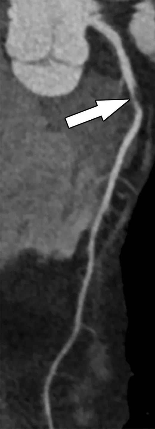
- Koroner BT anjiyografide koroner lümen ve darlık değerlendirilir.
- Kalsiyum blooming gerçek lümen çapını olduğundan küçük gösterebilir; yoğun kalsifik segmentte kesin derece güvenilmez olabilir.
- Hareket, gürültü, yetersiz kontrastlanma veya stent metal artefaktı varsa segmenti tanısal olmayan olarak işaretle.
Ölçütler ve sınıflamalar
- CAD-RADS 2.0 stenoz kategorileri
- Kaynaklarda desteklenen CAD-RADS stenoz kategorileri: 0: koroner hastalık bulgusu yok; 1: %1–24; 2: %25–49; 3: %50–69; 4: %70–99; 5: tam oklüzyon.
- CAD-RADS 2.0 stenoz derecesi
- CAD-RADS, hasta düzeyindeki görüntüleme sonucunu en ciddi koroner lezyona göre özetler. CAD-RADS 0 hem plak hem darlık yokluğunu ifade eder.
Normalde
CAD-RADS 0, koroner BT anjiyografide koroner arter hastalığına ilişkin bulgu olmadığını ifade eder. Koroner damarlar, eğrisel çok düzlemli rekonstrüksiyonda ardışık kesitlerle incelenebilir. CAD-RADS 0 için hem koroner plak hem de lümen darlığı bulunmamalıdır; yalnızca darlık görülmemesi, nonobstrüktif plağı dışlamaz.
Normal BT ile kıyasla
Aynı bölgenin, kaynak veri setinde patoloji içermediği belirtilen bir BT incelemesini kesit kesit kaydırın; organ sınırlarını açarak anatomiyi hasta görüntüsüyle karşılaştırın. Küçük rastlantısal bulgular tümüyle dışlanmamıştır.
Kaynak: TotalSegmentator v2.01 veri seti (Wasserthal ve ark., Radiology: Artificial Intelligence 2023), CC BY 4.0, vaka s0977; organ sınırları veri setinin otomatik segmentasyonundan, birleştirilerek sadeleştirildi. Görüntüler 2,5 mm kesit aralığına yeniden örneklendi.
Ayırıcı tanı
- Koroner spazm
- fokal daralma plak olmaksızın görülebilir; tek anlık BT görüntüsü spazmı aterosklerozdan kesin ayıramayabilir.
- Spontan koroner arter diseksiyonunda OCT'de intramural hematom ve yalancı lümen morfolojisi tanıyı destekler.
- Miyokard köprüsü
- özellikle LAD orta segmentinde intramiyokardiyal seyir ve sistolik sıkışma görünümü verir.
- Koroner arter anomalisi
- beklenmedik çıkış veya seyir darlık gibi izlenebilir; damar ostiumu ve üç boyutlu seyri takip edilir.
Tuzaklar
- Yoğun kalsifik plağın blooming artefaktı lümeni örter ve stenozu fazla tahmin ettirebilir; bu segmentte kesin yüzde yerine değerlendirme kısıtını raporla.
- Koroner BT anjiyografide kalsiyum bloomingi ve sınırlı uzaysal çözünürlük, stenoz derecesinin fazla tahmin edilmesine yol açabilir.
- Hareket basamaklanması küçük koroner dallarda lümen sınırını bozabilir; kesinti tek düzlemdeyse ardışık faz ve rekonstrüksiyonları kontrol et.
- Anatomik darlık, iskemiyle eş anlamlı değildir; CAD-RADS stenoz kategorisini fizyolojik test sonucuyla karıştırma.
Kendini dene
Koroner BT anjiyografide aşırı vasküler kalsifikasyonlar hangi artefakte yol açar ve bunun etkisi nedir?
Cevabı göster
B) Blooming artefaktı, stenozu abartabilir. S2 kaynağında, aşırı kalsifikasyonların kalsiyum blooming artefaktına neden olarak pozitif prediktif değeri düşürdüğü ve stenoz derecesinin abartılabileceği ifade edilmiştir.
Koroner BT anjiyografinin anatomik değerlendirmesini işlevsel değerlendirmeyle tamamlayabilen teknikler hangileridir?
Cevabı göster
CT-MPI ve CT-FFR. CT-MPI ve CT-FFR, koroner BT anjiyografiye ek olarak miyokard perfüzyonunu veya lezyonun fizyolojik sonuçlarını değerlendirebilir.
İlgili konular
- CAD-RADS 2.0 raporlama kavramı
- Koroner plak tipleri: kalsifiye, kısmen kalsifiye ve kalsifiye olmayan
Kaynaklar
Bu sayfadaki 37 cümle ve 2 quiz sorusu aşağıdaki kaynaklardan alıntılarla otomatik olarak karşılaştırıldı; 10 cümle bu karşılaştırmada düzeltildi (2026-10-09).
- Coronary Artery Disease in Male Athletes—Is Sport Healthy in the Long Run?pmc.ncbi.nlm.nih.gov
- Diagnostic Performance of Photon-Counting CT Angiography in Vascular Stenosis Assessment: A Systematic Review and Meta-Analysispmc.ncbi.nlm.nih.gov
- The imaging spectrum of myocardial infarction and its associated complications: a contemporary pictorial review of computed tomography and magnetic resonance imagingpmc.ncbi.nlm.nih.gov
- Accuracy of deep learning in the differential diagnosis of coronary artery stenosis: a systematic review and meta-analysispmc.ncbi.nlm.nih.gov
- Cardiac Computed Tomography: Technological Developments and Clinical Applicationspmc.ncbi.nlm.nih.gov
- Coronary Artery Calcification: Current Concepts and Clinical Implications.pubmed.ncbi.nlm.nih.gov
- Histopathological validation of optical coherence tomography findings of the coronary arteries.pubmed.ncbi.nlm.nih.gov
- Cardiac hybrid imaging.pubmed.ncbi.nlm.nih.gov
- Long-term Management of the Arterial Switch Patient.pubmed.ncbi.nlm.nih.gov
- The performance of non-invasive tests to rule-in and rule-out significant coronary artery stenosis in patients with stable angina: a meta-analysis focused on post-test disease probability.pubmed.ncbi.nlm.nih.gov
- Role of cardiac CTA in estimating left ventricular volumes and ejection fraction.pubmed.ncbi.nlm.nih.gov
- Acute coronary syndrome on non-electrocardiogram-gated contrast-enhanced computed tomography.pubmed.ncbi.nlm.nih.gov
- Role of invasive imaging in acute and long-term assessment of bioresorbable scaffold technology.pubmed.ncbi.nlm.nih.gov
- 15O-Water PET MPI: Current Status and Future Perspectives.pubmed.ncbi.nlm.nih.gov
- Noninvasive physiologic assessment of coronary stenoses using cardiac CT.pubmed.ncbi.nlm.nih.gov
- Computed tomographic evaluation of myocardial ischemia.pubmed.ncbi.nlm.nih.gov
- Update of 18 F-flurpiridaz.pubmed.ncbi.nlm.nih.gov
- Noninvasive imaging in coronary artery disease.pubmed.ncbi.nlm.nih.gov
- CT-based fractional flow reserve: development and expanded application.pubmed.ncbi.nlm.nih.gov
- Diffusely impaired myocardial perfusion in patients with suspected coronary artery disease: Clinical utility of relative perfusion.pubmed.ncbi.nlm.nih.gov
- Low- versus high-concentration iodine contrast media for coronary computed tomography angiography and dynamic stress computed tomography perfusion: a study protocol for a prospective randomized trial.pubmed.ncbi.nlm.nih.gov
- Diffuse Pericoronary Soft-Tissue Cuffing on Coronary Computed Tomography Angiography in a Patient with Unstable Angina: Possible IgG4-Related Coronary Periarteritis.pubmed.ncbi.nlm.nih.gov
- Advanced quantitative CT for coronary artery disease: integrating stenosis quantification, plaque quantification, and fractional flow reserve-CT.pubmed.ncbi.nlm.nih.gov
- Sleep Phenotype, Shift-Related Occupation, and Coronary Plaque Complexity in Young Adults: Association with CT-Derived Functional Ischemia.pubmed.ncbi.nlm.nih.gov
- Spectral computed tomography characterization of coronary atherosclerotic plaque: principles, imaging biomarkers, and clinical significance.pubmed.ncbi.nlm.nih.gov
- Diagnostic Accuracy of Quantitative Coronary Analysis as Compared with Instantaneous Wave-Free Ratio in Intermediate Coronary Lesions: A Prospective Vessel-Level Study.pubmed.ncbi.nlm.nih.gov
- Coronary Arterial Anomalies in Adults with Congenital Heart Disease: From Detection to Management Strategies.pubmed.ncbi.nlm.nih.gov
- Exertional Syncope Caused by Hemodynamically Significant Myocardial Bridging With Coronary Vasospasm.pubmed.ncbi.nlm.nih.gov
- Case Report: A rare case of multi-vessel coronary artery-coronary sinus fistula combined with multiple coronary aneurysms initially presenting with frequent premature ventricular complexes.pubmed.ncbi.nlm.nih.gov
- South African Flag Sign During Atrial Fibrillation: Physician Recognition of Acute Proximal Left Anterior Descending Artery Occlusion Despite No Automated ST-Segment Elevation Myocardial Infarction Alert.pubmed.ncbi.nlm.nih.gov
- Refractory Kawasaki Disease in Infants Complicated with Giant Coronary Artery Aneurysms and Takayasu Arteritis: A Rare Case Report and Literature Review.pubmed.ncbi.nlm.nih.gov
- A novel multimodal imaging approach for working diagnosis of acute myocardial infarction with non-obstructive coronary arteries: a promising diagnostic strategy.pubmed.ncbi.nlm.nih.gov
- From intravascular imaging to adaptive vascular care: intelligent photonics and digital twins in panvascular disease.pubmed.ncbi.nlm.nih.gov
- Cardiac Computed Tomography in Structural Heart Interventions: From Preprocedural Planning to Procedural Strategy.pubmed.ncbi.nlm.nih.gov
- Integrating Invasive and Noninvasive Imaging for Coronary Atherosclerosis: A Systematic Review With Pragmatic Algorithms From Anatomy to Physiology.pubmed.ncbi.nlm.nih.gov
- Emerging quantitative CCTA imaging biomarkers for cardiovascular risk stratification: a narrative review.pubmed.ncbi.nlm.nih.gov
- From anatomy to vulnerability: A comprehensive appraisal of coronary physiology and plaque characterization.pubmed.ncbi.nlm.nih.gov
- Coronary plaque burden assessed by coronary CT angiography in individuals with type 1 diabetes and healthy controls.pubmed.ncbi.nlm.nih.gov
- Clinical benefits and current challenges of photon-counting detector CT in vascular imaging.pubmed.ncbi.nlm.nih.gov
- CAD-RADS: A Standardized Reporting System for Coronary CT Angiography.pubmed.ncbi.nlm.nih.gov
- Clinical Applications of Artificial Intelligence in Cardiac CT: From Coronary CT Angiography to CT-Derived Fractional Flow Reserve and Myocardial Perfusion Imaging.pubmed.ncbi.nlm.nih.gov
- Radiation dose optimization in coronary CT angiography clinical application of absolute phase gating in patients with atrial fibrillation.pubmed.ncbi.nlm.nih.gov
- Age-Dependent Efficacy of Incidental Coronary Artery Calcium on Pre-CAG Chest CT: Potentially Missed Prevention Opportunities in Chinese Population.pubmed.ncbi.nlm.nih.gov
- Coronary CT Angiography in PCI Planning: Advances, Clinical Applications, and Challenges.pubmed.ncbi.nlm.nih.gov
- Intracoronary imaging for left main percutaneous coronary intervention: a clinical consensus statement of the European Association of Percutaneous Cardiovascular Interventions (EAPCI) of the ESC and the European Bifurcation Club (EBC).pubmed.ncbi.nlm.nih.gov
- Rationale and Design of the "PRECISION-CT" Study-A Prospective Evaluation of Coronary CTA Integration for Strategy Improvement and Optimization of PCI in Chronic Coronary Syndrome.pubmed.ncbi.nlm.nih.gov
- Severe arterial hypertension, a silent complication of Williams-Beuren syndrome: A case report with literature review.pubmed.ncbi.nlm.nih.gov
- FFR-CT: Technical Advances and Implementation in Clinical Practice.pubmed.ncbi.nlm.nih.gov
- CT - derived fractional flow reserve can predict recurrent ischemia in patients with MCA stenosis.pubmed.ncbi.nlm.nih.gov
- Simulation of calcium blooming artifacts in coronary CT angiography from digital phantoms.pubmed.ncbi.nlm.nih.gov
- Ultra-High-Resolution Photon-Counting CT Angiography for Noninvasive Detection of Neurovascular In-Stent Restenosis.pubmed.ncbi.nlm.nih.gov
- Coronary Computed Tomography Angiography Demonstrates a High Burden of Coronary Artery Disease Despite Low-Risk Nuclear Studies in Pre-Liver Transplant Evaluation.pubmed.ncbi.nlm.nih.gov
- Deep learning-based assessment of coronary artery disease using curved-multiplanar reconstruction: comprehensive evaluation of stenosis, calcification, and plaque.pubmed.ncbi.nlm.nih.gov
- Quantifying Carotid Stenosis: History, Current Applications, Limitations, and Potential: How Imaging Is Changing the Scenario.pubmed.ncbi.nlm.nih.gov
- Determinants of Non-calcified Low-Attenuation Coronary Plaque Burden in Patients Without Known Coronary Artery Disease: A Coronary CT Angiography Study.pubmed.ncbi.nlm.nih.gov
- Pictorial review: a step-by-step guide to coronary CT angiography with photon-counting detector CT.pubmed.ncbi.nlm.nih.gov
- Cannabis-Associated Spontaneous Coronary Artery Dissection in a Community Hospital: A Case-Based Focused Literature Review.pubmed.ncbi.nlm.nih.gov
- Optical Coherence Tomography: Case Series and Review on Differentiating Spontaneous Coronary Artery Dissection From Recanalized Thrombus.pubmed.ncbi.nlm.nih.gov
- Beyond Angiography: Cardiac CT for Planning Complex PCI in Calcified Coronary Lesions.pubmed.ncbi.nlm.nih.gov
- Comparative diagnostic performance and stability of deep learning- and CFD-based CT-FFR across vessels, cardiac phases, and centers.pubmed.ncbi.nlm.nih.gov
- Clinical Management Across CAD-RADS Categories in Patients with Intermediate CT-FFR.pubmed.ncbi.nlm.nih.gov
- Comparative evaluation of large language models for generating CAD-RADS 2.0-compliant diagnostic conclusions in cardiac CT reports.pubmed.ncbi.nlm.nih.gov
- Multimodality Non-Invasive Imaging Approach in Acute Coronary Syndrome: Diagnostic and Prognostic Assessment.pubmed.ncbi.nlm.nih.gov
- Lipoprotein(a) is associated with coronary atheroma progression: analysis from a serial coronary computed tomography angiography study.pubmed.ncbi.nlm.nih.gov
- Kounis Syndrome in the Modern Era: A Comprehensive Review of Allergic Acute Coronary Syndromes.pubmed.ncbi.nlm.nih.gov
- Diagnostic Advancements in MINOCA: Do They Translate to a Better Clinical Outcome? A Review of the Literature.pubmed.ncbi.nlm.nih.gov
- Beyond Stenosis: Mechanism-Based Multimodality Imaging and Invasive Coronary Function Testing for Endotype Definition in ANOCA/INOCA.pubmed.ncbi.nlm.nih.gov
- Operative Procedures for Ultrasound Assessment of Extracranial Artery Disease: A Narrative Review by the Italian Society for Vascular Investigation (SIDV).pubmed.ncbi.nlm.nih.gov
- Mechanisms of aortic dissection: From pathological changes to experimental and <i>in silico</i> models.pubmed.ncbi.nlm.nih.gov
- Protruding Aortic Plaque and Coronary Plaque Vulnerability.pubmed.ncbi.nlm.nih.gov
- Intramyocardial Left Anterior Descending Artery Extending Toward the Right Ventricle Demonstrated on Coronary CT Angiography.pubmed.ncbi.nlm.nih.gov
- Coronary Artery Anomalies Revisited: Description of the Types, Pathophysiology and Treatment Options Based on Latest Guidelines.pubmed.ncbi.nlm.nih.gov
- Single Coronary Artery in the Context of Vascular Disease: Anatomy, Development, Multimodality Imaging, and Clinical Interpretation.pubmed.ncbi.nlm.nih.gov
- Quadricuspid Aortic Valve: Imaging, Diagnosis, and Prognosis.pubmed.ncbi.nlm.nih.gov
- Therapeutic Management of Anomalous Coronary Arteries Originating From the Opposite Sinus of Valsalva: Current Evidence, Proposed Approach, and the Unknowing.pubmed.ncbi.nlm.nih.gov
- CT angiography after 20 years: a transformation in cardiovascular disease characterization continues to advance.pubmed.ncbi.nlm.nih.gov
- Subtraction fractional flow reserve with computed tomography and pericoronary fat attenuation index enhances the identification of revascularization needs in patients.pubmed.ncbi.nlm.nih.gov
- Ultrasound-Based Image Analysis for Predicting Carotid Artery Stenosis Risk: A Comprehensive Review of the Problem, Techniques, Datasets, and Future Directions.pubmed.ncbi.nlm.nih.gov
- Long-term safety and effectiveness of the "OptEase" vena cava filter.pubmed.ncbi.nlm.nih.gov
- Case Report: Iatrogenic left main coronary artery dissection during premature ventricular contraction ablation managed with emergent coronary artery bypass grafting.pubmed.ncbi.nlm.nih.gov
- Coronary Computed Tomography Angiography for the Diagnosis and Revascularization Guidance of Coronary Bifurcation Lesions: A Contemporary Review.pubmed.ncbi.nlm.nih.gov
- Multimodal ultrasound and computed tomography imaging may improve the discrimination between symptomatic and asymptomatic patients with carotid stenosis.pubmed.ncbi.nlm.nih.gov
- Diagnostic performance of coronary computed tomography (CT) angiography without electrocardiographic (ECG)-gating: comparison with invasive coronary angiography.pubmed.ncbi.nlm.nih.gov
- AI-driven radiomics and functional imaging integration for intelligent identification of coronary high-risk plaques and prediction of cardiovascular events.pubmed.ncbi.nlm.nih.gov
- Artificial Intelligence in Cardiovascular CT and MR Imaging.pubmed.ncbi.nlm.nih.gov
- Artificial Intelligence Advancements in the Cardiovascular Imaging of Coronary Atherosclerosis.pubmed.ncbi.nlm.nih.gov
- The current state of artificial intelligence-based invasive coronary angiography image analysis: a systematic review.pubmed.ncbi.nlm.nih.gov
- Artificial Intelligence to Assist in Exclusion of Coronary Atherosclerosis During CCTA Evaluation of Chest Pain in the Emergency Department: Preparing an Application for Real-world Use.pubmed.ncbi.nlm.nih.gov
- Standards for quantitative assessments by coronary computed tomography angiography (CCTA): An expert consensus document of the society of cardiovascular computed tomography (SCCT).pubmed.ncbi.nlm.nih.gov
- Diagnostic Performance of Photon-Counting CT Angiography in Vascular Stenosis Assessment: A Systematic Review and Meta-Analysis.pubmed.ncbi.nlm.nih.gov
- AI-Driven Quantitative Coronary CT Angiography in Suspected Coronary Artery Disease: Multicenter CONFIRM2 Registry.pubmed.ncbi.nlm.nih.gov
- Deep Learning-based Monoenergetic Imaging for Calcified Coronary Stenosis Assessment at Energy-integrating Detector CT.pubmed.ncbi.nlm.nih.gov
- Prospective comparison of ultra-high-resolution photon-counting detector CT coronary angiography versus conventional CT.pubmed.ncbi.nlm.nih.gov
- Total laparoscopic radical hysterectomy for cervical cancer in prolapsed uterus.pubmed.ncbi.nlm.nih.gov
- Commentary: an eye on PET quantification.pubmed.ncbi.nlm.nih.gov
- Left main coronary artery stenosis assessment on coronary CT angiography.pubmed.ncbi.nlm.nih.gov
- UK Kidney Association Clinical Practice Guideline on vascular access for haemodialysis.pubmed.ncbi.nlm.nih.gov
- Computational Analysis of Intravascular OCT Images for Future Clinical Support: A Comprehensive Review.pubmed.ncbi.nlm.nih.gov
- Interrater Variability of ML-Based CT-FFR in Patients without Obstructive CAD before TAVR: Influence of Image Quality, Coronary Artery Calcifications, and Location of Measurement.pubmed.ncbi.nlm.nih.gov
- Coronary computed tomography angiography for clinical practice.pubmed.ncbi.nlm.nih.gov
- Comparative diagnostic performance of coronary CT angiography and invasive coronary angiography in patients with suspected coronary artery disease.pubmed.ncbi.nlm.nih.gov
- The Diagnostic and Prognostic Impact of Cardiovascular Magnetic Resonance in Ischemic Heart Disease: A Systematic Review.pubmed.ncbi.nlm.nih.gov
- Case Report: Fabry disease mimicking coronary artery disease and hypertrophic cardiomyopathy-a 15-year diagnostic delay.pubmed.ncbi.nlm.nih.gov
- Technical Recommendations for Computed Tomography Guidance of Intervention in the Right Ventricular Outflow Tract: Native RVOT, Conduits and Bioprosthetic Valves: A White Paper of the Society of Cardiovascular Computed Tomography (SCCT), Congenital Heart Surgeons' Society (CHSS), and Society for Cardiovascular Angiography & Interventions (SCAI).pubmed.ncbi.nlm.nih.gov
- Technical Recommendations for Computed Tomography Guidance of Intervention in the Right Ventricular Outflow Tract: Native RVOT, Conduits, and Bioprosthetic Valves.pubmed.ncbi.nlm.nih.gov
- Diffuse coronary artery disease among other atherosclerotic plaque characteristics by coronary computed tomography angiography for predicting coronary vessel-specific ischemia by fractional flow reserve.pubmed.ncbi.nlm.nih.gov
- A Prospective Evaluation of Optical Coherence Tomography in Below-the-Knee Endovascular Interventions.pubmed.ncbi.nlm.nih.gov
- Ultrasound imaging for risk assessment in atherosclerosis.pubmed.ncbi.nlm.nih.gov
- Coronary computed tomographic angiography (CCTA) in community hospitals: "current and emerging role".pubmed.ncbi.nlm.nih.gov
- A Comparison of Coupling Strategies for 1D-3D Simulations of Coronary Hemodynamics.pubmed.ncbi.nlm.nih.gov
- Society for Cardiovascular Magnetic Resonance 2024 Cases of SCMR case series.pubmed.ncbi.nlm.nih.gov
- Coronary angiography: a review of the state of the art and the evolution of angiography in cardio therapeutics.pubmed.ncbi.nlm.nih.gov
- Deciphering age- and sex-specific patterns of coronary artery atherosclerosis from a large Chinese cohort.pubmed.ncbi.nlm.nih.gov
- Idiopathic IgG4-negative perivascular fibrosis with extensive thoracic aortic, coronary artery, and supra-aortic branch involvement: A case report.pubmed.ncbi.nlm.nih.gov
- Clinical implications of vessel wall imaging-State-of-the-art review.pubmed.ncbi.nlm.nih.gov
Eğitim amaçlıdır; hasta başında tanı ve tedavi kararının yerine geçmez, tıbbi tavsiye değildir. Metinler yapay zekâ desteğiyle yazıldı ve otomatik bir adımda kaynak alıntılarıyla karşılaştırıldı; bu bir tıbbi doğrulama değildir ve içerik uzman radyolog incelemesinden geçmedi. Klinik kararlarda güncel kılavuzları, kurum protokollerini ve radyoloji raporunu esas alın.