Özet
Aort çapının doğru ölçümü, aort hastalıklarının kılavuzlara dayalı yönetimi için önemlidir. Aort kökü kalp hareketine duyarlıdır; EKG kapılaması hareket artefaktını azaltıp kenar görünümünü iyileştirerek ölçüm değişkenliğini düşürür ve merkez çizgiye dik rekonstrüksiyonlar çap ölçümünde kullanılabilir. Kontrastlı BT, aortayı ve dallarını kısa sürede görüntüleyerek duvar ile lümenin ayırt edilmesine ve belirlenmiş anatomik seviyelerde çap ölçümüne olanak verir. Standartlaştırılmamış ölçüm yöntemleri aort çapının olduğundan büyük ya da küçük sınıflandırılmasına ve büyüme hızının hatalı hesaplanmasına yol açabilir.
Faz ve pencere
- BTA tanısal
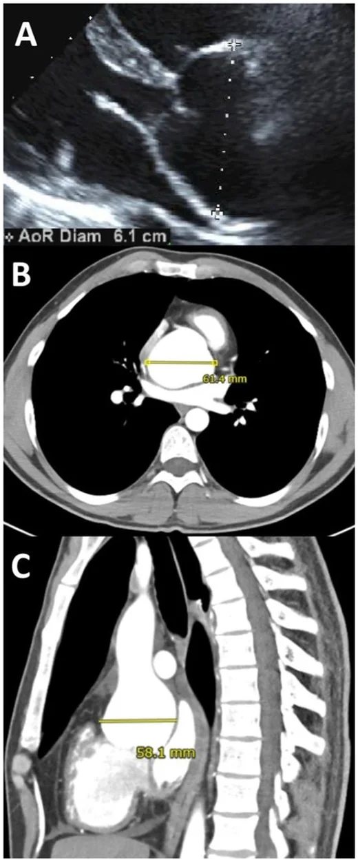
- EKG kapılamalı görüntüler aort kökü değerlendirmesinde hareket artefaktını azaltıp kenar görünümünü iyileştirir; aort çapları merkez çizgiye dik kesitlerde ölçülebilir. Aort kökü sinüs-komissür yöntemiyle ölçülür; çap, merkez çizgiye dik ölçüm düzleminde dış sınırdan değerlendirilir ve kalın duvar trombüsü ölçümün doğruluğunu etkileyebilir.
Önerilen pencereler: Anjiyo (G 600 / M 150), Mediasten (G 350 / M 50).
BT bulguları
- Aort çapı merkez çizgiye dik MPR düzleminde ölçülür; abdominal aort anevrizmasında standartlaştırılmamış ölçüm yöntemleri çapın sınıflandırılmasında ve büyüme hızının hesaplanmasında hataya yol açabilir.
- Kök asimetrisi — biküspit aort kapağında kök dairesel olmayabilir ve maksimum çapın yönü kapak açıklığının yönünden farklı olabilir.
- EKG kapılama etkisi — kök ve çıkan aortada hareket bulanıklığı azaldıkça lümen kenarı daha güvenilir seçilir.
- Dış çap ölçümü — aort çapı, merkez çizgiye dik ölçüm düzleminde en büyük dış çap olarak tanımlanabilir.
- Aynı seviye eşleştirmesi — sinüsler, sinotübüler bileşke, çıkan aorta ve inen aorta ölçümleri seri tetkikte aynı anatomik noktalardan alınır.
- Lümen ile dış çap farkı — kalın duvar trombüsü bulunan segmentte otomatik çap ölçümü hatalı olabilir; ölçüm bu bölgede dikkatle değerlendirilmelidir.
Normalde
Aort değerlendirmesinde sinüs Valsalva, sinotübüler bileşke, çıkan aorta, arkus ve inen aorta gibi anatomik seviyeler ayrı ayrı ölçülebilir. Ölçüm düzleminin aort merkez çizgisine dik olmasına dikkat edin; biküspit aort kapağında kök asimetrisi maksimum çapın yönünü etkileyebileceğinden kök ölçümünü yorumlarken bu özelliği göz önünde bulundurun.
Normal BT ile kıyasla
Aynı bölgenin, kaynak veri setinde patoloji içermediği belirtilen bir BT incelemesini kesit kesit kaydırın; organ sınırlarını açarak anatomiyi hasta görüntüsüyle karşılaştırın. Küçük rastlantısal bulgular tümüyle dışlanmamıştır.
Kaynak: TotalSegmentator v2.01 veri seti (Wasserthal ve ark., Radiology: Artificial Intelligence 2023), CC BY 4.0, vaka s0115; organ sınırları veri setinin otomatik segmentasyonundan, birleştirilerek sadeleştirildi. Görüntüler 2,5 mm kesit aralığına yeniden örneklendi.
Ayırıcı tanı
- Gerçek aort dilatasyonu
- centerline'a dik düzlemde de geniş çap korunur ve komşu segmentlerle geçişli ya da fokal genişleme görülür.
- Oblik kesit yalancı genişliği
- standart aksiyel görüntüde aort uzun ekseni kesite eğik girer; dik MPR'de çap küçülür.
- Kök asimetrisi
- sinüs-sinüs ölçümleri farklı olabilir; tek kısa çap kök boyutunu olduğundan küçük gösterebilir.
- Pulsasyon bulanıklığı
- kapılanmamış çıkan aortada kenar çiftlenir; aynı düzeyde kapılanmış rekonstrüksiyon daha keskin sınır verir.
Tuzaklar
- Kavisli çıkan aortayı aksiyel düzlemde ölçmek lümeni elips gibi gösterip çapı fazla hesaplatabilir; aynı seviyeye centerline'a dik çift eğik MPR yerleştirin.
- Kökün tek çapını raporlamak asimetrik sinüs geometrisini örter; sinüs-sinüs düzlemini ve gerekiyorsa kısa-uzun ekseni birlikte kaydedin.
- Duvar hastalığında iç sınırdan ölçmek dışa doğru kalınlaşmayı dışarıda bırakır; hematom veya ateromlu segmentte duvarın dış marjını izleyin.
Kendini dene
Aksiyel BT'de çıkan aorta geniş görünüyor; centerline'a dik MPR'de çap küçülüyor. En olası açıklama?
Cevabı göster
Oblik kesit etkisi. Eğri aortaya dik olmayan aksiyel düzlem çapı uzun eksen boyunca abartabilir; dik MPR gerçek kesit boyutunu verir. Gerçek anevrizma dik düzlemde de geniş kalır; hematom duvarı kalınlaştırır, psödoanevrizma ise sınırlı sakküler dışa çıkıntıdır.
Aort kökü ölçümünde kaynakta belirtilen yöntem hangisidir?
Cevabı göster
Sinüs-komissür yöntemi. Aort kökü, kaynakta sinüs-komissür yöntemiyle ölçülmüştür.
Kaynaklar
Bu sayfadaki 33 cümle ve 2 quiz sorusu aşağıdaki kaynaklardan alıntılarla otomatik olarak karşılaştırıldı; 10 cümle bu karşılaştırmada düzeltildi (2026-10-09).
- Evaluation of clinical performance of artificial intelligence-assisted measurement of thoracic aortic diameter on non-contrast CTpmc.ncbi.nlm.nih.gov
- Moderate aortic stenosis: diagnostic pitfalls and the role of multimodality imaging in risk stratification and clinical managementpmc.ncbi.nlm.nih.gov
- Nutcracker syndrome with pelvic congestion syndrome: A case reportpmc.ncbi.nlm.nih.gov
- Correlation Between Aortic Valve Area Measured by Cardiac Computed Tomography and Transthoracic Echocardiography in Patients with Aortic Stenosispmc.ncbi.nlm.nih.gov
- Haemodynamic Predictors of Early Aortic Growth in Uncomplicated Type B Dissection: A Cohort Studypmc.ncbi.nlm.nih.gov
- Multimodality imaging in bicuspid aortic valve.pubmed.ncbi.nlm.nih.gov
- Automatic aortic root landmark detection in CTA images for preprocedural planning of transcatheter aortic valve implantation.pubmed.ncbi.nlm.nih.gov
- Importance of vascular calcification in kidney transplant recipients.pubmed.ncbi.nlm.nih.gov
- Vascular Deformation Mapping of Abdominal Aortic Aneurysm.pubmed.ncbi.nlm.nih.gov
- Aortic volume determines global end-diastolic volume measured by transpulmonary thermodilution.pubmed.ncbi.nlm.nih.gov
- Ascending aortic diameter and metabolic syndrome in Korean men.pubmed.ncbi.nlm.nih.gov
- Outer Wall Segmentation of Abdominal Aortic Aneurysm by Variable Neighborhood Search Through Intensity and Gradient Spaces.pubmed.ncbi.nlm.nih.gov
- Automatic 3D aortic annulus sizing by computed tomography in the planning of transcatheter aortic valve implantation.pubmed.ncbi.nlm.nih.gov
- Predicting the degree of difficulty of the trans-radial approach in cerebral angiography.pubmed.ncbi.nlm.nih.gov
- Aortic Root Anatomy Is Related to the Bicuspid Aortic Valve Phenotype.pubmed.ncbi.nlm.nih.gov
- Principles and applications of dual source CT.pubmed.ncbi.nlm.nih.gov
- Measurement of arterial activity on routine FDG PET/CT images improves prediction of risk of future CV events.pubmed.ncbi.nlm.nih.gov
- On the assessment of arterial compliance from carotid pressure waveform.pubmed.ncbi.nlm.nih.gov
- Predictive Model for Thrombus Formation After Transcatheter Valve Replacement.pubmed.ncbi.nlm.nih.gov
- Determining the normal aorta size in children.pubmed.ncbi.nlm.nih.gov
- Clinical benefits and risks of the wall-push technique in endovascular repair of abdominal aortic aneurysm with severely angulated neck: a case report.pubmed.ncbi.nlm.nih.gov
- Free-breathing, blood-flow-independent, three-dimensional relaxation-enhanced angiography without contrast and triggering (REACT) for aorta measurement and diagnosis in patients with aortic disease: a comparison with computed tomography angiography.pubmed.ncbi.nlm.nih.gov
- Undiagnosed Thoracic Aortic Aneurysms: Prevalence, Risk Factors, Screening Modalities.pubmed.ncbi.nlm.nih.gov
- Preprocedural computed tomography angiography aortic root geometry and 1-year left ventricular reverse remodeling after transcatheter aortic valve replacement.pubmed.ncbi.nlm.nih.gov
- Predicting procedural outcomes in transcatheter aortic valve implantation: a scoping review of numerical patient-specific simulations.pubmed.ncbi.nlm.nih.gov
- CoreValve vs. Sapien 3 Transcatheter Aortic Valve Replacement: A Finite Element Analysis Study.pubmed.ncbi.nlm.nih.gov
- Tricuspid regurgitation: standardized stepwise work-up from referral to expert heart valve centre.pubmed.ncbi.nlm.nih.gov
- Feasibility and reproducibility of semi-automated magnetic resonance angiography measurements of the thoracic aorta using commercial software.pubmed.ncbi.nlm.nih.gov
- Isotropic, high-resolution, whole-chest inversion recovery contrast-enhanced magnetic resonance angiography in under 4.5 min using image-based navigator fluoro trigger.pubmed.ncbi.nlm.nih.gov
- From Bits to Bedside: From Data to Knowledge in Vascular Surgery.pubmed.ncbi.nlm.nih.gov
- Thoracic aortic disease in young patients: from genetics to management.pubmed.ncbi.nlm.nih.gov
- The Role of Echocardiography in the Assessment of Epicardial Adipose Tissue: A Systematic Review and Meta-analysis.pubmed.ncbi.nlm.nih.gov
- Magnetic Resonance Imaging and Computed Tomography for the Noninvasive Assessment of Arterial Aging: A Review by the VascAgeNet COST Action.pubmed.ncbi.nlm.nih.gov
- Computed Tomography Derived Coronary Triangulated Orifice Area-Deduction of a New Parameter for Follow-up After Surgical Correction of Anomalous Aortic Origin of Coronary Arteries and Call for Validation.pubmed.ncbi.nlm.nih.gov
- Artificial intelligence-powered solutions for automated aortic diameter measurement in computed tomography: a narrative review.pubmed.ncbi.nlm.nih.gov
- 2022 ACC/AHA guideline for the diagnosis and management of aortic disease: A report of the American Heart Association/American College of Cardiology Joint Committee on Clinical Practice Guidelines.pubmed.ncbi.nlm.nih.gov
- 2022 ACC/AHA Guideline for the Diagnosis and Management of Aortic Disease: A Report of the American Heart Association/American College of Cardiology Joint Committee on Clinical Practice Guidelines.pubmed.ncbi.nlm.nih.gov
- Coronary Computed Tomography Angiography to Guide Percutaneous Coronary Intervention: Expert Opinion from a SCAI/SCCT Roundtable.pubmed.ncbi.nlm.nih.gov
- Abdominal aortic aneurysm measurement at CT/MRI: potential clinical ramifications of non-standardized measurement technique and importance of multiplanar reformation.pubmed.ncbi.nlm.nih.gov
- The Systolic Anterior Motion After Mitral Valve Repair: A Narrative Review of Current Therapeutic Algorithms in the Operating Room.pubmed.ncbi.nlm.nih.gov
- Paravalvular regurgitation after transcatheter aortic valve replacement: incidence, quantification, and prognostic impact.pubmed.ncbi.nlm.nih.gov
- Unraveling Bicuspid Aortic Valve Enigmas by Multimodality Imaging: Clinical Implications.pubmed.ncbi.nlm.nih.gov
- Society for Cardiovascular Magnetic Resonance reference values ("normal values") in cardiovascular magnetic resonance: 2025 update.pubmed.ncbi.nlm.nih.gov
- Novel Techniques in Imaging Congenital Heart Disease: JACC Scientific Statement.pubmed.ncbi.nlm.nih.gov
- Fluid transport in the brain.pubmed.ncbi.nlm.nih.gov
- Operative Procedures for Ultrasound Assessment of Extracranial Artery Disease: A Narrative Review by the Italian Society for Vascular Investigation (SIDV).pubmed.ncbi.nlm.nih.gov
- Cardiac Imaging in the Diagnosis and Management of Heart Failure.pubmed.ncbi.nlm.nih.gov
- Regional variation in biomechanical properties of ascending thoracic aortic aneurysms.pubmed.ncbi.nlm.nih.gov
- Myval Beyond the Aortic Valve: Device-Anatomy Interaction, Procedural Strategy, and Clinical Evidence in Mitral, Tricuspid, and Pulmonary Positions.pubmed.ncbi.nlm.nih.gov
- Mitral Regurgitation in Contemporary Practice: From Diagnosis to Management.pubmed.ncbi.nlm.nih.gov
- Ascending Aortic Aneurysms: From Pathophysiology to Surgical Repair.pubmed.ncbi.nlm.nih.gov
- Risk factors of postoperative complications after F/B-TEVAR for aortic arch lesions: a multicenter retrospective analysis.pubmed.ncbi.nlm.nih.gov
- Early Outcomes of a Curvature-Guided Strategy for Dual-Branch Revascularization in Zone 1 TEVAR.pubmed.ncbi.nlm.nih.gov
- Dedicated Single-Branch Platforms for Totally Endovascular Zone 2 TEVAR with LSA Revascularization: A Comparison of Castor/Cratos and Gore TAG Thoracic Branch Endoprosthesis.pubmed.ncbi.nlm.nih.gov
- Advanced 3D echocardiographic assessment of the aortic valve and aortic root complex: a repair-oriented perspective.pubmed.ncbi.nlm.nih.gov
- Bicuspid Aortic Valve Disease-Associated Aortopathy in Pediatric Subjects-From Traditional Assessment to Current Advances and Future Perspectives: A Narrative Review.pubmed.ncbi.nlm.nih.gov
- Cardiac Computed Tomography in Structural Heart Interventions: From Preprocedural Planning to Procedural Strategy.pubmed.ncbi.nlm.nih.gov
- Biomechanical Variation of the Vessel Wall Along the Length of the Healthy Aorta-Linking Geometric, Flow-, and Pressure-Mediated Adaptations.pubmed.ncbi.nlm.nih.gov
- Mechanisms of aortic dissection: From pathological changes to experimental and <i>in silico</i> models.pubmed.ncbi.nlm.nih.gov
- Transcatheter Aortic Valve Therapy for Bicuspid Aortic Valve Stenosis.pubmed.ncbi.nlm.nih.gov
- Role of Echocardiography Before Transcatheter Aortic Valve Implantation (TAVI).pubmed.ncbi.nlm.nih.gov
- Effect of blending Jersey and Holstein-Friesian milk on Cheddar cheese processing, composition, and quality.pubmed.ncbi.nlm.nih.gov
- The role of computed tomography in pre-procedural planning of cardiovascular surgery and intervention.pubmed.ncbi.nlm.nih.gov
- Commentary: an eye on PET quantification.pubmed.ncbi.nlm.nih.gov
- CT and MRI in the Evaluation of Thoracic Aortic Diseases.pubmed.ncbi.nlm.nih.gov
- Assessment of Coronary Arteries during the Prenatal and Postnatal Periods using Two- and Three-dimensional Ultrasound.pubmed.ncbi.nlm.nih.gov
- Reference ranges ("normal values") for cardiovascular magnetic resonance (CMR) in adults and children: 2020 update.pubmed.ncbi.nlm.nih.gov
- The pivotal role of echocardiography in structural heart interventions: from planning to success.pubmed.ncbi.nlm.nih.gov
- MDCT evaluation of dynamic changes in aortic root parameters during the cardiac cycle in patients with aortic regurgitation.pubmed.ncbi.nlm.nih.gov
- Advanced quantitative CT for coronary artery disease: integrating stenosis quantification, plaque quantification, and fractional flow reserve-CT.pubmed.ncbi.nlm.nih.gov
- A novel physics-based model for fast computation of blood flow in coronary arteries.pubmed.ncbi.nlm.nih.gov
- Society for Cardiovascular Magnetic Resonance/European Society of Cardiovascular Imaging/American Society of Echocardiography/Society for Pediatric Radiology/North American Society for Cardiovascular Imaging Guidelines for the Use of Cardiac Magnetic Resonance in Pediatric Congenital and Acquired Heart Disease: Endorsed by The American Heart Association.pubmed.ncbi.nlm.nih.gov
- CT diagnosis and destiny of acute aortic intramural hematoma.pubmed.ncbi.nlm.nih.gov
- Diagnostic Management of Gastroenteropancreatic Neuroendocrine Neoplasms: Technique Optimization and Tips and Tricks for Radiologists.pubmed.ncbi.nlm.nih.gov
- SCMR Position Paper (2020) on clinical indications for cardiovascular magnetic resonance.pubmed.ncbi.nlm.nih.gov
- Imaging of the aortic root on high-pitch non-gated and ECG-gated CT: awareness is the key!pubmed.ncbi.nlm.nih.gov
- Society for Cardiovascular Magnetic Resonance/European Society of Cardiovascular Imaging/American Society of Echocardiography/Society for Pediatric Radiology/North American Society for Cardiovascular Imaging Guidelines for the use of cardiovascular magnetic resonance in pediatric congenital and acquired heart disease : Endorsed by The American Heart Association.pubmed.ncbi.nlm.nih.gov
- Clinical value of non-electrocardiogram-gated computed tomography angiography for planning transcatheter aortic valve implantation: A retrospective single-centre study.pubmed.ncbi.nlm.nih.gov
- An Overview of the Diagnostic and Treatment Strategies for Aortic Disease.pubmed.ncbi.nlm.nih.gov
- Construction and validation of risk prediction models for new- onset conduction block complications after transcatheter aortic valve replacement.pubmed.ncbi.nlm.nih.gov
- High-affinity alphavbeta3 integrin targeted optical probe as a new imaging biomarker for early atherosclerosis: initial studies in Watanabe rabbits.pubmed.ncbi.nlm.nih.gov
- Moderate aortic stenosis: diagnostic pitfalls and the role of multimodality imaging in risk stratification and clinical management.pubmed.ncbi.nlm.nih.gov
- Computed tomographic Characteristics of Presumed Normal Canine Iliosacral Lymph Nodes.pubmed.ncbi.nlm.nih.gov
- Evaluation of clinical performance of artificial intelligence-assisted measurement of thoracic aortic diameter on non-contrast CT.pubmed.ncbi.nlm.nih.gov
- The Correlates of Recoarctation After Repair in Pediatric Patients with Aortic Coarctation: A Real-World Descriptive Study.pubmed.ncbi.nlm.nih.gov
- CT-Centered Multimodality Imaging of Arterial Wall Fragility in Acute Aortic Syndromes: A Narrative Review of Imaging Markers and Clinical Implications.pubmed.ncbi.nlm.nih.gov
- Arterial-Venous Balance in the Human Renal Vasculature: Renal-Aortic and Renal-Caval Ratios and Factors on Contrast-Enhanced Computed Tomography.pubmed.ncbi.nlm.nih.gov
- From Modified Haller Index to a Novel Patented Anatomical Measurement Device: Engineering Development, Validation, and Clinical Applications of Non-Invasive Thoracic Morphometry.pubmed.ncbi.nlm.nih.gov
- Pericoronary adipose tissue imaging: quantitative assessment, artificial intelligence integration, and therapeutic modulation.pubmed.ncbi.nlm.nih.gov
- The Association of Coloproctology of Great Britain and Ireland consensus guidelines in emergency colorectal surgery.pubmed.ncbi.nlm.nih.gov
- Long-term safety and effectiveness of the "OptEase" vena cava filter.pubmed.ncbi.nlm.nih.gov
- A systematic approach for analysis, interpretation, and reporting of coronary CTA studies.pubmed.ncbi.nlm.nih.gov
- Chest Discomfort: Could Coronary Pathology Extend Beyond Atherosclerosis?pubmed.ncbi.nlm.nih.gov
- Aortic Arch Incision and Closure Technique (AICT) for Proximal Fixation of the Frozen Elephant Trunk.pubmed.ncbi.nlm.nih.gov
- Transesophageal Echocardiography in Transcatheter Mitral Valve Replacement.pubmed.ncbi.nlm.nih.gov
- Continuous-wave Doppler interrogation in valvular heart disease: pearls and pitfalls.pubmed.ncbi.nlm.nih.gov
- MDCT Imaging of Non-Traumatic Thoracic Aortic Emergencies and Its Impact on Diagnosis and Management-A Reappraisal.pubmed.ncbi.nlm.nih.gov
- Too Young, Too Silent: Painless Stanford Type A Aortic Dissection in a Young Adult.pubmed.ncbi.nlm.nih.gov
- Tertiary Syphilitic Aortitis Manifesting as Ascending Aortic Aneurysm and Dissection.pubmed.ncbi.nlm.nih.gov
- Role of Cardiac CT in the Evaluation of Valvular Heart Disease.pubmed.ncbi.nlm.nih.gov
- Transseptal Access to the Left Atrium: A Narrative Review of Techniques, Indications, and Device Innovations.pubmed.ncbi.nlm.nih.gov
- Strategies for Aortic Root Measurement in Patients Undergoing Surveillance for Thoracic Aortic Disease.pubmed.ncbi.nlm.nih.gov
- A difference that matters: the aortic root anatomy of large animal models vs. humans.pubmed.ncbi.nlm.nih.gov
- Imaging and Surveillance of Chronic Aortic Dissection: A Scientific Statement From the American Heart Association.pubmed.ncbi.nlm.nih.gov
- A simple patient-tailored aortic arch tangential angle measuring method to achieve better clinical results for thoracic endovascular repair of type B aortic dissection.pubmed.ncbi.nlm.nih.gov
- Cardiovascular CT in Bicuspid Aortic Valve Disease: A State-of-the-Art Narrative Review of Advances, Clinical Integration, and Future Directions.pubmed.ncbi.nlm.nih.gov
- Anatomical features of aortic root in patients with aortic stenosis treated by TAVR: an observational study.pubmed.ncbi.nlm.nih.gov
Eğitim amaçlıdır; hasta başında tanı ve tedavi kararının yerine geçmez, tıbbi tavsiye değildir. Metinler yapay zekâ desteğiyle yazıldı ve otomatik bir adımda kaynak alıntılarıyla karşılaştırıldı; bu bir tıbbi doğrulama değildir ve içerik uzman radyolog incelemesinden geçmedi. Klinik kararlarda güncel kılavuzları, kurum protokollerini ve radyoloji raporunu esas alın.