Özet
Aort kökü hareket artefaktı, özellikle EKG-gated olmayan toraks BT anjiyografisinde kalp atımının keskin aort duvarını bulanıklaştırmasıyla oluşur. Kısa lineer yalancı flep çıkan aort diseksiyonunu taklit edebileceğinden acil kararları ve rapor güvenini etkiler. Aksiyel kesitleri komşu düzlemler ve mümkünse EKG-gated rekonstrüksiyonla karşılaştırmak artefaktın süreksizliğini açığa çıkarır. Gerçek diseksiyonu artefakt saymak ölümcül tanıyı geciktirebilir; yalancı flep ise gereksiz girişimlere yol açabilir.
Faz ve pencere
- BTA tanısal
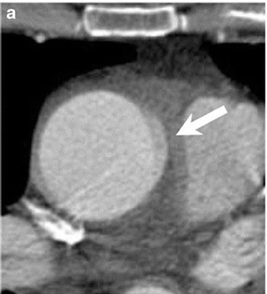
- Arteriyel kontrastla opak çıkan aortada hareket kaynaklı çizgi veya çift kontur, çoğunlukla aort kökü-sinotübüler bileşke çevresinde görülür; çizgi komşu kesitlerde ve ortogonal MPR’de damar duvarı boyunca süreklilik göstermez.
Önerilen pencereler: Anjiyo (G 600 / M 150), Mediasten (G 350 / M 50).
BT bulguları
- Kısa lineer yalancı flep — çıkan aorta lümeninde birkaç kesitte belirip devam etmeyen ince hipodens çizgi.
- Çift duvar konturu — hareket bulanıklığı nedeniyle aort kökünün ön veya sol-ön kenarında paralel kontur.
- Kesitler arasında kaybolma — çizgi kraniokaudal takipte kesilir ve aort boyunca gerçek bir intimal membran gibi uzanmaz.
- Ortogonal düzlem uyumsuzluğu — aksiyelde görülen çizgi sagittal veya koronal oblik planda devamlı bir flep oluşturmaz.
- Gated seride düzelme — aynı seviyedeki EKG-gated görüntüde yalancı kontur kaybolur veya belirgin azalır.
- Duvar devamlılığının korunması — aort dış konturu düzgün kalır; gerçek iki lümen, giriş yırtığı veya eşlik eden periaortik hematom gösterilmez.
Normalde
Normal çıkan aortanın kontrastlı lümeni homojen, iç sınırı tek ve düzgün konturludur; aort kökü ile pulmoner arter sınırları keskin görünür. Aynı seviyedeki komşu kesitlerde ve koronal-sagittal oblik MPR’de şüpheli çizginin devam edip etmediğini, dış duvarın kesintisiz olup olmadığını ve gated seriyle eşleşmesini karşılaştırın.
Normal BT ile kıyasla
Aynı bölgenin, kaynak veri setinde patoloji içermediği belirtilen bir BT incelemesini kesit kesit kaydırın; organ sınırlarını açarak anatomiyi hasta görüntüsüyle karşılaştırın. Küçük rastlantısal bulgular tümüyle dışlanmamıştır.
Kaynak: TotalSegmentator v2.01 veri seti (Wasserthal ve ark., Radiology: Artificial Intelligence 2023), CC BY 4.0, vaka s0977; organ sınırları veri setinin otomatik segmentasyonundan, birleştirilerek sadeleştirildi. Görüntüler 2,5 mm kesit aralığına yeniden örneklendi.
Ayırıcı tanı
- Gerçek aort diseksiyonu
- intimomedial flep birden çok kesitte ve düzlemde sürer, iki lümeni ayırabilir ve giriş yırtığı ya da dal damar ilişkisi gösterebilir.
- Aort intramural hematomu
- kontrastsız BT’de duvar içinde hilal biçimli hiperdens kalınlaşma görülür; lümen içi kısa hareket çizgisi değildir.
- SVC kaynaklı streak artefaktı
- yüksek yoğunluklu venöz kontrasttan çıkan radyal parlak-koyu çizgiler aortayı kesebilir.
- Aort kökü kalsifikasyonu
- yüksek atenüasyonlu odakların duvar boyunca yerleşmesiyle oluşur ve hareket artefaktıyla karıştırılabilir.
- Aort kapağı veya koroner ostium düzeyinde hareket artefaktı çift kontur ya da yalancı diseksiyon görünümü oluşturabilir.
Tuzaklar
- Hareket çizgisi aksiyel tek kesitte gerçek flep izlenimi verir; aortayı koronal-sagittal oblik MPR’de baştan sona izleyin.
- Hareket artefaktının yokluğu için tüm EKG-gated seriye değil, şüpheli kök düzeyindeki eşleşen gated görüntüye bakın.
- Kök yakınındaki çizgiyi otomatik olarak artefakt saymayın; devamlı intimal flep veya duvar hematomu varsa gerçek akut aort sendromunu araştırın.
Kendini dene
EKG-gated olmayan toraks BT’de kalp ve aort hareketi hangi yalancı görünümü oluşturabilir?
Cevabı göster
Diseksiyonu taklit eden yalancı flep veya çift kontur. Hareket artefaktları görüntülerin yorumlanmasını güçleştirebilir ve diseksiyonu taklit eden yalancı flep ya da çift kontur oluşturabilir.
Aort kökü içindeki çizgi gated CTA’da da sürüyor ve iki lümeni ayırıyor. Hangi tanı daha olası?
Cevabı göster
Aort diseksiyonu. İntimal membranın devam etmesi ve iki lümenin varlığı diseksiyonu destekler. Hareket çizgisi gated görüntüde azalır; vena cava superior kaynaklı streak artefaktı ışınsal çizgiler biçimindedir.
Bu konunun yer aldığı turlar
İlgili konular
Kaynaklar
Bu sayfadaki 33 cümle ve 2 quiz sorusu aşağıdaki kaynaklardan alıntılarla tek tek karşılaştırıldı; 5 cümle bu karşılaştırmada düzeltildi (2026-10-08).
- Single-Bolus Sequential Triple-Rule-Out CT Angiography: Image Quality and Radiation Dose on Wide-Area Detector and Dual-Source CTpmc.ncbi.nlm.nih.gov
- Aortic root motion artifacts on non-electrocardiographic-gated chest CT: A comparison of five CT scanner protocols including an ultrafast wide-detector CT systempmc.ncbi.nlm.nih.gov
- Aortic Dissection Mimicry Under Extracorporeal Membrane Oxygenation (ECMO) After Cardiac Arrest: A Case Report of Emergency Imaging Dilemmaspmc.ncbi.nlm.nih.gov
- Sequential Helical–Axial–Helical Triple-Rule-Out CT Angiography: Technical Feasibility and Territory-Specific Image Quality in the Emergency Departmentpmc.ncbi.nlm.nih.gov
- Evaluation of clinical performance of artificial intelligence-assisted measurement of thoracic aortic diameter on non-contrast CTpmc.ncbi.nlm.nih.gov
- ECG-gated Versus Non-ECG-gated High-pitch Dual-source CT for Whole Body CT Angiography (CTA).pubmed.ncbi.nlm.nih.gov
- Role of advanced imaging techniques in cardiac surgery: Aortic dissection.pubmed.ncbi.nlm.nih.gov
- High-pitch coronary CT angiography with third generation dual-source CT: limits of heart rate.pubmed.ncbi.nlm.nih.gov
- Computed Tomography Imaging Artifact Simulating Type A Aortic Dissection.pubmed.ncbi.nlm.nih.gov
- Motion correction for routine X-ray lung CT imaging.pubmed.ncbi.nlm.nih.gov
- Grade I traumatic aortic injury progression.pubmed.ncbi.nlm.nih.gov
- Role of Computed Tomography in Assessment of the Thoracic Aorta.pubmed.ncbi.nlm.nih.gov
- Impact of Thoracic Endovascular Aortic Repair on Pulsatile Circumferential and Longitudinal Strain in Patients With Aneurysm.pubmed.ncbi.nlm.nih.gov
- Influence of virtual monochromatic spectral image at different energy levels on motion artifact correction in dual-energy spectral coronary CT angiography.pubmed.ncbi.nlm.nih.gov
- [Contained rupture of the non-coronary sinus of Valsalva aneurysm into the right atrium (Sakakibara type IV) treated by surgery].pubmed.ncbi.nlm.nih.gov
- Imaging of the aortic root on high-pitch non-gated and ECG-gated CT: awareness is the key!pubmed.ncbi.nlm.nih.gov
- 128-slice CT angiography of the aorta without ECG-gating: efficacy of faster gantry rotation time and iterative reconstruction in terms of image quality and radiation dose.pubmed.ncbi.nlm.nih.gov
- Pitfalls in suspected acute aortic syndrome: impact of appropriate and if required repeated imaging.pubmed.ncbi.nlm.nih.gov
- Further Improving Image Quality of Cardiovascular Computed Tomography Angiography for Children With High Heart Rates Using Second-Generation Motion Correction Algorithm.pubmed.ncbi.nlm.nih.gov
- Single-Bolus Sequential Triple-Rule-Out CT Angiography: Image Quality and Radiation Dose on Wide-Area Detector and Dual-Source CT.pubmed.ncbi.nlm.nih.gov
- Aortic Dissection Mimicry Under Extracorporeal Membrane Oxygenation (ECMO) After Cardiac Arrest: A Case Report of Emergency Imaging Dilemmas.pubmed.ncbi.nlm.nih.gov
- Aortic root motion artifacts on non-electrocardiographic-gated chest CT: A comparison of five CT scanner protocols including an ultrafast wide-detector CT system.pubmed.ncbi.nlm.nih.gov
- Society for Cardiovascular Magnetic Resonance 2024 Cases of SCMR case series.pubmed.ncbi.nlm.nih.gov
- Artificial intelligence in aortic CT angiography: current applications and future perspectives.pubmed.ncbi.nlm.nih.gov
- Misdiagnosis of Aortic Dissection Due to Streak Artifact in the Descending Aorta.pubmed.ncbi.nlm.nih.gov
- MDCT Imaging of Non-Traumatic Thoracic Aortic Emergencies and Its Impact on Diagnosis and Management-A Reappraisal.pubmed.ncbi.nlm.nih.gov
- Multimodality Imaging Approach in Diagnosis and Follow-Up of Aortic Coarctation in Adulthood.pubmed.ncbi.nlm.nih.gov
- Heart in Focus: Advancing Pericardial Effusion Diagnosis With Point-of-Care Ultrasound.pubmed.ncbi.nlm.nih.gov
- Multimodality Imaging in the Evaluation of Hypertrophic Cardiomyopathy.pubmed.ncbi.nlm.nih.gov
- Mechanisms of aortic dissection: From pathological changes to experimental and <i>in silico</i> models.pubmed.ncbi.nlm.nih.gov
- Echocardiographic guidance in transcatheter tricuspid valve interventions.pubmed.ncbi.nlm.nih.gov
- Acute Traumatic Aortic Injury: What the Radiologist Needs to Know.pubmed.ncbi.nlm.nih.gov
- Transcatheter Tricuspid Valve Replacement for His Bundle Pacing Lead-Associated Tricuspid Regurgitation.pubmed.ncbi.nlm.nih.gov
- Sequential Helical-Axial-Helical Triple-Rule-Out CT Angiography: Technical Feasibility and Territory-Specific Image Quality in the Emergency Department.pubmed.ncbi.nlm.nih.gov
- CT-Centered Multimodality Imaging of Arterial Wall Fragility in Acute Aortic Syndromes: A Narrative Review of Imaging Markers and Clinical Implications.pubmed.ncbi.nlm.nih.gov
- CT Imaging Features of Acute Aortic Syndrome: A Case Series.pubmed.ncbi.nlm.nih.gov
- An Overview of the Diagnostic and Treatment Strategies for Aortic Disease.pubmed.ncbi.nlm.nih.gov
- Single-stage repair of congenital complete sternal agenesis with PFO-type interatrial communication and aortic arch variant complicated by an unexpected ascending aortic pseudoaneurysm: a case report.pubmed.ncbi.nlm.nih.gov
- Reversed Midgut Rotation in an Adult With Atypical Presentation as Upper Gastrointestinal Bleeding: A Case Report.pubmed.ncbi.nlm.nih.gov
- TotalSegmentator-AI for aortic segmentation in CT: Reliable performance in normal anatomy but limited utility in pathological aortic disease.pubmed.ncbi.nlm.nih.gov
- Complete abdominal aortic occlusion following seat-belt trauma: a proposal for early recognition.pubmed.ncbi.nlm.nih.gov
- Aortic Syndrome in an Elderly Female: A Case of Type A Intramural Hematoma.pubmed.ncbi.nlm.nih.gov
- Craniocervical arterial dissections with cerebral infarction: a five-case series with diagnostic application of vessel wall MRI.pubmed.ncbi.nlm.nih.gov
- Computed Tomography Imaging of Thoracic Aortic Surgery: Distinguishing Life-Saving Repairs from Life-Threatening Complications.pubmed.ncbi.nlm.nih.gov
- High-pressure-resistant totally implantable venous power ports for improving image quality in contrast-enhanced computed tomography.pubmed.ncbi.nlm.nih.gov
- Weight-based flow rate triphasic contrast injection protocol for pediatric cardiac computed tomography.pubmed.ncbi.nlm.nih.gov
- Pictorial review: a step-by-step guide to coronary CT angiography with photon-counting detector CT.pubmed.ncbi.nlm.nih.gov
- Four-Dimensional Cine Cinematic Rendering of Structural Heart and Mechanical Circulatory Support Devices: An Illustrative Technical Experience.pubmed.ncbi.nlm.nih.gov
- Case Report: Fenestration-free non-obstructive left atrial membrane overlapping with the cor triatriatum spectrum: multimodality imaging and three-year follow-up.pubmed.ncbi.nlm.nih.gov
- Case Report: Diagnostic pitfalls in localized aortic root dissection: two cases and a literature review.pubmed.ncbi.nlm.nih.gov
- Clinical implications of vessel wall imaging-State-of-the-art review.pubmed.ncbi.nlm.nih.gov
- High-frequency ultrasound for microcirculation in metastatic mouse lymph node: contrast-free versus contrast-enhanced imaging.pubmed.ncbi.nlm.nih.gov
- Type A Aortic Intramural Hematoma Without Identifiable Intimal Tear Presenting With Minimal Initial Findings.pubmed.ncbi.nlm.nih.gov
- Diagnostic value of a combined model for acute aortic syndrome based on internal migration of calcification on non-contrast computed tomography.pubmed.ncbi.nlm.nih.gov
- Tertiary Syphilitic Aortitis Manifesting as Ascending Aortic Aneurysm and Dissection.pubmed.ncbi.nlm.nih.gov
- Real-life clinical validation of artificial intelligence-assisted echocardiography for aortic root and left ventricular outflow tract measurements against human readers and cardiac magnetic resonance.pubmed.ncbi.nlm.nih.gov
- "Roundabout" Bailout Stenting for Subacute Right Coronary Artery Obstruction After Self-Expandable Transcatheter Aortic Valve Replacement.pubmed.ncbi.nlm.nih.gov
- Free-breathing, blood-flow-independent, three-dimensional relaxation-enhanced angiography without contrast and triggering (REACT) for aorta measurement and diagnosis in patients with aortic disease: a comparison with computed tomography angiography.pubmed.ncbi.nlm.nih.gov
- Too Young, Too Silent: Painless Stanford Type A Aortic Dissection in a Young Adult.pubmed.ncbi.nlm.nih.gov
- Feasibility and reproducibility of semi-automated magnetic resonance angiography measurements of the thoracic aorta using commercial software.pubmed.ncbi.nlm.nih.gov
- Hemodynamic Impact of the Aberrant Subclavian Artery: A CFD Investigation.pubmed.ncbi.nlm.nih.gov
- CT Imaging for Valvular Interventions.pubmed.ncbi.nlm.nih.gov
- Cardiac Computed Tomography in Structural Heart Interventions: From Preprocedural Planning to Procedural Strategy.pubmed.ncbi.nlm.nih.gov
- Cardiovascular Computed Tomography Angiographic Assessment of Simple Cardiac Shunts in Adults.pubmed.ncbi.nlm.nih.gov
- Tricuspid regurgitation: standardized stepwise work-up from referral to expert heart valve centre.pubmed.ncbi.nlm.nih.gov
- Cardiac CT in the Era of Precision Cardiology: From Calcium Scoring to Comprehensive Risk Profiling.pubmed.ncbi.nlm.nih.gov
- Targeting modulated vascular smooth muscle cells in atherosclerosis via FAP-directed immunotherapy.pubmed.ncbi.nlm.nih.gov
- Clinical Imaging and Innovations in Aortic Aneurysm and Dissection.pubmed.ncbi.nlm.nih.gov
- Pregnancy-Related Cardiovascular Diseases: A Radiological Overview.pubmed.ncbi.nlm.nih.gov
- Advanced Cardiac Imaging and Women's Chest Pain: A Question of Gender.pubmed.ncbi.nlm.nih.gov
- Guidelines on the Use of Therapeutic Apheresis in Clinical Practice-Evidence-Based Approach From the Writing Committee of the American Society for Apheresis: The Tenth Special Issue.pubmed.ncbi.nlm.nih.gov
- Special Issue: EACR 2026 Congress: Innovative Cancer Science, 8-11 June 2026.pubmed.ncbi.nlm.nih.gov
- 2.5-mm articulated endoluminal forceps using a compliant mechanism.pubmed.ncbi.nlm.nih.gov
- Direct carotid stenting with minimal predilation after embolic protection device crossing failure in cervical internal carotid artery dissection: A case report.pubmed.ncbi.nlm.nih.gov
- Role of CT in the Pre- and Postoperative Assessment of Conotruncal Anomalies.pubmed.ncbi.nlm.nih.gov
- Imaging of cardiac valves by computed tomography.pubmed.ncbi.nlm.nih.gov
- Medical hyperspectral imaging: an updated review of technology advancements and biomedical applications.pubmed.ncbi.nlm.nih.gov
- Vascular computed tomography angiography technique and indications.pubmed.ncbi.nlm.nih.gov
- 2022 ACC/AHA guideline for the diagnosis and management of aortic disease: A report of the American Heart Association/American College of Cardiology Joint Committee on Clinical Practice Guidelines.pubmed.ncbi.nlm.nih.gov
- 2022 ACC/AHA Guideline for the Diagnosis and Management of Aortic Disease: A Report of the American Heart Association/American College of Cardiology Joint Committee on Clinical Practice Guidelines.pubmed.ncbi.nlm.nih.gov
- 2022 ACC/AHA Guideline for the Diagnosis and Management of Aortic Disease: A Report of the American Heart Association/American College of Cardiology Joint Committee on Clinical Practice Guidelines.pubmed.ncbi.nlm.nih.gov
- Spontaneous Intracranial Vertebral Artery Dissection: A Rare Cause of Ischemic Stroke.pubmed.ncbi.nlm.nih.gov
- Assessment of the thoracic aorta after aortic root replacement and/or ascending aortic surgery using 3D relaxation-enhanced angiography without contrast and triggering.pubmed.ncbi.nlm.nih.gov
- Dynamic contrast-enhanced MRA of the aorta using a Golden-angle RAdial Sparse Parallel (GRASP) sequence: comparison with conventional time-resolved cartesian MRA (TWIST).pubmed.ncbi.nlm.nih.gov
- Intracranial Migration of Silicone Delaying Life Saving Surgical Management: A Mimicker of Hemorrhage.pubmed.ncbi.nlm.nih.gov
- Radiation dose optimization in coronary CT angiography clinical application of absolute phase gating in patients with atrial fibrillation.pubmed.ncbi.nlm.nih.gov
- Optimizing acute chest pain diagnosis: Efficacy of 64-channel multi-slice CT with Snap-Shot Freeze technique in Triple-Rule-out CT angiography.pubmed.ncbi.nlm.nih.gov
- A Novel Reconstruction Technique to Reduce Stair-Step Artifacts in Sequential Mode Coronary CT Angiography.pubmed.ncbi.nlm.nih.gov
- Undiagnosed Thoracic Aortic Aneurysms: Prevalence, Risk Factors, Screening Modalities.pubmed.ncbi.nlm.nih.gov
- Value of cinematic rendering in cardiac and vascular imaging.pubmed.ncbi.nlm.nih.gov
- Acute type A aortic intramural hematoma and type A aortic dissection: correlation between the intimal tear features and pathogenesis.pubmed.ncbi.nlm.nih.gov
- Non-pulmonary postoperative complications of cardiothoracic surgery.pubmed.ncbi.nlm.nih.gov
- Imaging for Pediatric Head and Neck Emergencies: Critical Insights.pubmed.ncbi.nlm.nih.gov
- Contained Juxtarenal Aortic Perforation in the Setting of Severe Warfarin-Associated Coagulopathy.pubmed.ncbi.nlm.nih.gov
- Commentary: an eye on PET quantification.pubmed.ncbi.nlm.nih.gov
- The loss of microRNA-26b promotes aortic calcification through the regulation of cell-specific target genes.pubmed.ncbi.nlm.nih.gov
- Current updates in acute traumatic aortic injury: radiologic diagnosis and management.pubmed.ncbi.nlm.nih.gov
- Application of postmortem imaging modalities in cases of sudden death due to cardiovascular diseases-current achievements and limitations from a pathology perspective : Endorsed by the Association for European Cardiovascular Pathology and by the International Society of Forensic Radiology and Imaging.pubmed.ncbi.nlm.nih.gov
- Diagnosis and Management of Aortic Valve Stenosis: The Role of Non-Invasive Imaging.pubmed.ncbi.nlm.nih.gov
- Validation of aortic valve calcification quantification on contrast-enhanced computed tomography against ex vivo gravimetric analysis: comparison of fixed Hounsfield unit thresholds and deep learning segmentation.pubmed.ncbi.nlm.nih.gov
- Diffuse Pericoronary Soft-Tissue Cuffing on Coronary Computed Tomography Angiography in a Patient with Unstable Angina: Possible IgG4-Related Coronary Periarteritis.pubmed.ncbi.nlm.nih.gov
- Improvement in Image Quality and Efficiency of Non-contrast Thoracic MR Angiography: Comparison of a Highly Accelerated Dixon-based Technique with the Conventional Fat-suppressed Technique.pubmed.ncbi.nlm.nih.gov
- Atrial wall dissection: a contemporary overview for a proper decision-making approach.pubmed.ncbi.nlm.nih.gov
- High-affinity alphavbeta3 integrin targeted optical probe as a new imaging biomarker for early atherosclerosis: initial studies in Watanabe rabbits.pubmed.ncbi.nlm.nih.gov
Eğitim amaçlıdır; hasta başında tanı ve tedavi kararının yerine geçmez. İçerik yapay zekâ destekli bir süreçle güncel literatüre göre cümle cümle kaynaklarla karşılaştırıldı, ancak uzman radyolog incelemesinden geçmedi. Klinik kararlarda güncel kılavuzları, kurum protokollerini ve radyoloji raporunu esas alın.